



Abazistobart

Based on 1 Customer Validation
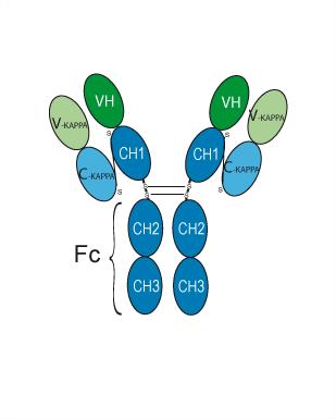
Abazistobart is an immunostimulant and antineoplastic agent targeting the programmed cell death protein 1 (PDCD1). Abazistobart is a chimeric and humanized monoclonal antibody that specifically binds to PDCD1 to block the relevant signaling pathway, thereby activating the immune system and exerting antineoplastic activity. Abazistobart is promising for research of cancers.
Nos produits utilisent uniquement pour la recherche. Nous ne vendons pas aux patients.
- Pureté : 96.045%
- CAS No.: 3002062-67-8
- Masse moléculaire:144.063 kDa
-
Stockage:
Please store the product under the recommended conditions in the Certificate of Analysis.


To place orders, for customer services and technical support, please contact: MedChemExpress USA
Tel: 609-228-6898 E-mail: [email protected] [email protected]


-
 Activité biologique
-
Chemical Information
- Protocole
- Pureté et documentation
- Références
-
 Help & FAQs




Activité biologique
Description
Isotype
Human IgG4 kappa
Recommend Isotype Controls
Species Reactivity
Human
IC50 & Target
PDCD1/PD-1/CD279
Gene ID
Accession
Conjugated
Unconjugated
Reconsititution
The product can be reconstituted/diluted with sterile PBS or saline.
Format
-
IgG4-kappa
Application
ELISA, FACS, Functional assay
Verified Bioactivity
-
 Immobilized PD-1 Protein, Human (147a.a, HEK293, His, HY-P70766) can bind Abazistobart. The EC50 for this effect is 126.9 ng/mL.


Chemical Information
-
CAS No. 3002062-67-8
-
Appearance Liquid
-
Masse moléculaire 144.063 kDa
-
Color Colorless to light yellow
-
SMILES
[Abazistobart]
-
Synonyms
609A
-
Livraison
Shipping with dry ice.
-
Formulation
Please refer to the lot-specific COA for specific buffer information.
-
Stockage
Please store the product under the recommended conditions in the Certificate of Analysis.


Pureté et documentation
-
Fiche technique (255 KB)
-
SDS (251 KB)
- English - EN (251 KB)
- Français - FR (251 KB)
- Deutsch - DE (251 KB)
- Norwegian - NO (251 KB)
- Español - ES (251 KB)
- Swedish - SV (251 KB)
- Italian - IT (251 KB)
- Korean - KR (251 KB)
- Portuguese - PT (251 KB)
-
Inhibitory Antibodies User Guide (603 KB)
Références

Calculators
Concentration (start) × Volume (start) = Concentration (final) × Volume (final)


Abazistobart Related Classifications
HY-P991113 Related Classifications
 Powered by Bioz