



Anti-Mouse/Rat/Human CD47/IAP Antibody (MIAP410)

Based on 1 Customer Validation
Anti-Mouse/Rat/Human CD47/IAP Antibody (MIAP410) is an anti-mouse/rat/human CD47/IAP IgG1 monoclonal antibody. Anti-Mouse/Rat/Human CD47/IAP Antibody (MIAP410) can effectively block CD47 signaling and enhance macrophage phagocytic function. Anti-Mouse/Rat/Human CD47/IAP Antibody (MIAP410) can increase the infiltration of immune cells. Anti-Mouse/Rat/Human CD47/IAP Antibody (MIAP410) may interfere with wound healing. Anti-Mouse/Rat/Human CD47/IAP Antibody (MIAP410) can be used for researches on cancer and inflammation conditions such as breast cancer and intestinal mucosal repair.
For research use only. We do not sell to patients.
- Purity : 99.57%
- Molecular Weight:150 kDa
-
Storage:
Please store the product under the recommended conditions in the Certificate of Analysis.

To place orders, for customer services and technical support, please contact: MedChemExpress USA
Tel: 609-228-6898 E-mail: [email protected] [email protected]


-
 Biological Activity
-
Chemical Information
- Protocols
- Purity & Documentation
- References
-
 Help & FAQs






Biological Activity
Description
Isotype
Mouse IgG1 kappa
Recommend Isotype Controls
Species Reactivity
Human/Mouse/Rat
IC50 & Target
CD47/IAP
In Vitro
Anti-Mouse/Rat/Human CD47/IAP Antibody (MIAP410) (10 μg/mL, 24 h) significantly inhibits epithelial cell migration and wound closure[1].
Anti-Mouse/Rat/Human CD47/IAP Antibody (MIAP410) (10-20 μg/mL, 2-4 h) significantly enhances the phagocytic activity of macrophages towards tumor cells[2].
MedChemExpress (MCE) has not independently confirmed the accuracy of these methods. They are for reference only. Further protocols information, click here.
-
Cell Line:Epithelial cells
-
Concentration:10 μg/mL
-
Incubation Time:24 h
-
Result:Significant delayed in cell migration and wound closure.
In Vivo
Anti-Mouse/Rat/Human CD47/IAP Antibody (MIAP410) (400 μg, intratumorally injection, once every 2 days, for 30 days) significantly inhibits tumor growth in FVB mice bearing MT1A2 tumors[2].
MedChemExpress (MCE) has not independently confirmed the accuracy of these methods. They are for reference only.
-
Animal Model:Male C57Bl/6J mice with a biopsy-based mucosal wound (8-12 weeks)[1]
-
Dosage:400 μg
-
Administration:Intraperitoneal injection (i.p.), single dose, 24 hours after the wound
-
Result:Caused delayed wound closure.
-
Animal Model:5×10⁴ MT1A2 cells injected FVB mice (6-8 weeks)[2]
-
Dosage:400 μg
-
Administration:Intratumorally injection, once every 2 days, for 30 days
-
Result:Significantly reduced tumor weight.
Caused a large infiltration of inflammatory cells around the tumor.
Did not cause significant toxicity.
Gene ID
Accession
Q61735-1
Conjugated
Unconjugated
Reconsititution
The product can be reconstituted/diluted with sterile PBS or saline.
Application
ELISA, FACS, Functional assay, Research in vivo
Verified Bioactivity
-
 Immobilized CD47 Protein, Mouse (HEK293, solution, HY-P74289) can bind Anti-Mouse/Rat/Human CD47/IAP Antibody (MIAP410). The EC50 for this effect is 179.7 ng/mL.
 -
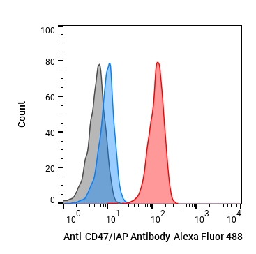 Flow cytometric analysis of 106 Jurkat cells with Anti-Mouse/Rat/Human CD47/IAP Antibody (MIAP410) (HY-P990132, red). Cells were fixed with 4% paraformaldehyde. Then stained with the primary antibody at 1/200 dilution for an hour at 4℃. Alexa Fluor® 488-conjugated AffiniPure Goat Anti-Mouse IgG H&L (HY-P8005) was used as the secondary antibody at 1/1,000 dilution for 30 minutes at 4℃. Mouse IgG1 kappa (HY-P99977, blue) was used as the isotype control, cells without incubation with primary antibody were used as the unlabeled control (black).


Chemical Information
-
Appearance Liquid
-
Molecular Weight 150 kDa
-
Color Colorless to light yellow
-
SMILES
[Anti-Mouse/Rat/Human CD47/IAP Antibody (MIAP410)]
-
Formulation
Please refer to the lot-specific COA for specific buffer information.
-
Storage
Please store the product under the recommended conditions in the Certificate of Analysis.

Protocols
-
Phagocytosis Functional Assay
A phagocytosis functional assay measures the ability of phagocytic cells, such as neutrophils, macrophages, monocytes, or microglia/macrophages, to bind and internalize particulate targets including bacteria, yeast particles, beads, or myelin particles. Fluorescent flow-cytometry assays detect target uptake as fluorescence associated with gated phagocytes, while pH-sensitive dyes such as pHrodo increase signal in acidic phagosomal compartments and therefore preferentially report internalized particles rather than particles remaining outside the cell. Microscopy or high-content imaging can be used to confirm intracellular localization and, in some protocols, to follow uptake kinetics.
-
Cell migration
Cell migration is a method that plays an important role in wound healing, cell differentiation, embryonic development, etc.
-
Research Protocol for Inflammation-related Diseases
The NLRP3 inflammasome is a cytosolic innate immune signaling platform that integrates priming signals and danger-signal activation to promote caspase-1 activation, maturation of IL-1β and IL-18, and gasdermin D-mediated pyroptotic cell death. The core experimental logic is to determine whether inflammatory disease phenotypes are driven by increased NLRP3 expression, ASC-containing inflammasome assembly, caspase-1 cleavage, GSDMD cleavage, and extracellular release of IL-1β/IL-18 rather than by nonspecific cell injury alone. The pathway is strongly linked to inflammation-related disease phenotypes because monosodium urate crystals activate NALP3/NLRP3 inflammasome signaling in gout-like crystal inflammation, cholesterol crystals activate NLRP3 inflammasomes in atherogenesis models, and DSS-induced intestinal inflammation has been reported to involve NLRP3 inflammasome activity. However, experimental colitis studies also show context-dependent protective effects of NLRP3 inflammasome co
-
Cell-Exclusion Zone Migration Assay
The Cell-Exclusion Zone (CEZ) migration assay is an in vitro 2D cell migration method in which a defined cell-free area is created using removable physical barriers such as silicone stoppers, allowing cells to be seeded around the barrier and subsequently migrate into the cleared zone after barrier removal. This approach enables quantification of collective cell migration by monitoring repopulation of the initially cell-free region over time using microscopy-based imaging. Compared with scratch-based wound healing assays, barrier-based exclusion methods are designed to avoid mechanical damage to the extracellular matrix and reduce injury-induced effects on boundary cells, thereby improving interpretability of migration behavior in vitro. The assay readout is typically the progressive reduction in the cell-free area or the number of cells invading the exclusion zone, reflecting coordinated cell motility relevant to physiological processes such as wound healing, epithelial repair, and ca
-
Breast Cancer Modeling
Breast cancer is a heterogeneous cancer, and it has been distinguished into four subtypes: luminal A, luminal B, HER2-positive and basal-like. Molecular mutations, epigenetic alterations, hormone exposure and immune microenvironment are related to the progression of breast cancer.


Purity & Documentation
-
Data Sheet (261 KB)
-
SDS (251 KB)
- English - EN (251 KB)
- Français - FR (251 KB)
- Deutsch - DE (251 KB)
- Norwegian - NO (251 KB)
- Español - ES (251 KB)
- Swedish - SV (251 KB)
- Italian - IT (251 KB)
- Korean - KR (251 KB)
- Portuguese - PT (251 KB)
-
Inhibitory Antibodies User Guide (603 KB)
References
[1]. Reed M, et al. Epithelial CD47 is critical for mucosal repair in the murine intestine in vivo. Nat Commun. 2019 Nov 1;10(1):5004. [Content Brief]
[2]. Willingham SB, et al. The CD47-signal regulatory protein alpha (SIRPa) interaction is a therapeutic target for human solid tumors. Proc Natl Acad Sci U S A. 2012 Apr 24;109(17):6662-7. [Content Brief]

Calculators
Concentration (start) × Volume (start) = Concentration (final) × Volume (final)


Anti-Mouse/Rat/Human CD47/IAP Antibody (MIAP410) Related Classifications
HY-P990132 Related Classifications
 Powered by Bioz