



Phospho-PKC delta (Ser299) Antibody (YA2628)
(Synonyms: Protein kinase C delta type; Tyrosine-protein kinase PRKCD; nPKC-delta)
Based on 1 Customer Validation
Phospho-PKC delta (Ser299) Antibody (YA2628) is a Rabbit-derived and non-conjugated IgG monoclonal antibody, targeting to Phospho-PKC delta (Ser299).
-
Host:
Rabbit
-
Isotype:
IgG
-
Application:
WB, IHC-P
-
Reactivity :
Human
-
Formulation:
Supplied in 50mM Tris-Glycine(pH 7.4), 0.15M NaCl, 40% Glycerol, 0.01% Sodium azide and 0.05% BSA
-
Conjugation:
Non-conjugated


To place orders, for customer services and technical support, please contact: MedChemExpress USA
Tel: 609-228-6898 E-mail: [email protected] [email protected]





Applications
| Application |
WB

WB: Western Blot
|
IHC-P
IHC-P: Immunohistochemistry-Paraffin
|
|---|---|---|
| Dilution Ratio | 1:1000-1:5000 | 1:1000-1:5000 |


Product Details
Phospho-PKC delta (Ser299) Antibody (YA2628) is a Rabbit-derived and non-conjugated IgG monoclonal antibody, targeting to Phospho-PKC delta (Ser299).
-
Host Rabbit

-
Clonality Recombinant,Monoclonal

-
Species ReactivityHuman
-
Observed Molecular WeightObserved band size: 78 kDa
Note: Due to possible protein modifications or aggregation, the molecular weight should be confirmed by actual measurement, and the predicted value is for reference only.

-
Calculated Molecular Weight Predicted band size: 78 kDa

A synthetic phosphopeptide corresponding to residues surrounding Ser299 of human PKC delta
Endogenous
Affinity Purified
Non-conjugated
Phosphorylated
IgG


Product Properties
-
Appearance
Solution
-
Formulation
Supplied in 50mM Tris-Glycine(pH 7.4), 0.15M NaCl, 40% Glycerol, 0.01% Sodium azide and 0.05% BSA
-
Concentration
Batch-dependent, Please check the COA for the concentration of each lot. Check Lot Concentration
-
Storage & Stability
Stored at -20°C for 1 year. Avoid repeated freeze / thaw cycles.
-
Shipping
Shipping with blue ice.


Verification Images
-
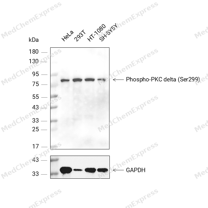 Western blot analysis was performed on protein extracts (25 μg) from HeLa (lane 2), 293T (lane 3), HT-1080 (lane 4), and SH-SY5Y (lane 5) using Phospho-PKC delta (Ser299) antibody. Proteins were transferred onto a 0.45 μm PVDF membrane using the Trans-Blot® Turbo™ system for 13 min. The membrane was then blocked with 5% nonfat milk in TBST (HY-K1025) for 1 h at room temperature. The primary antibody (1:1000) and loading control antibody GAPDH Antibody (HRP) (HY-P80954A) (1:5000) were diluted in 5% nonfat milk in TBST and incubated with the membrane overnight at 4°C. After washing, the membrane of primary antibody was incubated with HRP-conjugated goat anti-rabbit/mouse IgG secondary antibody (HY-P8001/HY-P8004) (1:5000) diluted in 5% nonfat milk in TBST for 1 h at room temperature. Protein bands were visualized using an Ultra High Sensitivity ECL detection kit (HY-K1005).


Background
-
Function
PKC delta is a Calcium-independent, phospholipid- and diacylglycerol (DAG)-dependent serine/threonine-protein kinase that plays contrasting roles in cell death and cell survival by functioning as a pro-apoptotic protein during DNA damage-induced apoptosis, but acting as an anti-apoptotic protein during cytokine receptor-initiated cell death, is involved in tumor suppression as well as survival of several cancers, is required for oxygen radical production by NADPH oxidase and acts as positive or negative regulator in platelet functional responses. Negatively regulates B cell proliferation and also has an important function in self-antigen induced B cell tolerance induction. Upon DNA damage, activates the promoter of the death-promoting transcription factor BCLAF1/Btf to trigger BCLAF1-mediated p53/TP53 gene transcription and apoptosis. In response to oxidative stress, interact with and activate CHUK/IKKA in the nucleus, causing the phosphorylation of p53/TP53. In the case of ER stress or DNA damage-induced apoptosis, can form a complex with the tyrosine-protein kinase ABL1 which trigger apoptosis independently of p53/TP53. In cytosol can trigger apoptosis by activating MAPK11 or MAPK14, inhibiting AKT1 and decreasing the level of X-linked inhibitor of apoptosis protein (XIAP), whereas in nucleus induces apoptosis via the activation of MAPK8 or MAPK9. Upon ionizing radiation treatment, is required for the activation of the apoptosis regulators BAX and BAK, which trigger the mitochondrial cell death pathway. Can phosphorylate MCL1 and target it for degradation which is sufficient to trigger for BAX activation and apoptosis. Is required for the control of cell cycle progression both at G1/S and G2/M phases. Mediates phorbol 12-myristate 13-acetate (PMA)-induced inhibition of cell cycle progression at G1/S phase by up-regulating the CDK inhibitor CDKN1A/p21 and inhibiting the cyclin CCNA2 promoter activity. In response to UV irradiation can phosphorylate CDK1, which is important for the G2/M DNA damage checkpoint activation. Can protect glioma cells from the apoptosis induced by TNFSF10/TRAIL, probably by inducing increased phosphorylation and subsequent activation of AKT1. Is highly expressed in a number of cancer cells and promotes cell survival and resistance against chemotherapeutic drugs by inducing cyclin D1 (CCND1) and hyperphosphorylation of RB1, and via several pro-survival pathways, including NF-kappa-B, AKT1 and MAPK1/3 (ERK1/2). Involved in antifungal immunity by mediating phosphorylation and activation of CARD9 downstream of C-type lectin receptors activation, promoting interaction between CARD9 and BCL10, followed by activation of NF-kappa-B and MAP kinase p38 pathways. Can also act as tumor suppressor upon mitogenic stimulation with PMA or TPA. In N-formyl-methionyl-leucyl-phenylalanine (fMLP)-treated cells, is required for NCF1 (p47-phox) phosphorylation and activation of NADPH oxidase activity, and regulates TNF-elicited superoxide anion production in neutrophils, by direct phosphorylation and activation of NCF1 or indirectly through MAPK1/3 (ERK1/2) signaling pathways. May also play a role in the regulation of NADPH oxidase activity in eosinophil after stimulation with IL5, leukotriene B4 or PMA. In collagen-induced platelet aggregation, acts a negative regulator of filopodia formation and actin polymerization by interacting with and negatively regulating VASP phosphorylation. Downstream of PAR1, PAR4 and CD36/GP4 receptors, regulates differentially platelet dense granule secretion; acts as a positive regulator in PAR-mediated granule secretion, whereas it negatively regulates CD36/GP4-mediated granule release. Phosphorylates MUC1 in the C-terminal and regulates the interaction between MUC1 and beta-catenin. The catalytic subunit phosphorylates 14-3-3 proteins (YWHAB, YWHAZ and YWHAH) in a sphingosine-dependent fashion. Phosphorylates ELAVL1 in response to angiotensin-2 treatment. Phosphorylates mitochondrial phospholipid scramblase 3 (PLSCR3), resulting in increased cardiolipin expression on the mitochondrial outer membrane which facilitates apoptosis. Phosphorylates SMPD1 which induces SMPD1 secretion[1][2][3][4][5][6][7][8][9][10][11].
-
Subcellular Localization
Cytoplasm; Cytoplasm, perinuclear region; Nucleus; Cell membrane; Peripheral membrane protein; Mitochondrion; Endomembrane system
-
Isoforms & Post-Translational Modification
Q05655 has 2 isomers: Q05655-1: 77505 Da (predicted); Q05655-2: 80953 Da (predicted).
Autophosphorylated and/or phosphorylated at Thr-507, within the activation loop; phosphorylation at Thr-507 is not a prerequisite for enzymatic activity (PubMed:19801500). Autophosphorylated at Ser-299, Ser-302 and Ser-304 (PubMed:17603046). Upon TNFSF10/TRAIL treatment, phosphorylated at Tyr-155; phosphorylation is required for its translocation to the endoplasmic reticulum and cleavage by caspase-3 (PubMed:15774464). Phosphorylated at Tyr-313, Tyr-334 and Tyr-567; phosphorylation of Tyr-313 and Tyr-567 following thrombin or zymosan stimulation potentiates its kinase activity (PubMed:17570831). Phosphorylated by protein kinase PDPK1; phosphorylation is inhibited by the apoptotic C-terminal cleavage product of PKN2 (PubMed:11781095). Phosphorylated at Tyr-313 through a SYK and SRC mechanism downstream of C-type lectin receptors activation, promoting its activation (By similarity);Proteolytically cleaved into a catalytic subunit and a regulatory subunit by caspase-3 during apoptosis which results in kinase activation -
Subunit
Interacts with PDPK1 (via N-terminal region) (PubMed:11781095). Interacts with RAD9A (PubMed:12628935). Interacts with CDCP1 (PubMed:15851033). Interacts with MUC1 (PubMed:11877440). Interacts with VASP (PubMed:16940418). Interacts with CAVIN3 (By similarity). Interacts with PRKD2 (via N-terminus and zing-finger domain 1 and 2) in response to oxidative stress; the interaction is independent of PRKD2 tyrosine phosphorylation (PubMed:28428613). Interacts with PLSC3; interaction is enhanced by UV irradiation (PubMed:12649167). Interacts with PRKCH upstream open reading frame 2; the interaction leads to inhibition of kinase activity (PubMed:34593629)
-
SwissProt ID
-
Synonyms
Protein kinase C delta type; Tyrosine-protein kinase PRKCD; nPKC-delta
-
Research Field
Signal Transduction


Documentation
-
Data Sheet (235 KB)
-
SDS (251 KB)
- English - EN (251 KB)
- Français - FR (251 KB)
- Deutsch - DE (251 KB)
- Norwegian - NO (251 KB)
- Español - ES (251 KB)
- Swedish - SV (251 KB)
- Italian - IT (251 KB)
- Korean - KR (251 KB)
- Portuguese - PT (251 KB)
-
User Guide for Antibodies (1077 KB)

 Powered by Bioz
