



PKC beta Antibody (YA9971)
(Synonyms: PKCA, PRKACA, PRKCA, Protein kinase C alpha type, PKC-A, PKC-alpha)PKC beta Antibody (YA9971) is a Rabbit-derived and non-conjugated IgG Monoclonal antibody, targeting to PKC beta Antibody.
-
Host:
Rabbit
-
Isotype:
IgG
-
Application:
WB, IHC-P, ICC/IF
-
Reactivity :
Human, Mouse, Rat
-
Formulation:
Supplied in PBS, containing 50% glycerol, 0.2% BSA and 0.01% sodium azide.
-
Conjugation:
Non-conjugated


To place orders, for customer services and technical support, please contact: MedChemExpress USA
Tel: 609-228-6898 E-mail: [email protected] [email protected]





Applications
| Application |
WB

WB: Western Blot
|
IHC-P
IHC-P: Immunohistochemistry-Paraffin
|
ICC/IF
ICC/IF: Immunocytochemistry/
Immunofluorescence |
|---|---|---|---|
| Dilution Ratio | 1:1000-2000 | 1:100-200 | 1:50-100 |


Product Details
PKC beta Antibody (YA9971) is a Rabbit-derived and non-conjugated IgG Monoclonal antibody, targeting to PKC beta Antibody.
-
Host Rabbit

-
Clonality Monoclonal

-
Species ReactivityHuman, Mouse, Rat
-
Observed Molecular WeightObserved band size: 77 kDa
Note: Due to possible protein modifications or aggregation, the molecular weight should be confirmed by actual measurement, and the predicted value is for reference only.

-
Calculated Molecular Weight Predicted band size: 77 kDa

Synthetic peptide corresponding to human PKC beta
Endogenous
affinity purified
Non-conjugated
Unmodified
IgG


Product Properties
-
Appearance
Solution
-
Formulation
Supplied in PBS, containing 50% glycerol, 0.2% BSA and 0.01% sodium azide.
-
Storage & Stability
Stored at -20°C for 1 year. Avoid repeated freeze / thaw cycles.
-
Shipping
Shipping with blue ice.


Verification Images
-
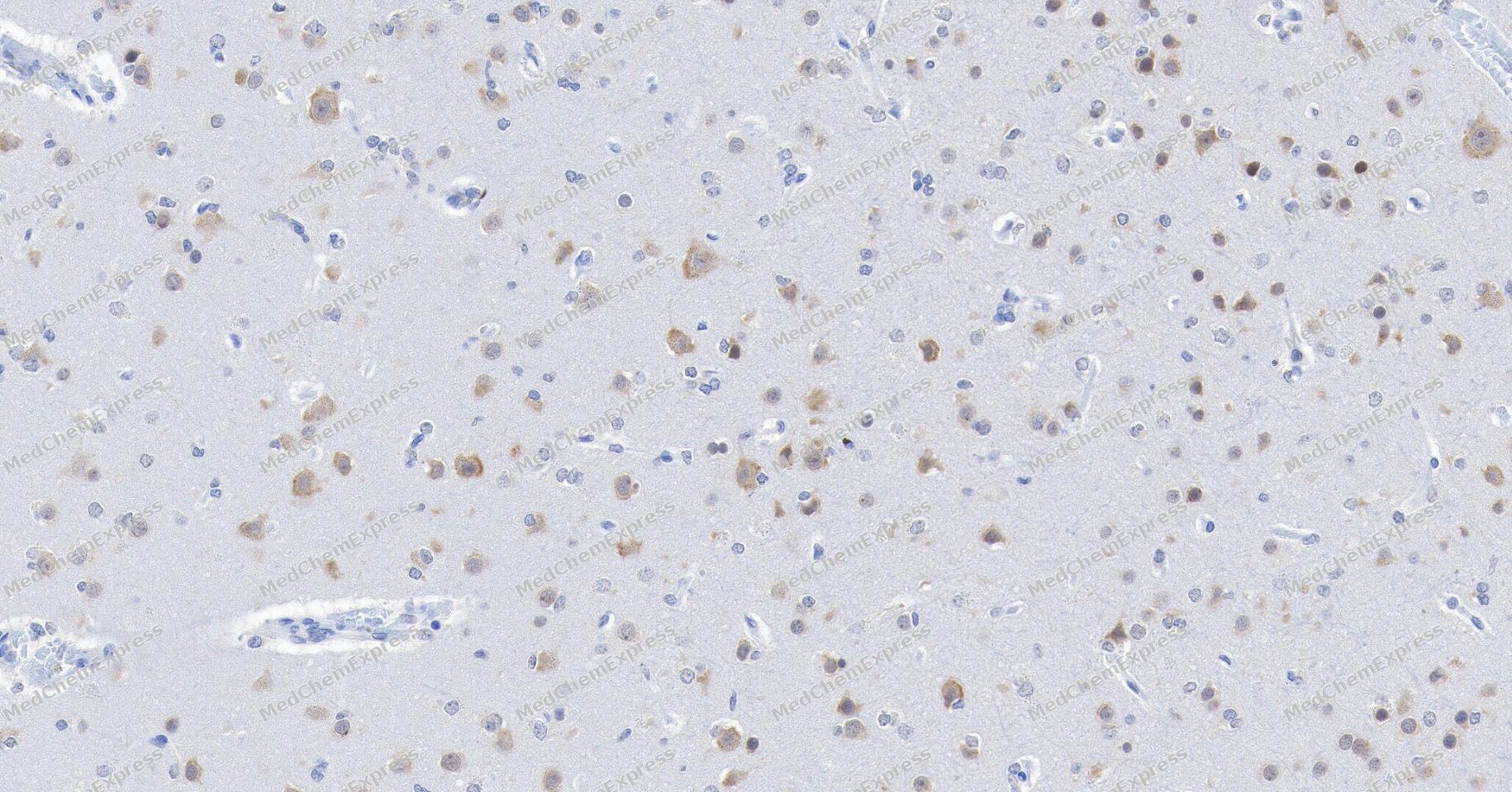 Immunohistochemical analysis of paraffin-embedded human brain tissue using PKC beta Antibody. The section was pre-treated using heat mediated antigen retrieval with Tris/EDTA buffer (pH 9.0) for 20 minutes. The tissues were probed with the primary antibody (HY-P810727, 1/200) overnight at 4℃. The detection was performed using an HRP conjugated compact polymer system (HY-P83652). DAB was used as the chromogen. Tissues were counterstained with hematoxylin and mounted with neutral balsam.
 -
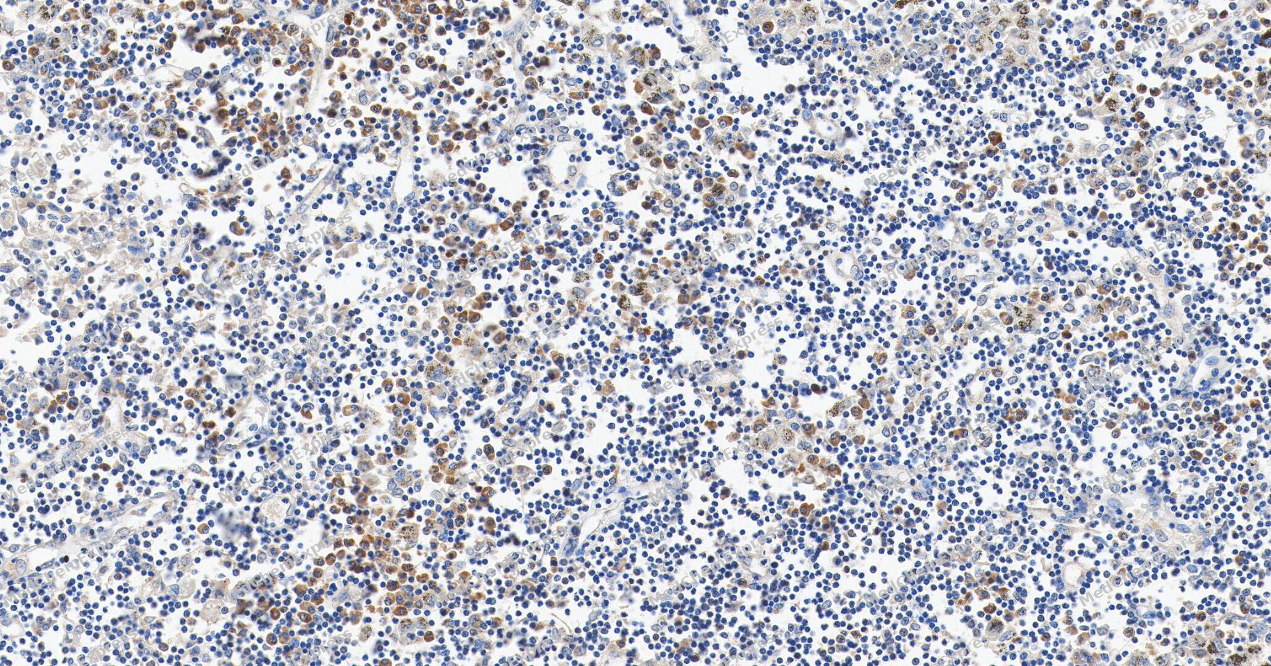 Immunohistochemical analysis of paraffin-embedded human pulmonary hilum lymph node tissue using PKC beta Antibody. The section was pre-treated using heat mediated antigen retrieval with Tris/EDTA buffer (pH 9.0) for 20 minutes. The tissues were probed with the primary antibody (HY-P810727, 1/100) overnight at 4℃. The detection was performed using an HRP conjugated compact polymer system (HY-P83652). DAB was used as the chromogen. Tissues were counterstained with hematoxylin and mounted with neutral balsam.
 -
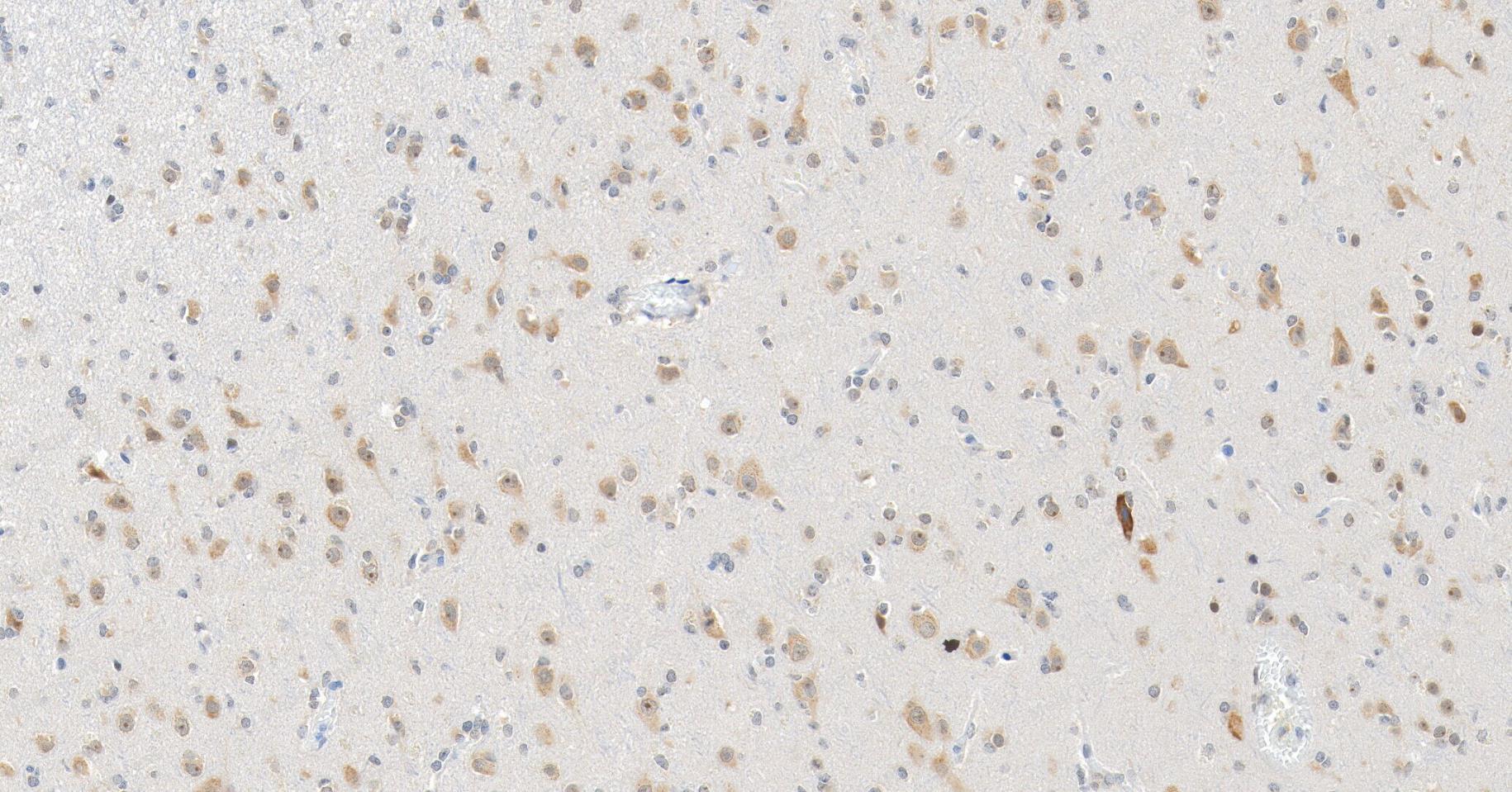 Immunohistochemical analysis of paraffin-embedded human brain tissue using PKC beta Antibody. The section was pre-treated using heat mediated antigen retrieval with Tris/EDTA buffer (pH 9.0) for 20 minutes. The tissues were probed with the primary antibody (HY-P810727, 1/100) overnight at 4℃. The detection was performed using an HRP conjugated compact polymer system (HY-P83652). DAB was used as the chromogen. Tissues were counterstained with hematoxylin and mounted with neutral balsam.
 -
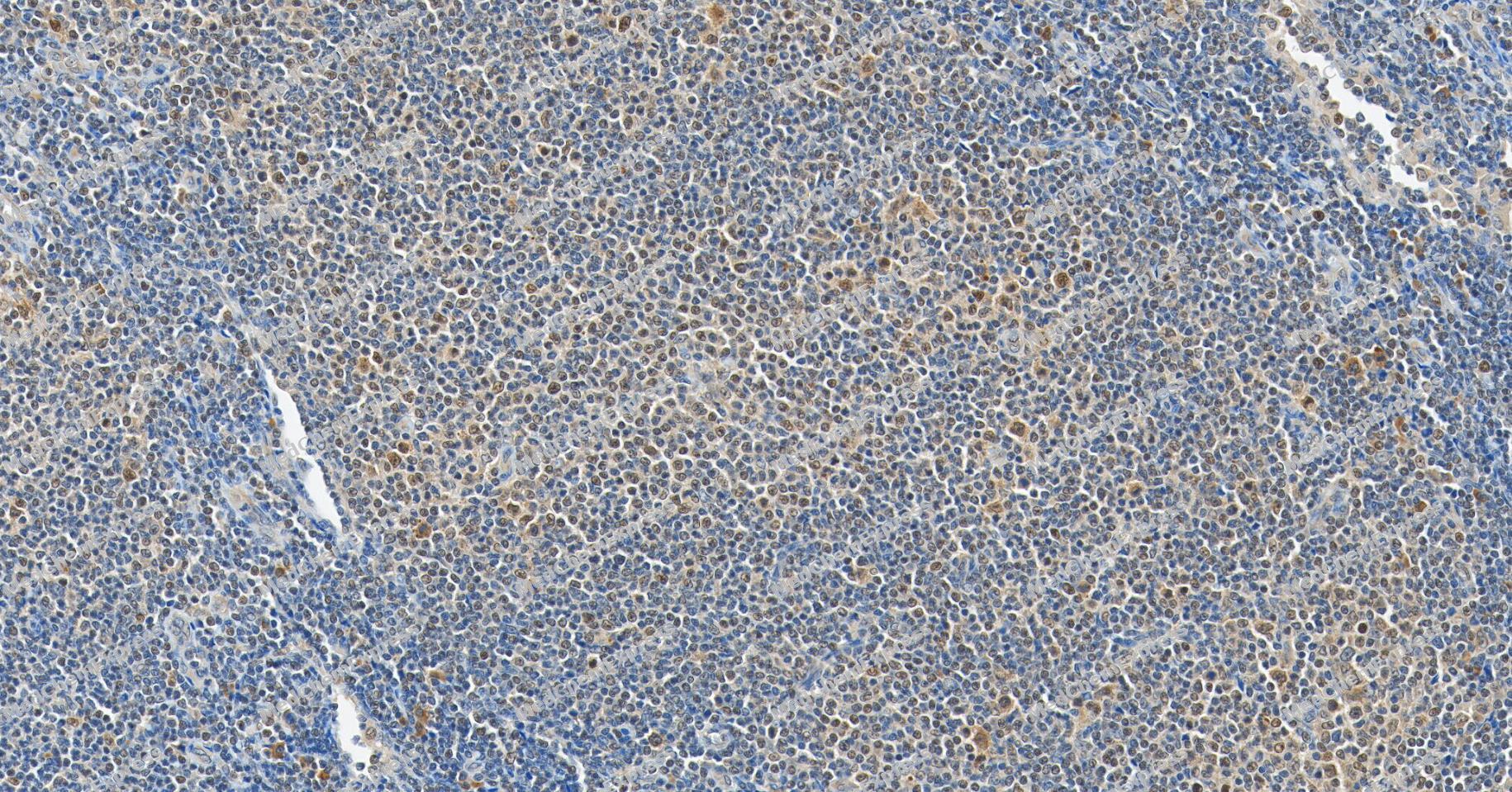 Immunohistochemical analysis of paraffin-embedded human follicular lymphoma tissue using PKC beta Antibody. The section was pre-treated using heat mediated antigen retrieval with Tris/EDTA buffer (pH 9.0) for 20 minutes. The tissues were probed with the primary antibody (HY-P810727, 1/100) overnight at 4℃. The detection was performed using an HRP conjugated compact polymer system (HY-P83652). DAB was used as the chromogen. Tissues were counterstained with hematoxylin and mounted with neutral balsam.
 -
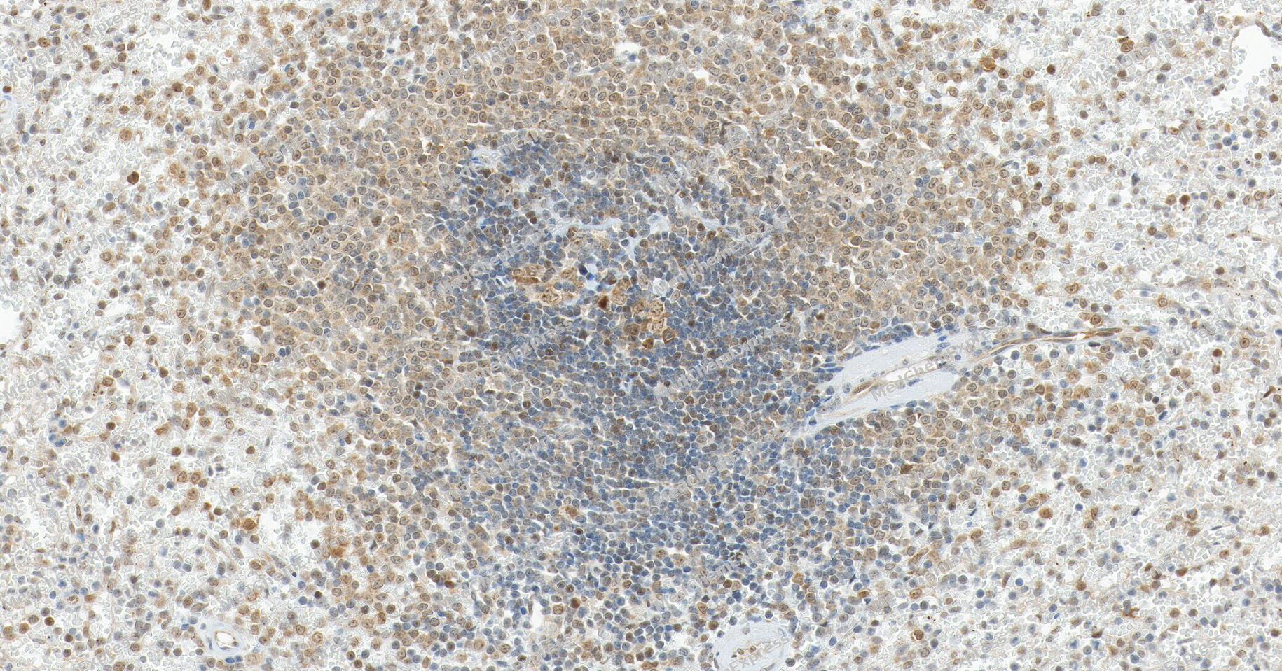 Immunohistochemical analysis of paraffin-embedded human spleen tissue using PKC beta Antibody. The section was pre-treated using heat mediated antigen retrieval with Tris/EDTA buffer (pH 9.0) for 20 minutes. The tissues were probed with the primary antibody (HY-P810727, 1/100) overnight at 4℃. The detection was performed using an HRP conjugated compact polymer system (HY-P83652). DAB was used as the chromogen. Tissues were counterstained with hematoxylin and mounted with neutral balsam.
 -
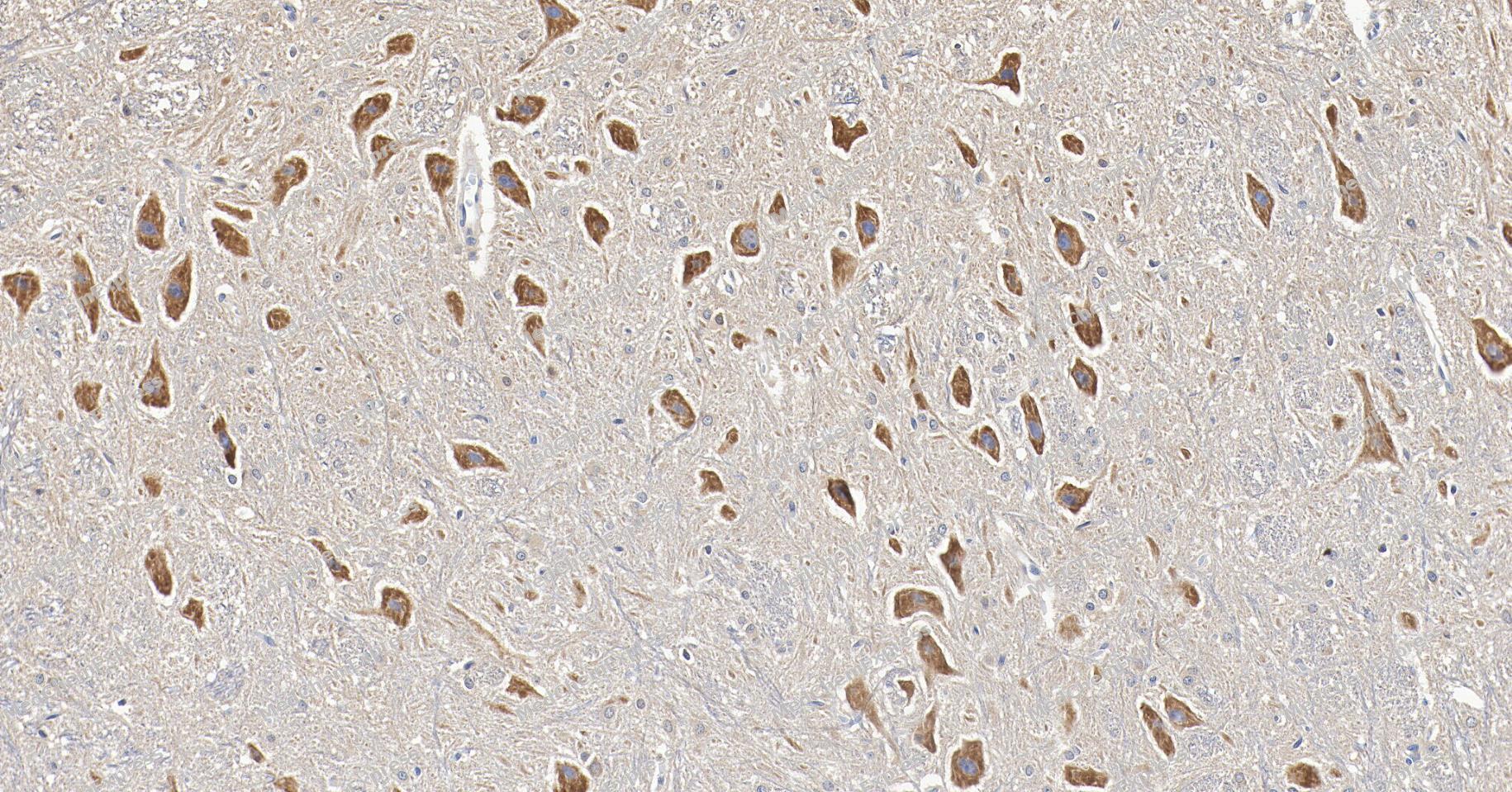 Immunohistochemical analysis of paraffin-embedded rat brain tissue using PKC beta Antibody. The section was pre-treated using heat mediated antigen retrieval with Tris/EDTA buffer (pH 9.0) for 20 minutes. The tissues were probed with the primary antibody (HY-P810727, 1/100) overnight at 4℃. The detection was performed using an HRP conjugated compact polymer system (HY-P83652). DAB was used as the chromogen. Tissues were counterstained with hematoxylin and mounted with neutral balsam.
 -
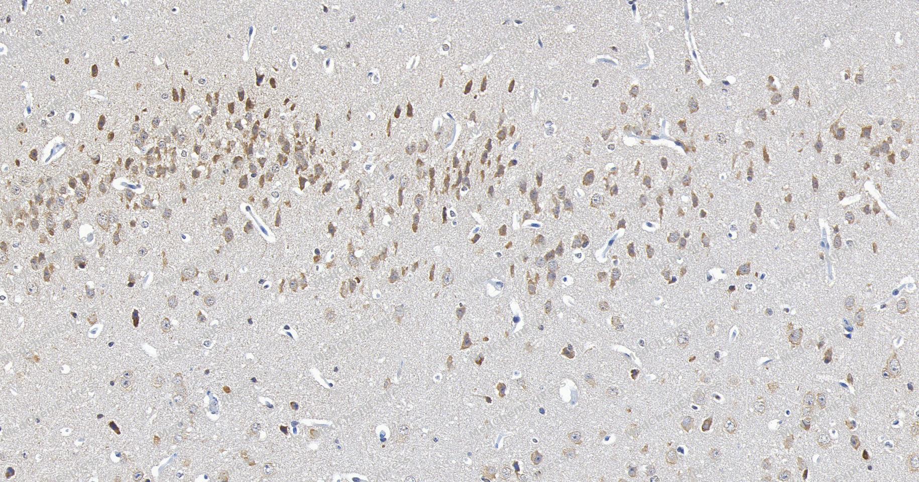 Immunohistochemical analysis of paraffin-embedded mouse brain tissue using PKC beta Antibody. The section was pre-treated using heat mediated antigen retrieval with Tris/EDTA buffer (pH 9.0) for 20 minutes. The tissues were probed with the primary antibody (HY-P810727, 1/100) overnight at 4℃. The detection was performed using an HRP conjugated compact polymer system (HY-P83652). DAB was used as the chromogen. Tissues were counterstained with hematoxylin and mounted with neutral balsam.


Background
-
Function
PKC beta is a Calcium-activated, phospholipid- and diacylglycerol (DAG)-dependent serine/threonine-protein kinase involved in various cellular processes such as regulation of the B-cell receptor (BCR) signalosome, oxidative stress-induced apoptosis, androgen receptor-dependent transcription regulation, insulin signaling and endothelial cells proliferation. Plays a key role in B-cell activation by regulating BCR-induced NF-kappa-B activation. Mediates the activation of the canonical NF-kappa-B pathway (NFKB1) by direct phosphorylation of CARD11/CARMA1 at 'Ser-559', 'Ser-644' and 'Ser-652'. Phosphorylation induces CARD11/CARMA1 association with lipid rafts and recruitment of the BCL10-MALT1 complex as well as MAP3K7/TAK1, which then activates IKK complex, resulting in nuclear translocation and activation of NFKB1. Plays a direct role in the negative feedback regulation of the BCR signaling, by down-modulating BTK function via direct phosphorylation of BTK at 'Ser-180', which results in the alteration of BTK plasma membrane localization and in turn inhibition of BTK activity. Involved in apoptosis following oxidative damage: in case of oxidative conditions, specifically phosphorylates 'Ser-36' of isoform p66Shc of SHC1, leading to mitochondrial accumulation of p66Shc, where p66Shc acts as a reactive oxygen species producer. Acts as a coactivator of androgen receptor (AR)-dependent transcription, by being recruited to AR target genes and specifically mediating phosphorylation of 'Thr-6' of histone H3 (H3T6ph), a specific tag for epigenetic transcriptional activation that prevents demethylation of histone H3 'Lys-4' (H3K4me) by LSD1/KDM1A. In insulin signaling, may function downstream of IRS1 in muscle cells and mediate insulin-dependent DNA synthesis through the RAF1-MAPK/ERK signaling cascade. Participates in the regulation of glucose transport in adipocytes by negatively modulating the insulin-stimulated translocation of the glucose transporter SLC2A4/GLUT4. Phosphorylates SLC2A1/GLUT1, promoting glucose uptake by SLC2A1/GLUT1. Under high glucose in pancreatic beta-cells, is probably involved in the inhibition of the insulin gene transcription, via regulation of MYC expression. In endothelial cells, activation of PRKCB induces increased phosphorylation of RB1, increased VEGFA-induced cell proliferation, and inhibits PI3K/AKT-dependent nitric oxide synthase (NOS3/eNOS) regulation by insulin, which causes endothelial dysfunction. Also involved in triglyceride homeostasis (By similarity). Phosphorylates ATF2 which promotes cooperation between ATF2 and JUN, activating transcription. Phosphorylates KLHL3 in response to angiotensin II signaling, decreasing the interaction between KLHL3 and WNK4. Phosphorylates and activates LRRK1, which phosphorylates RAB proteins involved in intracellular trafficking[1][2][3][4][5][6].
-
Subcellular Localization
Cytoplasm; Nucleus; Membrane
-
Isoforms & Post-Translational Modification
PKC beta has 2 isoforms, P05771-1: amino acid length is 671, molecular weight is 76869 Da (predicted); P05771-2: amino acid length is 673, molecular weight is 77012 Da (predicted).
Phosphorylation on Thr-500 within the activation loop renders it competent to autophosphorylate. Subsequent autophosphorylation of Thr-642 maintains catalytic competence, and autophosphorylation on Ser-661 appears to release the kinase into the cytosol. Autophosphorylation on other sites i.e. in the N-terminal and hinge regions have no effect on enzyme activity. -
Subunit
Interacts with PDPK1/PDK1 (By similarity).
-
SwissProt ID
-
Synonyms
PKCA, PRKACA, PRKCA, Protein kinase C alpha type, PKC-A, PKC-alpha


Documentation
References
[1]. Kang SW, et al. PKCbeta modulates antigen receptor signaling via regulation of Btk membrane localization. EMBO J. 2001 Oct 15;20(20):5692-702. [Content Brief]
[2]. Metzger E, et al. Phosphorylation of histone H3T6 by PKCbeta(I) controls demethylation at histone H3K4. Nature. 2010 Apr 1;464(7289):792-6. [Content Brief]
[3]. Lee EE, et al. A Protein Kinase C Phosphorylation Motif in GLUT1 Affects Glucose Transport and is Mutated in GLUT1 Deficiency Syndrome. Mol Cell. 2015 Jun 4;58(5):845-53. [Content Brief]
[4]. Yamasaki T, et al. Phosphorylation of Activation Transcription Factor-2 at Serine 121 by Protein Kinase C Controls c-Jun-mediated Activation of Transcription. J Biol Chem. 2009 Mar 27;284(13):8567-81. [Content Brief]
[5]. Shibata S, et al. Angiotensin II signaling via protein kinase C phosphorylates Kelch-like 3, preventing WNK4 degradation. Proc Natl Acad Sci U S A. 2014 Oct 28;111(43):15556-61. [Content Brief]
[6]. Malik AU, et al. PKC isoforms activate LRRK1 kinase by phosphorylating conserved residues (Ser1064, Ser1074 and Thr1075) within the CORB GTPase domain. Biochem J. 2022 Sep 30;479(18):1941-1965. [Content Brief]

 Powered by Bioz
