



Anticancer agent 109
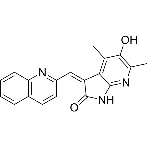
Anticancer agent 109 (compound 6-15) is an inhibitor of the Gas6-Axl axis with anti-cancer activity. Anticancer agent 109 inhibits the expression of Gas6 and Axl, and the expression p-PI3K and p-AKT in cancer cells, leads to G1 phase arrest and promotes cancer cells apoptosis, and inhibits tumor growth significantly in nude mouse tumor bearing models.
For research use only. We do not sell to patients.
- CAS No.: 2097497-16-8
- Formula: C19H15N3O2
- Molecular Weight:317.34
-
Storage:
Please store the product under the recommended conditions in the Certificate of Analysis.

To place orders, for customer services and technical support, please contact: MedChemExpress USA
Tel: 609-228-6898 E-mail: [email protected] [email protected]


-
 Biological Activity
-
Chemical Information
- Protocols
- Purity & Documentation
- References
-
 Help & FAQs




Biological Activity
Description
IC50 & Target
[1]|
Axl |
Cellular Effect
|
Cell Line

|
Type | Value | Description | References |
|---|---|---|---|---|
| A549 | IC50 |
4.2 μM
Compound: 6-15
|
Cytotoxicity against human A549 cells assessed as inhibition of cell growth incubated for 48 hrs by MTT assay
Cytotoxicity against human A549 cells assessed as inhibition of cell growth incubated for 48 hrs by MTT assay
|
[PMID: 36921529] |
| CHO-K1 | IC50 |
54.5 μM
Compound: 6-15
|
Cytotoxicity against CHO-K1 cells assessed as inhibition of cell growth incubated for 48 hrs by MTT assay
Cytotoxicity against CHO-K1 cells assessed as inhibition of cell growth incubated for 48 hrs by MTT assay
|
[PMID: 36921529] |
| DU-145 | IC50 |
1.1 μM
Compound: 6-15
|
Cytotoxicity against human DU-145 cells assessed as inhibition of cell growth incubated for 48 hrs by MTT assay
Cytotoxicity against human DU-145 cells assessed as inhibition of cell growth incubated for 48 hrs by MTT assay
|
[PMID: 36921529] |
| HEK293 | IC50 |
26.2 μM
Compound: 6-15
|
Cytotoxicity against HEK293 cells assessed as inhibition of cell growth incubated for 48 hrs by MTT assay
Cytotoxicity against HEK293 cells assessed as inhibition of cell growth incubated for 48 hrs by MTT assay
|
[PMID: 36921529] |
| HT-29 | IC50 |
4.6 μM
Compound: 6-15
|
Cytotoxicity against human HT-29 cells assessed as inhibition of cell growth incubated for 48 hrs by MTT assay
Cytotoxicity against human HT-29 cells assessed as inhibition of cell growth incubated for 48 hrs by MTT assay
|
[PMID: 36921529] |
| MCF7 | IC50 |
2 μM
Compound: 6-15
|
Cytotoxicity against human MCF7 cells assessed as inhibition of cell growth incubated for 48 hrs by MTT assay
Cytotoxicity against human MCF7 cells assessed as inhibition of cell growth incubated for 48 hrs by MTT assay
|
[PMID: 36921529] |
| MDA-MB-231 | IC50 |
2.8 μM
Compound: 6-15
|
Cytotoxicity against human MDA-MB-231 cells assessed as inhibition of cell growth incubated for 48 hrs by MTT assay
Cytotoxicity against human MDA-MB-231 cells assessed as inhibition of cell growth incubated for 48 hrs by MTT assay
|
[PMID: 36921529] |
| PANC-1 | IC50 |
4 μM
Compound: 6-15
|
Cytotoxicity against human PANC-1 cells assessed as inhibition of cell growth incubated for 48 hrs by MTT assay
Cytotoxicity against human PANC-1 cells assessed as inhibition of cell growth incubated for 48 hrs by MTT assay
|
[PMID: 36921529] |
| U-937 | IC50 |
6.7 μM
Compound: 6-15
|
Cytotoxicity against human U-937 cells assessed as inhibition of cell growth incubated for 48 hrs by MTT assay
Cytotoxicity against human U-937 cells assessed as inhibition of cell growth incubated for 48 hrs by MTT assay
|
[PMID: 36921529] |
In Vitro
Anticancer agent 109 (10 μM,48 h) inhibits of Gas6 and Axl in A549, inhibits of Gas6-Axl axis related proteins, increases the sub-G1 fraction and promotes of late stage apoptosis without altering DNA synthesis in PANC-1[1].
MedChemExpress (MCE) has not independently confirmed the accuracy of these methods. They are for reference only.
-
Cell Line:MCF-7; PANC-1; MDA-MB-231 ; HT-29 ; DU145 ; U937 ; A549 ; PANC-1
-
Concentration:30 μM
-
Incubation Time:48 h
-
Result:Inhibited the growth of cancer cells, and was up to 20-fold safer against normal cells and up to 5.4-fold more active than Sunitinib against the cancer cells.
Inhibited growth with IC50s of 2.0 μM (MCF-7); 2.8 μM (MDA-MB-231); 4.6 μM (HT-29); 1.1 μM (DU145); 6.7 μM (U937); 4.2 μM (A549); 4.0 μM (PANC-1).
-
Cell Line:PANC-1
-
Concentration:1 μM , 5 μM , 10 μM
-
Incubation Time:48 h
-
Result:Increased the sub-G1 fraction and induced late apoptosis.
-
Cell Line:A549; PANC-1
-
Concentration:10 μM
-
Incubation Time:48 h
-
Result:Inhibited Gas6 and Axl in A549 and PANC-1 cell, increased the expression ratio of Bax/Bcl-2 and inhibited p-PI3K and p-AKT in PANC-1 cell.
-
Cell Line:PANC-1
-
Concentration:3 μM ;5 μM 10 μM
-
Incubation Time:48 h
-
Result:Inhibited Gas6 and Axl.
In Vivo
MedChemExpress (MCE) has not independently confirmed the accuracy of these methods. They are for reference only.
-
Animal Model:A549 or PANC-1 xenografted in BALB/c-nu mice[1].
-
Dosage:1 mg/kg; 3 mg/kg
-
Administration:Intraperitoneal injection (i.p.) 6 times a week
-
Result:Promoted tumor regression to around a quarter with 1 mg/kg, smaller but not eliminated with 3 mg/kg in A549 models.
Promoted tumor regression to around a quarter with 3 mg/kg in PANC-1 models.
- RNA extraction experimental
- Detection of Apoptosis
- TUNEL staining for apoptotic DNA fragmentation
- Annexin V plus membrane-impermeant dye apoptosis staining
- Patient-Derived Xenograft (PDX)
- Apoptosis Solutions
- Patient-Derived Orthotopic Xenograft (PDOX)
- Subcutaneous Cell-Line-Derived Xenograft
- Orthotopic Cell-Line Xenograft


Chemical Information
-
CAS No. 2097497-16-8
-
Molecular Weight 317.34
-
Formula C19H15N3O2
-
SMILES
O=C(/C(C1=C2C)=C\C3=NC4=C(C=CC=C4)C=C3)NC1=NC(C)=C2O
-
Shipping
Room temperature in continental US; may vary elsewhere.
-
Storage
Please store the product under the recommended conditions in the Certificate of Analysis.


Purity & Documentation
References

Calculators
Concentration (start) × Volume (start) = Concentration (final) × Volume (final)


Anticancer agent 109 Related Classifications
HY-149092 Related Classifications
 Powered by Bioz