



Rhod-2 AM (solution)

Based on 1 Customer Validation
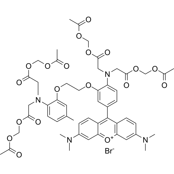
Rhod-2 (solution) is a high-affinity visible light excitation wavelength Ca2+ fluorescent probe, Rhod-2, AM is an acetyl methyl ester derivative of Rhod-2, which has cell membrane permeability and can easily enter cells with simple culture. Once it enters the cell, it is sheared by its lactesterase to produce Rhod-2 without membrane permeability, which remains in the cell to perform the corresponding physiological functions. Maximum excitation/emission wavelength: 549/578 nm.
Solvent and concentration: DMSO: 1 mM
Nur für Forschungszwecke. Wir verkaufen nicht an Patienten.
- CAS. Nr.: 145037-81-6
- Formel: C52H59BrN4O19
- Molecular Weight:1123.96
-
Speicherung:
Please store the product under the recommended conditions in the Certificate of Analysis.


To place orders, for customer services and technical support, please contact: MedChemExpress USA
Tel: 609-228-6898 E-mail: [email protected] [email protected]


-
 Biologische Aktivität
-
Chemical Information
- Protokoll
- Reinheit & Dokumentation
- Verweise
-
 Help & FAQs


-
HY-W009724
2-Aminoethyl diphenylborinateIP3R Inhibitor -
HY-D0989
Rhod-2 AMCa(2+) Indicator -
HY-D0083
DiIFluorescent Dye -
HY-125623
MitoPerOxFluorescent Probe -

HY-DY1073
MitoPerOx (solution)Fluorescent Probe -
HY-101891
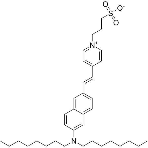
Di-8-ANEPPSVoltage-Sensitive Fluorescent Dye -

HY-DY1012
DiI (solution)Fluorescent Dye


Biologische Aktivität
Beschreibung
Solvent and concentration: DMSO: 1 mM
In Vitro
Guidelines (Following is our recommended protocol. This protocol only provides a guideline, and should be modified according to your specific needs).
1. Preparation of Rhod-2 AM working solution
Dilute the stock solution in serum-free cell culture medium or PBS. The corresponding stock solution can be diluted according to the actual situation. Note that if the solvent is DMSO, the cytotoxicity of DMSO must be considered, and a solvent control should be prepared; if the solvent is pure water, the working solution needs to be filtered and sterilized before adding cells.
Note: Please adjust the concentration of Rhod-2 AM working solution according to the actual situation.
2. Cell staining (Suspension cells)
2.1 Centrifuge at 1000 g at 4°C for 3-5 minutes and then discard the supernatant. Wash twice with PBS, 5 minutes each time. The cell density is 1×10⁶/mL.
2.2 Add 1 mL of working solution, and then incubate at room temperature for 5-30 minutes.
2.3 Centrifuge at 400 g at 4°C for 3-4 minutes and then discard the supernatant.
2.4 Wash twice with PBS, 5 minutes each time.
2.5 Resuspend cells with serum-free cell culture medium or PBS. Observation by fluorescence microscopy or flow cytometry.
3. Cell staining (Adherent cells)
3.1 Culture adherent cells on sterile coverslips.
3.2 Remove the coverslip from the medium and aspirate excess medium.
3.3 Add 100 μL of working solution, gently shake it to completely cover the cells, and then incubate at room temperature for 5-30 minutes.
3.4 Wash twice with medium, 5 minutes each time. Observation by fluorescence microscopy or flow cytometry.
MedChemExpress (MCE) has not independently confirmed the accuracy of these methods. They are for reference only.


Chemical Information
-
CAS. Nr. 145037-81-6
-
Appearance Liquid
-
Molecular Weight 1123.96
-
Formel C52H59BrN4O19
-
Color Purple to purplish red
-
SMILES
CN(C1=CC2=[O+]C3=C(C=CC(N(C)C)=C3)C(C4=CC=C(N(CC(OCOC(C)=O)=O)CC(OCOC(C)=O)=O)C(OCCOC5=CC(C)=CC=C5N(CC(OCOC(C)=O)=O)CC(OCOC(C)=O)=O)=C4)=C2C=C1)C.[Br-]
-
Versand
Room temperature in continental US; may vary elsewhere.
-
Speicherung
Please store the product under the recommended conditions in the Certificate of Analysis.


Reinheit & Dokumentation
-
Data Sheet (277 KB)
-
SDS (251 KB)
- English - EN (251 KB)
- Français - FR (251 KB)
- Deutsch - DE (251 KB)
- Norwegian - NO (251 KB)
- Español - ES (251 KB)
- Swedish - SV (251 KB)
- Italian - IT (251 KB)
- Korean - KR (251 KB)
- Portuguese - PT (251 KB)
-
Handling Instructions (2659 KB)
Verweise
[1]. Brisac C, et al. Calcium flux between the endoplasmic reticulum and mitochondrion contributes to poliovirus-induced apoptosis. J Virol. 2010 Dec;84(23):12226-35. [Content Brief]
[2]. Brisac C, et al. Calcium flux between the endoplasmic reticulum and mitochondrion contributes to poliovirus-induced apoptosis. J Virol. 2010 Dec;84(23):12226-35. [Content Brief]

Calculators
Konzentration (Stammlösung) × Volumen (Stammlösung) = Konzentration (Ziellösung) × Volumen (Ziellösung)


Rhod-2 AM (solution) Related Classifications
HY-DY1020 Related Classifications
 Powered by Bioz