



Atrosab
Based on 1 publication(s) in Google Scholar
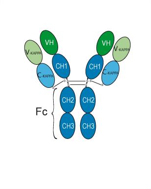
Atrosab is a humanized IgG1 antagonistic anti-TNFR1 antibody. Atrosab inhibits TNF-mediated Apoptosis induction and IL-6 and IL-8 production. Atrosab reduces neurological deficits. Atrosab can be used for research of inflammatory disease. The recommend isotype control is Human IgG1 kappa, Isotype Control (HY-P99001).
For research use only. We do not sell to patients.
- Purity : 99.94%
- CAS No.: 1365659-05-7
- Molecular Weight:145.837 kDa
-
Storage:
Please store the product under the recommended conditions in the Certificate of Analysis.


To place orders, for customer services and technical support, please contact: MedChemExpress USA
Tel: 609-228-6898 E-mail: [email protected] [email protected]


-
 Biological Activity
-
Chemical Information
- Protocols
- Purity & Documentation
- References
-
 Help & FAQs


-

HY-108847
EtanerceptTNF Inhibitor -

HY-P991192
BI-1808TNFR2 Inhibitor -

HY-P9908
AdalimumabTNF-α Inhibitor -
HY-13605
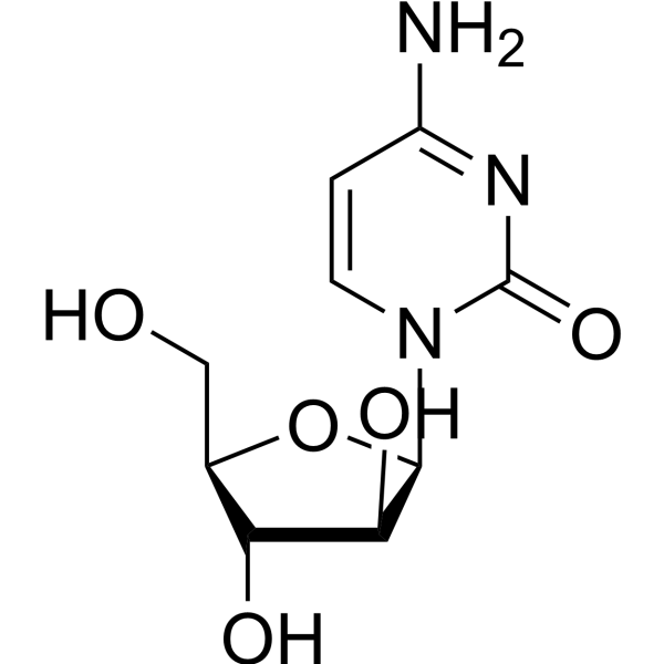
CytarabineDNA Synthesis Inhibitor -
HY-50856
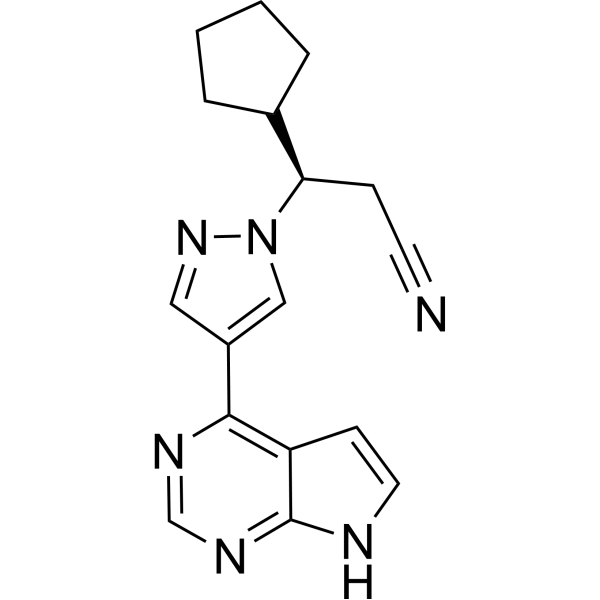
RuxolitinibJAK1/2 Inhibitor -

HY-P9970
InfliximabTNF-α Inhibitor -
HY-12848
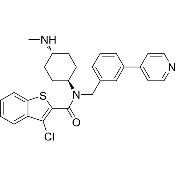
SAGSmoothened Agonist -

HY-P9904
AtezolizumabAnti-PD-L1 Antibody -
HY-P0201
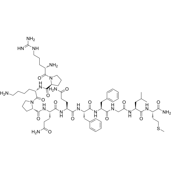
Substance PNeurotransmitter -
HY-12305
Q-VD-OPhPan-caspase Inhibitor -

HY-P9956
SiltuximabAnti-IL-6 mAb -

HY-P99001
Human IgG1 kappa, Isotype ControlIgG1κ Isotype Control
Publications Citing Use of MedChemExpress (MCE) Atrosab
More

Biological Activity
Description
Isotype
Human IgG1 kappa
Recommend Isotype Controls
Species Reactivity
Human
IC50 & Target
TNFR1
In Vitro
Atrosab (0.1-1000 nM, preincubate with antibody for 1 h, then add the stimulant TNF and co-incubate for 7 hours) inhibits in a dose-dependent manner the TNF-induced apoptosis of Kym-1 cells [1].
Atrosab (0.1-1000 nM, preincubate with antibody for 1 h, then add the stimulant TNF and co-incubate for 7 hours) inhibits release of IL-6 from HeLa cells and IL-8 from HT1080 cells induced by TNF dose-dependently[1].
Atrosab (100 μg/mL,24 h) reduces endothelial cell expression of adhesion molecules and T cell adhesion[3].
MedChemExpress (MCE) has not independently confirmed the accuracy of these methods. They are for reference only. Further protocols information, click here.
-
Cell Line:Kym-1 cells
-
Concentration:0.1-1 μM
-
Incubation Time:preincubate with antibody for 1 h, then add the stimulant TNF and co-incubate for 7 hours
-
Result:Exhibited half-maximal cytotoxicity at 60 nM in Kym-1 cells.
Inhibited IL-6 release from HeLa cells with an IC50 of 60 nM.
Inhibited IL-8 release from HT1080 cells with an IC50 of 60 nM.
In Vivo
Atrosab (20 mg/kg, i.p.) reduces immune cell infiltrates in humanized TNFR1 knock-in mice [3].
MedChemExpress (MCE) has not independently confirmed the accuracy of these methods. They are for reference only.
-
Animal Model:CIA rhesus monkey model[2].
-
Dosage:
-
Administration:i.v., twice weekly, 28 days
-
Result:Reduced of the acute-phase C-reactive protein (CRP) and IL-6 in serum.
Improved the clinical arthritis score, delayed the onset of symptoms, and inhibited cartilage degradation.
-
Animal Model:Human TNF transgenic/human TNFR1 Knock-in mice model (Tg197/hp55K/i) for arthritis and human TNFR1 Knock in mice (hp55K/i) EAE model for multiple sclerosis(MS)[2].
-
Dosage:
-
Administration:i.v., twice weekly, 28 days
-
Result:Significantly reduced arthritic pathology and the underlying histopathological lesions in rheumatoid arthritis (RA) mice.
Abolished the development of paralytic symptoms without affecting the overall health status of the animals in EAE mice model of MS.
Showed to be effective for treatment of inflammatory diseases such as RA and MS.
-
Animal Model:humanized TNFR1 knock-in mice[3].
-
Dosage:20 mg/kg
-
Administration:i.p., four times (on days 1, 4, 8 and 12 of manifest EAE)
-
Result:Significantly prevented EAE-associated weight loss.
Led less spinal cord demyelination.
Reduced accumulated amyloid precursor protein (APP), a marker of axonal injury.
Reduced numbers of CD3+ T cells, Mac-3+ activated microglia/macrophages and CD45R+ B cells.
Reduced immune cell infiltration.
Gene ID
Accession
Conjugated
Unconjugated
Reconsititution
The product can be reconstituted/diluted with sterile PBS or saline.
Format
-
Human IgG1 kappa
Application
ELISA, FACS, Functional assay
Verified Bioactivity
-
 Flow cytometric analysis of 1X10⁶ FaDu cells with Atrosab (HY-P990008, red). Cells were fixed with 4% paraformaldehyde. Then stained with the primary antibody at 1/200 dilution for an hour at 4℃. AF 488-conjugated AffiniPure Goat Anti-Human IgG H&L (HY-P83776) was used as the secondary antibody at 1/1,000 dilution for 30 minutes at 4℃. Human IgG1 kappa (HY-P99001, blue) was used as the isotype control, cells without incubation with primary antibody were used as the unlabeled control (black).
 -
 Immobilized Human TNFR1,His Tag can bind Atrosab. The EC50 for this effect is 8.161 ng/mL.
 -
 Loaded Atrosab on AHC2 biosensor, can bind TNFRSF1A Protein, Human (His, HY-P70801) with an affinity constant of 1.151E-08 M as determined in BLI assay.


Chemical Information
-
CAS No. 1365659-05-7
-
Appearance Liquid
-
Molecular Weight 145.837 kDa
-
Color Colorless to light yellow
-
SMILES
[Atrosab]
-
Shipping
Shipping with dry ice.
-
Formulation
Please refer to the lot-specific COA for specific buffer information.
-
Storage
Please store the product under the recommended conditions in the Certificate of Analysis.


Publications (1)
-
Journal Impact Factor
-
Most Recent
-
Cell Commun Signal
Helicobacter pylori infection promotes M1 macrophage polarization and gastric inflammation by activation of NLRP3 inflammasome via TNF/TNFR1 axis. [Abstract]2025 Jan 6;23(1):6. PMID: 39762835

Protocols
-
Apoptosis
Apoptosis, also called programmed cell death, is generally characterized by distinct morphological characteristics.
-
TUNEL staining for apoptotic DNA fragmentation
TUNEL staining detects DNA strand breaks by using terminal deoxynucleotidyl transferase to add labeled nucleotides to exposed 3′-OH DNA termini, generating either microscopic staining in fixed cells or tissue sections, or fluorescence/cytometric signal in cell suspensions. TUNEL positivity reflects DNA fragmentation but should not be interpreted alone as definitive apoptosis, because TUNEL can also label necrotic, autolytic, mechanically damaged, or DNA-repair-associated DNA breaks.
-
Annexin V plus membrane-impermeant dye apoptosis staining
Annexin V-based apoptosis assays rely on the detection of phosphatidylserine (PS) externalization from the inner leaflet of the plasma membrane to the outer leaflet, an early biochemical hallmark of apoptosis. Fluorescently labeled Annexin V binds PS in a calcium-dependent manner, enabling identification of early apoptotic cells by flow cytometry or fluorescence microscopy. When combined with a membrane-impermeant DNA-binding dye (e. g. , propidium iodide), this approach allows discrimination between viable (Annexin V−/dye−), early apoptotic (Annexin V+/dye−), and late apoptotic or necrotic (Annexin V+/dye+) cell populations by assessing membrane integrity and PS exposure.
-
Apoptosis Solutions
Apoptosis is a regulated, generally non-lytic cell-death pathway that removes unwanted, damaged, infected, or abnormal cells through coordinated morphological changes, caspase activation, DNA fragmentation, and membrane remodeling. The intrinsic apoptosis pathway is controlled mainly by mitochondrial outer membrane permeabilization, BCL-2 family proteins, cytochrome c release, apoptosome formation, caspase-9 activation, and downstream executioner caspase-3/7 activation. The extrinsic apoptosis pathway is initiated by death receptors such as Fas, TNFR, and TRAIL receptors, which recruit adaptor proteins and activate caspase-8 before engaging executioner caspases or mitochondrial amplification through BID cleavage. Apoptosis is linked to many phenotypes, including cancer cell killing, tissue homeostasis, immune regulation, neurodegeneration, infection response, and treatment-induced cytotoxicity; unresolved questions include how apoptosis interacts with necroptosis, pyroptosis, ferroptos
-
Research Protocol for Inflammation-related Diseases
The NLRP3 inflammasome is a cytosolic innate immune signaling platform that integrates priming signals and danger-signal activation to promote caspase-1 activation, maturation of IL-1β and IL-18, and gasdermin D-mediated pyroptotic cell death. The core experimental logic is to determine whether inflammatory disease phenotypes are driven by increased NLRP3 expression, ASC-containing inflammasome assembly, caspase-1 cleavage, GSDMD cleavage, and extracellular release of IL-1β/IL-18 rather than by nonspecific cell injury alone. The pathway is strongly linked to inflammation-related disease phenotypes because monosodium urate crystals activate NALP3/NLRP3 inflammasome signaling in gout-like crystal inflammation, cholesterol crystals activate NLRP3 inflammasomes in atherogenesis models, and DSS-induced intestinal inflammation has been reported to involve NLRP3 inflammasome activity. However, experimental colitis studies also show context-dependent protective effects of NLRP3 inflammasome co


Purity & Documentation
-
Data Sheet (266 KB)
-
SDS (251 KB)
- English - EN (251 KB)
- Français - FR (251 KB)
- Deutsch - DE (251 KB)
- Norwegian - NO (251 KB)
- Español - ES (251 KB)
- Swedish - SV (251 KB)
- Italian - IT (251 KB)
- Korean - KR (251 KB)
- Portuguese - PT (251 KB)
-
Inhibitory Antibodies User Guide (603 KB)
References
[1]. Zettlitz KA, et al. ATROSAB, a humanized antagonistic anti-tumor necrosis factor receptor one-specific antibody. MAbs. 2010 Nov-Dec;2(6):639-47. [Content Brief]
[2]. Zettlitz KA, et al. ATROSAB, a humanized antagonistic anti-tumor necrosis factor receptor one-specific antibody. [Content Brief]
[4]. Williams SK, et al. Anti-TNFR1 targeting in humanized mice ameliorates disease in a model of multiple sclerosis. Sci Rep. 2018 Sep 11;8(1):13628. [Content Brief]

Calculators
Concentration (start) × Volume (start) = Concentration (final) × Volume (final)


Atrosab Related Classifications
HY-P990008 Related Classifications
 Powered by Bioz