



Bcl-2-IN-10
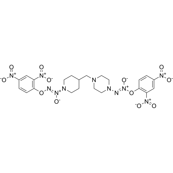
Bcl-2-IN-10 is an active Bcl-2 inhibitor that can release up to four nitric oxide (NO) molecules. Bcl-2-IN-10 has cytotoxic activities against cancer cells, such as human leukemia, breast cancer and lung cancer. Bcl-2-IN-10 induces cell apopotosis and arrest cell cycle of G2/M phase, and can be used in cancer-related research.
For research use only. We do not sell to patients.
- CAS No.: 2773354-28-0
- Formula: C22H25N11O12
- Molecular Weight:635.50
-
Storage:
Please store the product under the recommended conditions in the Certificate of Analysis.

To place orders, for customer services and technical support, please contact: MedChemExpress USA
Tel: 609-228-6898 E-mail: [email protected] [email protected]


-
 Biological Activity
-
Chemical Information
- Protocols
- Purity & Documentation
- References
-
 Help & FAQs




Biological Activity
Description
IC50 & Target
[1]|
Bax |
Cellular Effect
|
Cell Line

|
Type | Value | Description | References |
|---|---|---|---|---|
| A549 | IC50 |
11.3 μM
Compound: 1
|
Anticancer activity against human A549 cells assessed as inhibition of cell growth after 72 hrs by MTT or MTS assay
Anticancer activity against human A549 cells assessed as inhibition of cell growth after 72 hrs by MTT or MTS assay
|
[PMID: 32156499] |
| BT-474 | IC50 |
7.06 μM
Compound: 1
|
Anticancer activity against human BT474 cells assessed as inhibition of cell growth after 72 hrs by MTT or MTS assay
Anticancer activity against human BT474 cells assessed as inhibition of cell growth after 72 hrs by MTT or MTS assay
|
[PMID: 32156499] |
| CCRF-CEM | IC50 |
1.26 μM
Compound: 1
|
Anticancer activity against human CCRF-CEM cells assessed as inhibition of cell growth after 72 hrs by MTT or MTS assay
Anticancer activity against human CCRF-CEM cells assessed as inhibition of cell growth after 72 hrs by MTT or MTS assay
|
[PMID: 32156499] |
| Jurkat E6.1 | IC50 |
1.94 μM
Compound: 1
|
Anticancer activity against human Jurkat E6-1 cells assessed as inhibition of cell growth after 72 hrs by MTT or MTS assay
Anticancer activity against human Jurkat E6-1 cells assessed as inhibition of cell growth after 72 hrs by MTT or MTS assay
|
[PMID: 32156499] |
| MDA-MB-231 | IC50 |
17.86 μM
Compound: 1
|
Anticancer activity against human MDA-MB-231 cells assessed as inhibition of cell growth after 72 hrs by MTT or MTS assay
Anticancer activity against human MDA-MB-231 cells assessed as inhibition of cell growth after 72 hrs by MTT or MTS assay
|
[PMID: 32156499] |
In Vitro
Steroid sulfatase-IN-2 (compound 1, 0-20 μM approximately, 72 h) inhibits different cancer cells with IC50s ranging from 1.26 μM to 17.86 μM[1].
Steroid sulfatase-IN-2 (1 μM, 5 h) releases up to four molecules of nitric oxide[1].
Steroid sulfatase-IN-2 (8 μM, 8-72 h) induces leukemia cell CCRF-CEM apoptosis via MAPKs pathways, arrests cell in the G2/M phase, and increases ratio of Bax/Bcl-2[1].
MedChemExpress (MCE) has not independently confirmed the accuracy of these methods. They are for reference only.
-
Cell Line:CEM cells
-
Concentration:8 μM
-
Incubation Time:8, 24, 48 or 72 h
-
Result:Induced apoptosis in a time-dependent and dose-dependent manner.
-
Cell Line:CEM cells
-
Concentration:8 μM
-
Incubation Time:8, 24, 48 or 72 h
-
Result:Arrested cell in the G2/M phase.
-
Cell Line:CEM cells
-
Concentration:0-8 μM
-
Incubation Time:72 h
-
Result:Increased the levels of JNK and p38, and the levels of phosphorylated JNK and p38.
Decreased Bcl-2 level in a time- and dose-dependent manner, and increased pro-apoptotic Bax level.


Chemical Information
-
CAS No. 2773354-28-0
-
Molecular Weight 635.50
-
Formula C22H25N11O12
-
SMILES
O=[N+](C1=CC([N+]([O-])=O)=C(C=C1)O/N=[N+](N2CCC(CC2)CN3CCN(CC3)/N=[N+](OC4=C(C=C(C=C4)[N+]([O-])=O)[N+]([O-])=O)\[O-])\[O-])[O-]
-
Shipping
Room temperature in continental US; may vary elsewhere.
-
Storage
Please store the product under the recommended conditions in the Certificate of Analysis.


Purity & Documentation
References

Calculators
Concentration (start) × Volume (start) = Concentration (final) × Volume (final)


Bcl-2-IN-10 Related Classifications
HY-150540 Related Classifications
 Powered by Bioz