



BIM-26226

Based on 1 Customer Validation
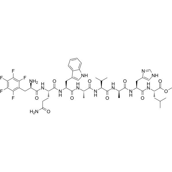
BIM-26226 is a selective gastrin-releasing peptide receptor (GRPR) (IC50 = 6 nM) and bombesin receptor (BN receptor) antagonist. BIM-26226 antagonizes BN- or GRP-stimulated amylase release with IC50 values of 0.3 nM and 0.2 nM, respectively. BIM-26226 is specific for the GRP-preferring BN receptor subtype with no interference with GRP receptor system. BIM-26226 can induce the synthesis of somatostatin receptor but has no significant effect on tumor growth.
For research use only. We do not sell to patients.
- Purity : 98.73%
- CAS No.: 136207-23-3
- Formula: C49H63F5N12O10
- Molecular Weight:1075.09
-
Storage:
Sealed storage, away from moisture.
Powder -80°C, 2 years , -20°C, 1 year* In solvent : -80°C, 6 months; -20°C, 1 month (sealed storage, away from moisture)

To place orders, for customer services and technical support, please contact: MedChemExpress USA
Tel: 609-228-6898 E-mail: [email protected] [email protected]


-
 Biological Activity
-
Chemical Information
-
Solvent & Solubility
- Protocols
- Purity & Documentation
- References
-
 Help & FAQs




Biological Activity
Description
IC50 & Target
bombesin receptor[1]
In Vitro
BIM-26226 (0.1 nM-1 μM, 24 h) significantly decreased [3H]thymidine incorporation in primary cultured pancreatic tumour cells[1].
BIM-26226 is specific for the GRP-preferring BN receptor subtype and is not able to inhibit binding of radio-labeled CCK-33, gastrin-17 or VIP in AR4-2J cells[2].
BIM-26226 (0.1 μM) is able to block the bombesin-induced increase of Ca2+ but fails to block the growth-stimulatory effect of bombesin[3].
MedChemExpress (MCE) has not independently confirmed the accuracy of these methods. They are for reference only. Further protocols information, click here.
In Vivo
BIM-26226 (100 μg/kg, s.c., once daily for 6 weeks) induces the synthesis of somatostatin receptor but has no effect on tumor growth in colon cancer rat model[3].
MedChemExpress (MCE) has not independently confirmed the accuracy of these methods. They are for reference only.
-
Animal Model:Azaserine (HY-B0919)-induced pancreatic acinar carcinoma model established in Lewis rats[1]
-
Dosage:30 and 100 μg/kg
-
Administration:Subcutaneous injection (s.c.), 3 times daily for 14 consecutive days
-
Result:Reduced the contents of protein, RNA, amylase and trypsin.
Did not significantly affect pancreatic growth parameters.
-
Animal Model:Colon cancer model induced by DHD/K12 tumor cell line established in male BDIX rats (12 weeks old) [3]
-
Dosage:100 μg/kg
-
Administration:Subcutaneous injection (s.c.), once daily for 6 weeks
-
Result:Significantly decreased gastrinemia.
Had no effect on bombesin binding sites kDs and affinity site number Bmax.


Chemical Information
-
CAS No. 136207-23-3
-
Appearance Solid
-
Molecular Weight 1075.09
-
Formula C49H63F5N12O10
-
Color White to off-white
-
Sequence
{D-F5 Phe}-Gln-Trp-{D-Ala}-Val-Ala-His-Leu-OMe
-
Sequence Shortening
{D-F5 Phe}-QW-{D-Ala}-VAHL-OMe
-
Shipping
Room temperature in continental US; may vary elsewhere.
-
Storage
Sealed storage, away from moisture
Powder -80°C 2 years -20°C 1 year * In solvent : -80°C, 6 months; -20°C, 1 month (sealed storage, away from moisture)


Solvent & Solubility
In Vitro:
DMSO : 100 mg/mL (93.02 mM; Need ultrasonic; Hygroscopic DMSO has a significant impact on the solubility of product, please use newly opened DMSO)
Please refer to the solubility information to select the appropriate solvent. Once prepared, please aliquot and store the solution to prevent product inactivation from repeated freeze-thaw cycles.
Storage method and period of stock solution: -80°C, 6 months; -20°C, 1 month (sealed storage, away from moisture). When stored at -80°C, please use it within 6 months. When stored at -20°C, please use it within 1 month.
Please refer to the solubility information to select the appropriate solvent. Once prepared, please aliquot and store the solution to prevent product inactivation from repeated freeze-thaw cycles.
Storage method and period of stock solution: -80°C, 6 months; -20°C, 1 month (sealed storage, away from moisture). When stored at -80°C, please use it within 6 months. When stored at -20°C, please use it within 1 month.
Concentration (start) × Volume (start) = Concentration (final) × Volume (final)

Protocols
-
Cell Cytotoxicity Assay
Cytotoxicity assays are usually based on the assessment of cell membrane damage, which can also be indirectly detected by measuring cell viability. Detection methods include MTT assay, CKK-8 assay, LDH assay and ATP assay, etc.
-
Patient-Derived Xenograft (PDX)
Patient-derived xenograft (PDX) models are generated by engrafting primary human tumor tissue directly into immunodeficient mice, allowing in vivo propagation of patient tumor biology without initial in vitro adaptation. These models are used to preserve key histopathological and molecular characteristics of the original tumor and enable assessment of tumor growth dynamics and therapeutic response in a living organism. The biological readout is tumor engraftment and subsequent growth in the murine host, which reflects the ability of human tumor cells to survive, vascularize, and expand in an immunocompromised microenvironment.
-
Patient-Derived Orthotopic Xenograft (PDOX)
Patient-derived orthotopic xenograft (PDOX) modeling implants fresh patient tumor tissue or patient-derived tumor cells into the anatomically corresponding organ or tissue site of immunodeficient mice, usually by surgical orthotopic implantation, to preserve patient tumor histology, local microenvironmental context, invasion, metastatic behavior, and treatment-response features better than subcutaneous implantation. PDOX readouts include tumor engraftment, orthotopic tumor growth, local invasion, metastasis, recurrence after resection, histologic similarity to the donor tumor, biomarker retention, molecular concordance, survival, and response or resistance to therapy. PDOX models are used for preclinical drug testing and individualized therapy evaluation, but engraftment success varies by tumor type and specimen quality.
-
Subcutaneous Cell-Line-Derived Xenograft
Subcutaneous cell-line-derived xenograft (CDX) models are established by implanting cultured human cancer cell lines into immunodeficient mice, where the injected cells form localized tumors that can be monitored in vivo as a measure of tumorigenic potential, growth kinetics, and treatment response. These models are widely used in oncology research because they allow reproducible tumor formation and enable comparative assessment of tumor growth between different cell lines or genetic manipulations in a controlled in vivo microenvironment. Subcutaneous implantation of cancer cells in immunodeficient mice is a standard approach for evaluating tumor growth behavior and therapeutic response across multiple cancer types, including prostate, esophageal, pancreatic, and colon cancer models.
-
Orthotopic Cell-Line Xenograft
Orthotopic cell-line xenograft models involve implantation of human cancer cell lines into the anatomically corresponding organ of immunodeficient mice to reproduce tumor growth within a native microenvironment, enabling more clinically relevant tumor behavior compared with subcutaneous models. These models are widely used because orthotopic placement better recapitulates tumor progression, including invasion and metastatic spread, which are often underrepresented in heterotopic implantation systems. Compared with conventional xenografts, orthotopic implantation is described as more technically complex but provides improved simulation of tumor-microenvironment interactions and metastatic behavior, making it particularly valuable for translational oncology research. Surgical orthotopic implantation approaches have been emphasized as enabling faithful reproduction of clinical cancer features, including metastasis and disease progression patterns that align with the tumor’s organ of origi


Purity & Documentation
-
Data Sheet (276 KB)
-
SDS (252 KB)
- English - EN (252 KB)
- Français - FR (252 KB)
- Deutsch - DE (252 KB)
- Norwegian - NO (252 KB)
- Español - ES (252 KB)
- Swedish - SV (252 KB)
- Italian - IT (252 KB)
- Korean - KR (252 KB)
- Portuguese - PT (252 KB)
-
Handling Instructions (2659 KB)
References
[1]. Dietrich JB, et, al. Effects of BIM26226, a potent and specific bombesin receptor antagonist, on amylase release and binding of bombesin-like peptides to AR4-2J cells. Regul Pept. 1994 Oct 21;53(3):165-73. [Content Brief]
[2]. Damgé C, et, al. Effect of the gastrin-releasing peptide antagonist BIM 26226 and lanreotide on an acinar pancreatic carcinoma. Eur J Pharmacol. 1998 Apr 17;347(1):77-86. [Content Brief]
[3]. Gouyon B, et al. In-vivo effect of somatostatin analog, lanreotide, and/or grp antagonist, bim-26226, on the growth of colon-cancer peritoneal carcinomatosis in the rat. Int J Oncol. 1995 Nov;7(5):1167-73. [Content Brief]
[4]. Bold RJ, et al. Bombesin stimulates the in vitro growth of a human gastric cancer cell line. J Cell Physiol. 1994 Dec;161(3):519-25. [Content Brief]


Complete Stock Solution Preparation Table
Please refer to the solubility information to select the appropriate solvent. Once prepared, please aliquot and store the solution to prevent product inactivation from repeated freeze-thaw cycles.
Storage method and period of stock solution: -80°C, 6 months; -20°C, 1 month (sealed storage, away from moisture). When stored at -80°C, please use it within 6 months. When stored at -20°C, please use it within 1 month.
| Optional Solvent | Concentration Solvent Mass | 1 mg | 5 mg | 10 mg | 25 mg |
|---|---|---|---|---|---|
| DMSO | 1 mM | 0.9302 mL | 4.6508 mL | 9.3015 mL | 23.2539 mL |
| 5 mM | 0.1860 mL | 0.9302 mL | 1.8603 mL | 4.6508 mL | |
| 10 mM | 0.0930 mL | 0.4651 mL | 0.9302 mL | 2.3254 mL | |
| 15 mM | 0.0620 mL | 0.3101 mL | 0.6201 mL | 1.5503 mL | |
| 20 mM | 0.0465 mL | 0.2325 mL | 0.4651 mL | 1.1627 mL | |
| 25 mM | 0.0372 mL | 0.1860 mL | 0.3721 mL | 0.9302 mL | |
| 30 mM | 0.0310 mL | 0.1550 mL | 0.3101 mL | 0.7751 mL | |
| 40 mM | 0.0233 mL | 0.1163 mL | 0.2325 mL | 0.5813 mL | |
| 50 mM | 0.0186 mL | 0.0930 mL | 0.1860 mL | 0.4651 mL | |
| 60 mM | 0.0155 mL | 0.0775 mL | 0.1550 mL | 0.3876 mL | |
| 80 mM | 0.0116 mL | 0.0581 mL | 0.1163 mL | 0.2907 mL |


BIM-26226 Related Classifications
HY-P0039 Related Classifications
 Powered by Bioz