



Rinatabart

Based on 1 Customer Validation
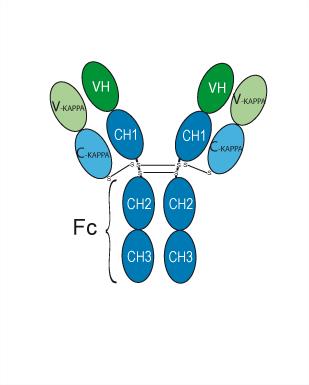
Rinatabart (F131) is an IgG1-κ human antibody targeting FOLR1, with its corresponding isotype control being Human IgG1 kappa, Isotype Control (HY-P99001). The Fc region of Rinatabart is engineered to reduce antibody-dependent cellular cytotoxicity (ADCC) and complement-dependent cytotoxicity (CDC) activities. The KD values of Rinatabart binding to human FOLR1 in two sets of BLI assays are 1.528 nM and 5.296 nM, respectively. Rinatabart shows significantly lower affinity for FOLR2 and mouse FOLR1, and no reliable specific binding to FOLR3 and rat FOLR1. Rinatabart can be used in studies of FOLR1 targeting and antibody-drug conjugates.
For research use only. We do not sell to patients.
- Purity : 99.47%
- CAS No.: 2853518-94-0
- Molecular Weight:145.2 kDa
-
Storage:
Please store the product under the recommended conditions in the Certificate of Analysis.


To place orders, for customer services and technical support, please contact: MedChemExpress USA
Tel: 609-228-6898 E-mail: [email protected] [email protected]


-
 Biological Activity
-
Chemical Information
- Protocols
- Purity & Documentation
- References
-
 Help & FAQs





Biological Activity
Description
Isotype
Human IgG1 kappa
Recommend Isotype Controls
Species Reactivity
Human
IC50 & Target
[1]|
FOLR1 1.528 nM (Kd, 物种交叉反应) |
FOLR1 5.296 nM (Kd, FOLR 家族选择性) |
In Vitro
Rinatabart (F131) exhibits KD values of 1.528 nM and 8.148 nM for binding to human and cynomolgus monkey FOLR1, respectively, and shows significantly weaker or negligible binding to human FOLR2, human FOLR3, mouse FOLR1, and rat FOLR1[1].
Rinatabart binds to FOLR1-positive JEG-3 cells with an EC50 of 1.549 nM, but does not specifically bind to FOLR1-negative PC-3 cells[1].
Rinatabart (10 μg/mL; 0-4 h) is rapidly internalized into FOLR1-positive OVCAR-3, KB, JEG-3, NCI-H441 and OV90 cells, but not into FOLR1-negative PC-3 cells[1].
Rinatabart (10 μg/mL; 0-23 h) undergoes time-dependent internalization into HeLa and RPTEC/TERT1 cells, and exhibits strong internalization ability among the tested anti-FOLR1 antibodies[1].
MedChemExpress (MCE) has not independently confirmed the accuracy of these methods. They are for reference only.
Gene ID
Accession
Conjugated
Unconjugated
Reconsititution
The product can be reconstituted/diluted with sterile PBS or saline.
Format
-
IgG1-kappa
Application
ELISA, FACS, Functional assay
Verified Bioactivity
-
 Immobilized Human FOLR1, His Tag can bind Rinatabart. The EC50 for this effect is 8.83 ng/mL.
 -
 Flow cytometric analysis of 1X10⁶ MCF-7 cells with Rinatabart (HY-P990941, red). Cells were fixed with 4% paraformaldehyde. Then stained with the primary antibody at 1/200 dilution for an hour at 4℃. Alexa Fluor 488-conjugated AffiniPure Goat Anti-Human IgG H&L (AF488) (HY-P83776) was used as the secondary antibody at 1/1,000 dilution for 30 minutes at 4℃. Human IgG1 kappa (HY-P99001, blue) was used as the isotype control, cells without incubation with primary antibody were used as the unlabeled control (black).


Chemical Information
-
CAS No. 2853518-94-0
-
Appearance Liquid
-
Molecular Weight 145.2 kDa
-
Color Colorless to light yellow
-
SMILES
[Rinatabart]
-
Synonyms
F131; GEN1184 antibody
-
Shipping
Shipping with dry ice.
-
Formulation
Please refer to the lot-specific COA for specific buffer information.
-
Storage
Please store the product under the recommended conditions in the Certificate of Analysis.


Purity & Documentation
-
Data Sheet (257 KB)
-
SDS (251 KB)
- English - EN (251 KB)
- Français - FR (251 KB)
- Deutsch - DE (251 KB)
- Norwegian - NO (251 KB)
- Español - ES (251 KB)
- Swedish - SV (251 KB)
- Italian - IT (251 KB)
- Korean - KR (251 KB)
- Portuguese - PT (251 KB)
-
Inhibitory Antibodies User Guide (603 KB)
References

Calculators
Concentration (start) × Volume (start) = Concentration (final) × Volume (final)


Rinatabart Related Classifications
HY-P990941 Related Classifications
 Powered by Bioz