



Lipase (MS grade)
Based on 1 publication(s) in Google Scholar
Lipase (MS grade) catalyzes the hydrolysis of triacylglycerols to release long-chain fatty acids in a site-specific manner. Lipase (MS grade) is involved in a variety of biological processes, from fat metabolism to cell signaling and inflammation, and can be used to study diseases such as pancreatic insufficiency, celiac disease and cystic fibrosis.
Nos produits utilisent uniquement pour la recherche. Nous ne vendons pas aux patients.
- CAS No.: 9001-62-1
- Masse moléculaire:48000.00
-
Stockage:
Please store the product under the recommended conditions in the Certificate of Analysis.


To place orders, for customer services and technical support, please contact: MedChemExpress USA
Tel: 609-228-6898 E-mail: [email protected] [email protected]


-
 Activité biologique
-
Chemical Information
- Protocole
- Pureté et documentation
- Références
-
 Help & FAQs


-

HY-Y1422D
Lipase,Candida antarctica (Immobilized)Immobilized Lipase -

HY-Y1422I
Lipase,Pseudomonas fluorescens (Immobilized)Triacylglycerol Carboxylic Ester Bonds Catalyst, Short to Middle-Chain Fatty Acid Methyl-Ester Ester Bonds Catalyst, Triglyceride Ester Bonds Catalyst, Triolein 1,3-Ester Bonds Preferential Cleaver, EPA- and DHA-Containing Fats and Oils Decomposer -

HY-Y1422H
Lipase,Candida cylindracea (Immobilized)Biocatalyst -
HY-Y1422
Triacylglycerol lipaseTriglyceride Hydrolase -
HY-P2902
Glucose oxidaseOxidoreductase Enzyme -

HY-Y1422E
Lipase,Aspergillusniger (Immobilized)Biocatalyst -

HY-Y1422J
Lipase,Burkholderiacepacia (Immobilized)Catalyzing Enzyme -
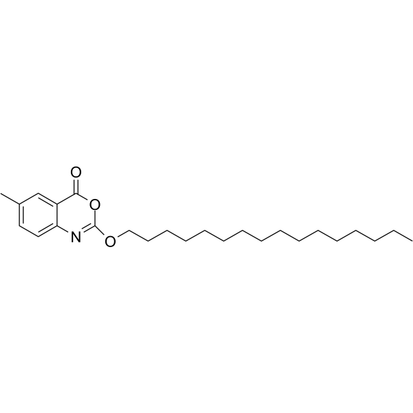
HY-14471
CetilistatLipase Inhibitor -

HY-14471R
Cetilistat (Standard)Lipase Inhibitor
Publications Citing Use of MedChemExpress (MCE) Lipase (MS grade)
MoreVoir tous les produits spécifiques à Isoform Endogenous Metabolite
More

Activité biologique
Description


Chemical Information
-
CAS No. 9001-62-1
-
Appearance Solid
-
Masse moléculaire 48000.00
-
Color White to off-white
-
SMILES
[Lipase (MS grade)]
-
Livraison
Room temperature in continental US; may vary elsewhere.
-
Stockage
Please store the product under the recommended conditions in the Certificate of Analysis.


Publications (1)
-
Journal Impact Factor
-
Most Recent
-
J Biomed Mater Res A
Injectable Butyl Glycidyl Ether-Modified Methylcellulose Thermosensitive Hydrogel for Topical Delivery of Vancomycin Hydrochloride in the Treatment of Osteomyelitis. [Abstract]2026 Feb;114(2):e70031. PMID: 41586469

Protocole
-
Fibrosis/Collagen Morphometry
Fibrosis and collagen morphometry is based on the quantitative visualization of fibrillar collagen deposition in tissue sections using histochemical stains such as Sirius Red (Picrosirius Red) or Masson's trichrome, followed by image-based or polarization-enhanced analysis to estimate collagen proportional area as a surrogate of extracellular matrix accumulation during fibrotic remodeling. Sirius Red combined with polarized light microscopy enhances detection of collagen fibers due to birefringence properties, enabling more specific visualization of collagen type I and III fibrils compared to conventional bright-field histology, while whole-section or region-restricted digital morphometry reduces field-selection bias in fibrosis assessment. Alternative quantitative approaches include second harmonic generation (SHG) and two-photon excited fluorescence microscopy, which enable label-free detection of fibrillar collagen and have been validated against histological staining and biochemica
-
Collagen: Sirius Red Staining
Sirius Red or picrosirius red staining is a histochemical method for visualizing collagen-rich extracellular matrix in tissue sections, and collagen fibers are detected as red-stained structures under bright-field microscopy with enhanced birefringence under polarized light. Picrosirius red is useful for assessing total collagen organization, distribution, and fibrosis burden, but polarized color should not be interpreted as a definitive collagen type I versus type III readout because color is affected by fiber orientation, thickness, and packing.
-
Connective Tissue: Masson's Trichrome/Collagen Trichrome Staining
Masson’s Trichrome (collagen/trichrome staining) is a histological technique that differentially stains tissue compartments using sequential acidic dyes to distinguish collagen from muscle and cytoplasmic components based on dye affinity and tissue permeability differences, enabling visualization of fibrosis and connective tissue architecture in histological sections. The classical formulation typically uses Weigert's iron hematoxylin for nuclear staining, Biebrich scarlet-acid fuchsin for cytoplasm and muscle, and aniline blue (or light green variants) for collagen, producing a characteristic blue/green collagen signal contrasted against red cytoplasm and dark nuclei. The staining principle relies on selective displacement of smaller dye molecules by larger anionic dyes in collagen-rich regions under controlled acidified conditions, which enhances collagen-specific dye retention. This property makes the method widely used for fibrosis assessment in organs such as heart, liver, lung, a
-
Research Protocol for Inflammation-related Diseases
The NLRP3 inflammasome is a cytosolic innate immune signaling platform that integrates priming signals and danger-signal activation to promote caspase-1 activation, maturation of IL-1β and IL-18, and gasdermin D-mediated pyroptotic cell death. The core experimental logic is to determine whether inflammatory disease phenotypes are driven by increased NLRP3 expression, ASC-containing inflammasome assembly, caspase-1 cleavage, GSDMD cleavage, and extracellular release of IL-1β/IL-18 rather than by nonspecific cell injury alone. The pathway is strongly linked to inflammation-related disease phenotypes because monosodium urate crystals activate NALP3/NLRP3 inflammasome signaling in gout-like crystal inflammation, cholesterol crystals activate NLRP3 inflammasomes in atherogenesis models, and DSS-induced intestinal inflammation has been reported to involve NLRP3 inflammasome activity. However, experimental colitis studies also show context-dependent protective effects of NLRP3 inflammasome co


Pureté et documentation
-
Fiche technique (266 KB)
-
SDS (251 KB)
- English - EN (251 KB)
- Français - FR (251 KB)
- Deutsch - DE (251 KB)
- Norwegian - NO (251 KB)
- Español - ES (251 KB)
- Swedish - SV (251 KB)
- Italian - IT (251 KB)
- Korean - KR (251 KB)
- Portuguese - PT (251 KB)
-
Instruction de manipulation (2659 KB)
Références

Calculators
Concentration (start) × Volume (start) = Concentration (final) × Volume (final)


Lipase (MS grade) Related Classifications
HY-Y1422B Related Classifications
 Powered by Bioz