



Neridronate

Based on 1 Customer Validation
Neridronate is a bisphosphonate. Bisphosphonates initiate the Apoptotic process. Neridronate sodium reduces the levels of bone resorption, bone turnover markers, the degree of back pain, and the risk of fractures. Neridronate inhibits capillary tube formation. Neridronate itself has weak anticancer activity, but liposomal encapsulation enhances this activity. Neridronate can be used in research related to demineralizing metabolic bone diseases, thalassemia-associated osteoporosis, chronic inflammatory diseases, cancer, and osteogenesis imperfecta.
Nos produits utilisent uniquement pour la recherche. Nous ne vendons pas aux patients.
- Pureté : 98.0%
- CAS No.: 79778-41-9
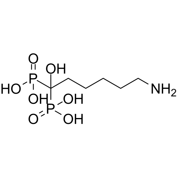
- Formule: C6H17NO7P2
- Masse moléculaire:277.15
-
Stockage:Powder -20°C, 3 years , 4°C, 2 years ; In solvent -80°C, 6 months , -20°C, 1 month

To place orders, for customer services and technical support, please contact: MedChemExpress USA
Tel: 609-228-6898 E-mail: [email protected] [email protected]


-
 Activité biologique
-
Chemical Information
-
Solvant et solubilité
- Protocole
- Pureté et documentation
- Références
-
 Help & FAQs




Activité biologique
Description
Cellular Effect
|
Cell Line

|
Type | Value | Description | References |
|---|---|---|---|---|
| HFF | IC50 |
59.7 μM
Compound: 34
|
In vitro inhibitory concentration against the growth of Toxoplasma gondii in human foreskin fibroblast monolayer cells (HFF cells)
In vitro inhibitory concentration against the growth of Toxoplasma gondii in human foreskin fibroblast monolayer cells (HFF cells)
|
[PMID: 15857119] |
| T-cell | IC50 |
1.96 μM
Compound: 45
|
Inhibitory activity, for stimulation of TNF-alpha release in gamma-delta T cells, using individual observed maximum TNF-alpha release
Inhibitory activity, for stimulation of TNF-alpha release in gamma-delta T cells, using individual observed maximum TNF-alpha release
|
[PMID: 14711309] |
| T-cell | IC50 |
503 μM
Compound: 45
|
Inhibitory activity, for stimulation of TNF-alpha release in gamma-delta T cells, using a constrained maximum TNF-alpha release of 2700 pg/mL
Inhibitory activity, for stimulation of TNF-alpha release in gamma-delta T cells, using a constrained maximum TNF-alpha release of 2700 pg/mL
|
[PMID: 14711309] |
| Vero | IC50 |
>200 μM
Compound: 10
|
Inhibition of Trypanosoma cruzi Amastigotes was determined in Vero cells culture and fetal calf serum
Inhibition of Trypanosoma cruzi Amastigotes was determined in Vero cells culture and fetal calf serum
|
[PMID: 11300872] |
In Vitro
Neridronate alters the biosynthetic activity of osteoblasts in vitro, which may affect bone turnover rate[2].
Neridronate (1-50 μM; 24 h) inhibits the proliferation of human umbilical vein endothelial cells (HUVEC) in a dose-dependent manner, with the inhibitory effect peaking at 30 μM and reaching a plateau at 50 μM[3].
Neridronate (30 μM; 18 h) disrupts the formation of capillary-like tubes by umbilical vein endothelial cells (HUVECs) cultured on Matrigel, which is induced by fibroblast growth factor-2 (FGF-2)[3].
Liposomal neridronate (0.27-17.56 μM; 24-72 h) potently inhibits the viability of MDA-MB-231, U87-MG and Caco2 cancer cells, with an EC50 of 1.7 μM after 72 h of incubation in MDA-MB-231 cells; in contrast, free neridronate (16 μM-1 mM; 72 h) exerts weak inhibitory effects on these cell lines[4].
MedChemExpress (MCE) has not independently confirmed the accuracy of these methods. They are for reference only.
-
Cell Line:human umbilical vein endothelial cells (HUVECs)
-
Concentration:1 μM, 3 μM, 10 μM, 30 μM, 50 μM
-
Incubation Time:24 h
-
Result:Reduced HUVEC growth in a dose-dependent fashion, with the maximum inhibitory effect observed at 30 μM; the effect plateaued at 50 μM.
Significantly inhibited HUVEC proliferation at 10 μM, 30 μM, and 50 μM relative to lower concentrations.
-
Cell Line:MDA-MB-231 human breast carcinoma cells, U87-MG human brain carcinoma cells, Caco2 human colon carcinoma cells
-
Concentration:16 μM-1 mM (free neridronate); 0.27 μM-17.56 μM (liposomal neridronate)
-
Incubation Time:72 h (free neridronate); 24 h, 48 h, 72 h (liposomal neridronate)
-
Result:Achieved maximum growth inhibition of 46% in MDA-MB-231 cells (EC50 = 95 μM), 52% in U87-MG cells (EC50 = 14 μM), and 37% in Caco2 cells (EC50 = 130 μM) after 72 h of free neridronate treatment.
Achieved maximum growth inhibition of 100% in MDA-MB-231 cells (EC50 = 1.7 μM), 70% in U87-MG cells (EC50 = 1 μM), and 62% in Caco2 cells (EC50 = 4 μM) after 72 h of liposomal neridronate treatment.
Achieved 100% growth inhibition in MDA-MB-231 cells with an EC50 of 2.4 μM after 48 h of liposomal neridronate treatment.
In Vivo
MedChemExpress (MCE) has not independently confirmed the accuracy of these methods. They are for reference only.
-
Animal Model:White Leghorn (fertilized embryos)[3]
-
Dosage:50 μM/embryo
-
Administration:local administration via gelatin sponge implant; single dose
-
Result:Reduced mean number of vessels entering the sponge to 15, compared to the FGF-2-only control mean of 30.
Essai clinique
| NCT Number | Sponsor | Condition | Start Date |
Phase
|
|---|---|---|---|---|
| NCT01329991 | 2011-05 | PHASE1 |


Chemical Information
-
CAS No. 79778-41-9
-
Appearance Solid
-
Masse moléculaire 277.15
-
Formule C6H17NO7P2
-
Color White to off-white
-
SMILES
OC(P(O)(O)=O)(CCCCCN)P(O)(O)=O
-
Livraison
Room temperature in continental US; may vary elsewhere.
-
Stockage
Powder -20°C 3 years 4°C 2 years In solvent -80°C 6 months -20°C 1 month


Solvant et solubilité
In Vitro:
H2O : 5 mg/mL (18.04 mM; ultrasonic and warming and adjust pH to 8 with NaOH and heat to 60°C)
Please refer to the solubility information to select the appropriate solvent. Once prepared, please aliquot and store the solution to prevent product inactivation from repeated freeze-thaw cycles.
Storage method and period of stock solution: -80°C, 6 months; -20°C, 1 month. When stored at -80°C, please use it within 6 months. When stored at -20°C, please use it within 1 month.
* Note: If you choose water as the stock solution, please dilute it to the working solution, then filter and sterilize it with a 0.22 μm filter before use.
Please refer to the solubility information to select the appropriate solvent. Once prepared, please aliquot and store the solution to prevent product inactivation from repeated freeze-thaw cycles.
Storage method and period of stock solution: -80°C, 6 months; -20°C, 1 month. When stored at -80°C, please use it within 6 months. When stored at -20°C, please use it within 1 month.
* Note: If you choose water as the stock solution, please dilute it to the working solution, then filter and sterilize it with a 0.22 μm filter before use.
Concentration (start) × Volume (start) = Concentration (final) × Volume (final)
In Vivo:
For the following dissolution methods, please prepare the working solution directly:
It is recommended to prepare fresh solutions and use them promptly within a short period of time.
The percentages shown for the solvents indicate their volumetric ratio in the final prepared solution. If precipitation or phase separation occurs during preparation, heat and/or sonication can be used to aid dissolution.
Add each solvent one by one: PBS
Solubility: 2 mg/mL (7.22 mM); Clear solution; Need ultrasonic and warming and heat to 60°C


Pureté et documentation
-
Fiche technique (278 KB)
-
SDS (396 KB)
- English - EN (396 KB)
- Français - FR (396 KB)
- Deutsch - DE (396 KB)
- Norwegian - NO (396 KB)
- Español - ES (396 KB)
- Swedish - SV (396 KB)
- Italian - IT (396 KB)
- Korean - KR (396 KB)
- Portuguese - PT (396 KB)
-
Instruction de manipulation (2659 KB)
Références
[1]. Nicolin V, et al. Effects of neridronic acid on osteoclasts derived by physiological dual-cell cultures. Acta Histochem. 2007;109(5):397-402. [Content Brief]
[2]. Forni GL, et al. Neridronate improves bone mineral density and reduces back pain in β-thalassaemia patients with osteoporosis: results from a phase 2, randomized, parallel-arm, open-label study. Br J Haematol. 2012 Jul;158(2):274-282. [Content Brief]
[3]. Ribatti D, et al. Neridronate inhibits angiogenesis in vitro and in vivo. Clin Rheumatol. 2007 Jul;26(7):1094-8. [Content Brief]
[4]. Chebbi I, et al. In vitro assessment of liposomal neridronate on MDA-MB-231 human breast cancer cells. Int J Pharm. 2010 Jan 4;383(1-2):116-22. [Content Brief]
[5]. Gatti D, et al. Intravenous neridronate in children with osteogenesis imperfecta: a randomized controlled study. J Bone Miner Res. 2005 May;20(5):758-63. [Content Brief]


Complete Stock Solution Preparation Table
Please refer to the solubility information to select the appropriate solvent. Once prepared, please aliquot and store the solution to prevent product inactivation from repeated freeze-thaw cycles.
Storage method and period of stock solution: -80°C, 6 months; -20°C, 1 month. When stored at -80°C, please use it within 6 months. When stored at -20°C, please use it within 1 month.
| Optional Solvent | Concentration Solvent Mass | 1 mg | 5 mg | 10 mg | 25 mg |
|---|---|---|---|---|---|
| H2O | 1 mM | 3.6082 mL | 18.0408 mL | 36.0815 mL | 90.2039 mL |
| 5 mM | 0.7216 mL | 3.6082 mL | 7.2163 mL | 18.0408 mL | |
| 10 mM | 0.3608 mL | 1.8041 mL | 3.6082 mL | 9.0204 mL | |
| 15 mM | 0.2405 mL | 1.2027 mL | 2.4054 mL | 6.0136 mL |
* Note: If you choose water as the stock solution, please dilute it to the working solution, then filter and sterilize it with a 0.22 μm filter before use.


Neridronate Related Classifications
HY-119443 Related Classifications
 Powered by Bioz