



Canine Fibrinogen
Based on 2 publication(s) in Google Scholar
Canine Fibrinogen is a natural fibrinogen derived from canine plasma. Canine Fibrinogen specifically binds to the host SpsL, but shows weak binding affinity to fibrinogen from other mammals including humans. Canine Fibrinogen binds to the N2N3 subdomain of SpsL via the "anchor-lock-latch" model, and interacts with multiple sites in its α-chain C domain. The binding of Canine Fibrinogen to SpsL promotes bacterial aggregation, biofilm formation, and enhances bacterial resistance to neutrophil phagocytosis. Canine Fibrinogen can be used in studies related to canine skin infections.
For research use only. We do not sell to patients.
- CAS No.: 9001-32-5
-
Storage:
Please store the product under the recommended conditions in the Certificate of Analysis.


To place orders, for customer services and technical support, please contact: MedChemExpress USA
Tel: 609-228-6898 E-mail: [email protected] [email protected]


-
 Biological Activity
-
Chemical Information
- Purity & Documentation
- References
-
 Help & FAQs


-

HY-D0842A
BSA Protein (Low Endotoxin,Fatty acid free) -

HY-D0842
BSA Protein -
HY-W250978
Ovalbumins -
HY-P1956
HSA Protein -
HY-125864
Fibrinogen (Bovine) -

HY-NP010
Type I Collagen, from rat tail -
HY-P2995
Hemoglobin -
HY-P2738
Collagen, Type I, from bovine achilles tendon -
HY-NP003
Collagen, Type II, from bovine snout membrane -

HY-W250978A
Ovalbumin, low endotoxin -

HY-125865
Casein -

HY-P1956A
HSA Protein (Cell culture grade, Endotoxin<0.125 EU/mg)
Publications Citing Use of MedChemExpress (MCE) Canine Fibrinogen
More Customer Validation & Images

-
2D/3D Cell Culture and Differentiation
-
Cell Imaging/Staining
-
Cell Imaging/Staining
-
Cell Imaging/Staining
-
Histological Imaging/Staining


Biological Activity
Description
In Vitro
Canine fibrinogen (2-10 μg/mL; 2 h) is preferentially bound by Staphylococcus pseudintermedius ED99 wild-type cells compared to bovine, human, and ovine fibrinogen when incubated at concentrations up to 10 μg/mL for 2 h at 37°C[1].
Canine fibrinogen exhibits higher binding probability and stronger adhesion forces (up to 2077 pN) for single Staphylococcus pseudintermedius cells expressing SpsL A+SD than for human fibrinogen in single-cell force spectroscopy assays[1].
Canine fibrinogen (2-10 μg/mL) is preferentially bound by the surface-expressed SpsL N121+N2N3+SD chimeric protein in Staphylococcus pseudintermedius ED99ΔspsLΔspsD compared to bovine, human, and ovine fibrinogen at concentrations up to 10 μg/mL[1].
Canine fibrinogen (100 nM; overnight coating, 24 h bacterial incubation) promotes enhanced biofilm formation by full-length SpsL expressed in Staphylococcus pseudintermedius on its coated surfaces compared to bovine, human, or uncoated surfaces[1].
Canine fibrinogen (50 nM; 15 min opsonization, 15 min neutrophil incubation) inhibits human neutrophil phagocytosis when opsonized with full-length SpsL expressed in Staphylococcus pseudintermedius[1].
MedChemExpress (MCE) has not independently confirmed the accuracy of these methods. They are for reference only.


Chemical Information
-
CAS No. 9001-32-5
-
SMILES
[Canine Fibrinogen]
-
Shipping
Room temperature in continental US; may vary elsewhere.
-
Storage
Please store the product under the recommended conditions in the Certificate of Analysis.


Publications (2)
-
Journal Impact Factor
-
Most Recent
-
Foods
Glycosylation Remodeling and Thermal Denaturation Dictate the Functional Diversification of Protein Z. [Abstract]2026 Mar 4;15(5):853. PMID: 41829126 -
Microvasc Res
DDIT4 knockdown suppresses venous malformation progression by inhibiting NF-κB signaling as a potential therapeutic target. [Abstract]2025 Sep:161:104833. PMID: 40571189
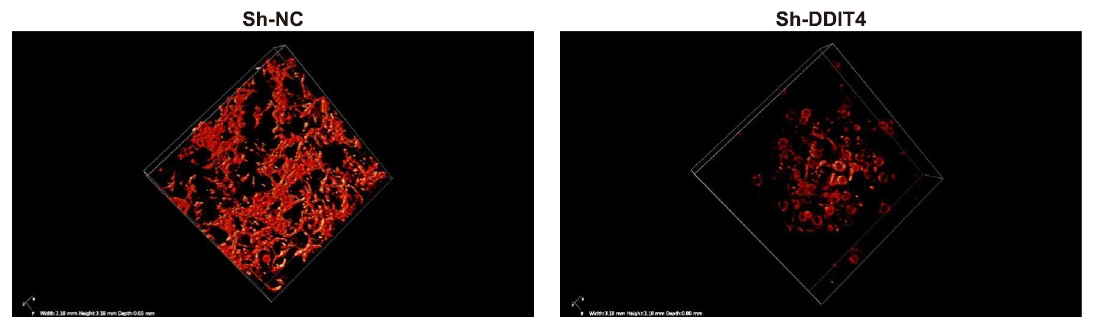
Canine Fibrinogen purchased from MedChemExpress. Usage Cited in: Microvasc Res. 2025 Sep:161:104833. [Abstract]
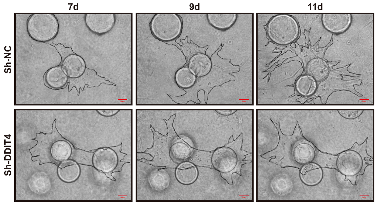
Fibrinogen (2 mg/mL; DPBS) was used to construct the VMs 3D cell model with or without DDIT4 knockdown.
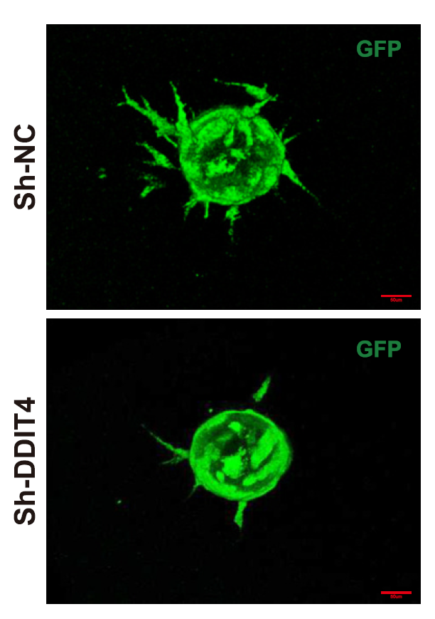
Canine Fibrinogen purchased from MedChemExpress. Usage Cited in: Microvasc Res. 2025 Sep:161:104833. [Abstract]
Fibrinogen (2 mg/mL; DPBS). Vascular sprouting in the VMs 3D cell model after 7 days of culture.
Canine Fibrinogen purchased from MedChemExpress. Usage Cited in: Microvasc Res. 2025 Sep:161:104833. [Abstract]
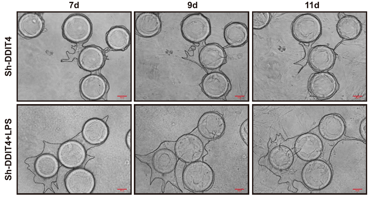
Fibrinogen (2 mg/mL; DPBS). Time-lapse analysis on days 7, 9, and 11 showed the angiogenesis rate in the VMs 3D cell model.
Canine Fibrinogen purchased from MedChemExpress. Usage Cited in: Microvasc Res. 2025 Sep:161:104833. [Abstract]
Fibrinogen (2 mg/mL; DPBS). Time-lapse analysis on days 7, 9, and 11 showed the angiogenesis rate in the DDIT4 knockdown VMs 3D cell model.
Canine Fibrinogen purchased from MedChemExpress. Usage Cited in: Microvasc Res. 2025 Sep:161:104833. [Abstract]
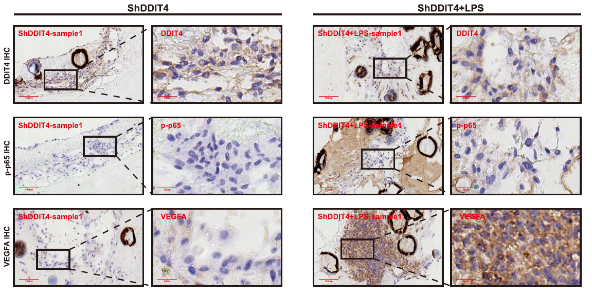
Fibrinogen (2 mg/mL; DPBS). Immunohistochemical (IHC) staining of DDIT4, phospho-p65, and VEGFA in the LPS-stimulated DDIT4 knockdown VMs 3D cell model was performed.


Purity & Documentation
References

Calculators
Concentration (start) × Volume (start) = Concentration (final) × Volume (final)


Canine Fibrinogen Related Classifications
HY-125864A Related Classifications
 Powered by Bioz