



CC-90001
Based on 3 publication(s) in Google Scholar
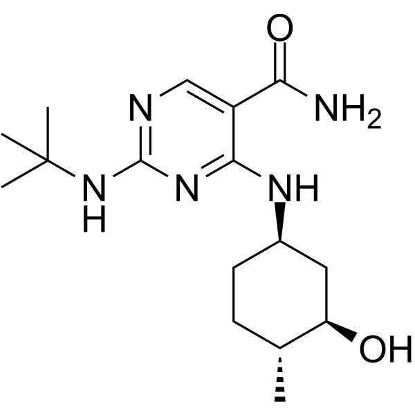
CC-90001 is a potent, selective and orally active inhibitor of c-Jun N-terminal kinase (JNK). CC-90001 shows 12.9-fold selectivity for JNK1 over JNK2 in a cell-based model. CC-90001 can be used for the research of idiopathic pulmonary fibrosis.
Nur für Forschungszwecke. Wir verkaufen nicht an Patienten.
- Reinheit : 99.98%
- CAS. Nr.: 1403859-14-2
- Formel: C16H27N5O2
- Molecular Weight:321.42
-
Speicherung:Powder -20°C, 3 years ; In solvent -80°C, 6 months , -20°C, 1 month

To place orders, for customer services and technical support, please contact: MedChemExpress USA
Tel: 609-228-6898 E-mail: [email protected] [email protected]


-
 Biologische Aktivität
-
Chemical Information
-
Lösungsmittel & Löslichkeit
- Protokoll
- Reinheit & Dokumentation
- Verweise
-
 Help & FAQs


-
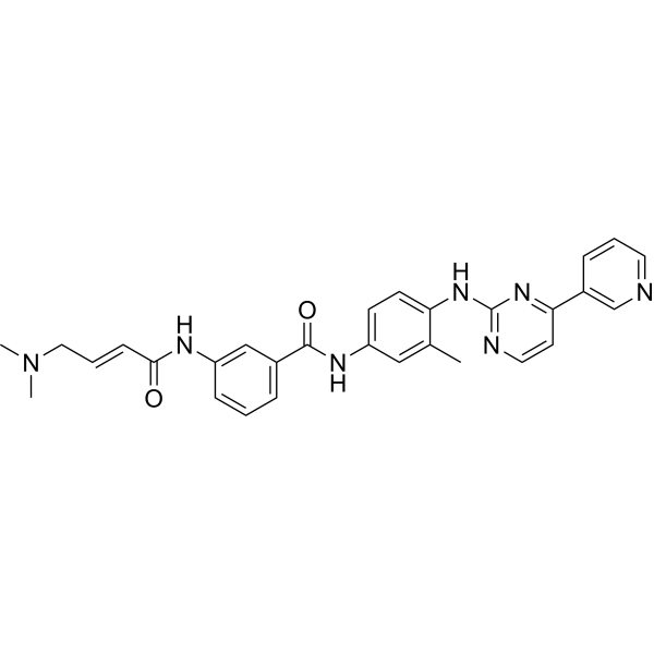
HY-13319
JNK-IN-8JNK Inhibitor -
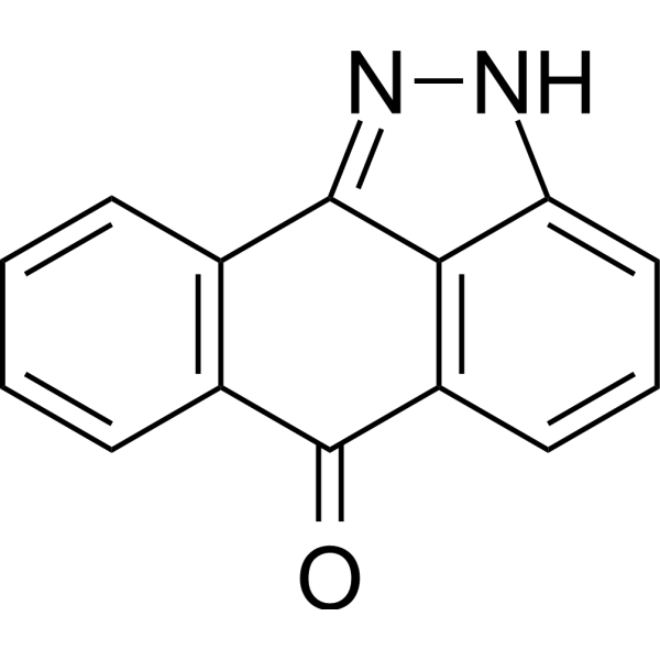
HY-12041
SP600125JNK Inhibitor -
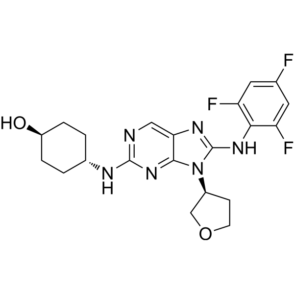
HY-15495
Tanzisertib -
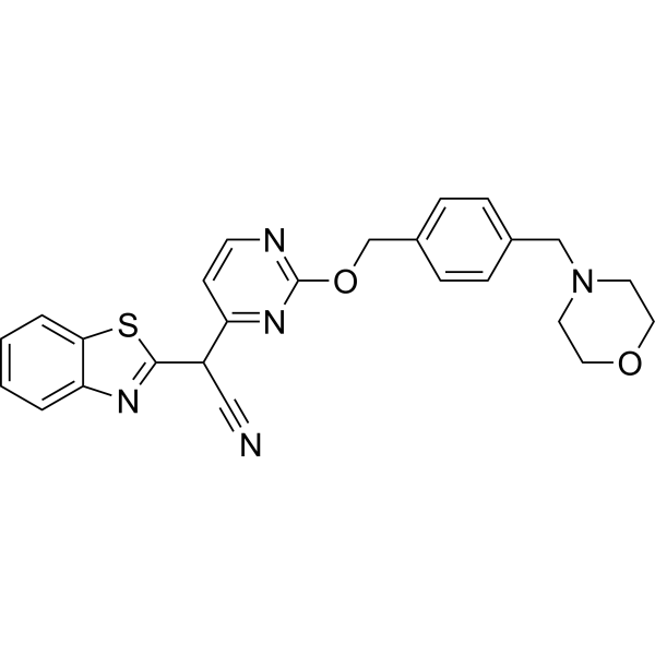
HY-14761
BentamapimodJNK Inhibitor -
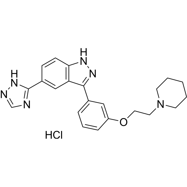
HY-13022
CC-401 hydrochlorideJNK Inhibitor -
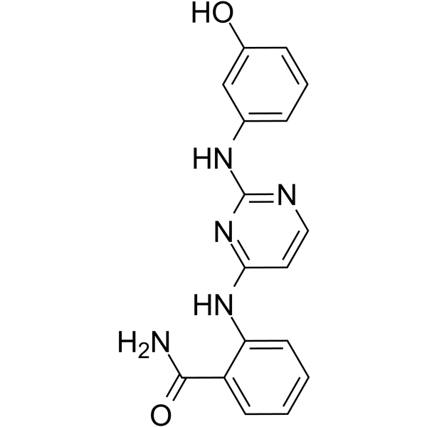
HY-15737
DB07268JNK Inhibitor -
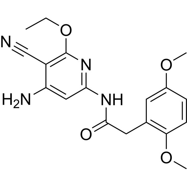
HY-107598
JNK Inhibitor VIIIJNK Inhibitor -
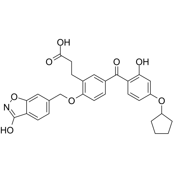
HY-12270
T-5224c-Fos/AP-1 Inhibitor -
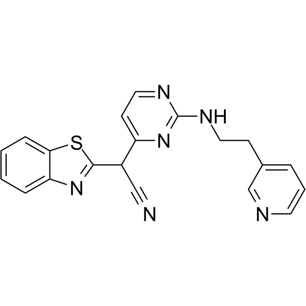
HY-11010
AS601245JNK Inhibitor -
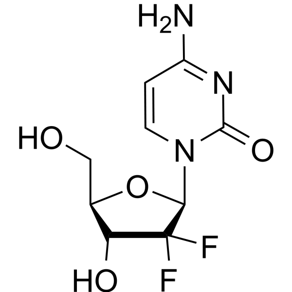
HY-17026
GemcitabineDNA Synthesis Inhibitor -

HY-10182
LaduviglusibGSK-3 Inhibitor -
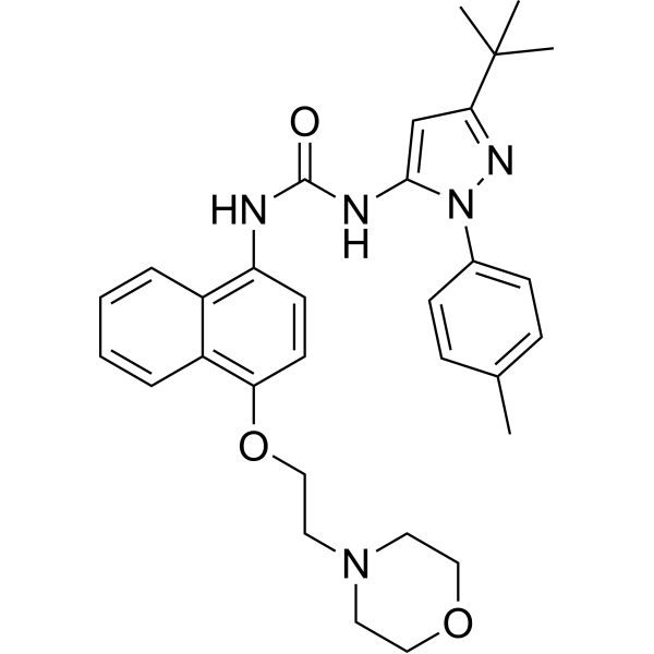
HY-10320
Doramapimodp38 MAPK Inhibitor
Publications Citing Use of MedChemExpress (MCE) CC-90001
More

Biologische Aktivität
Beschreibung
IC50 & Target
[1]|
JNK1 |
JNK2 |
In Vitro
CC-90001 blocks LPS-induced c-jun phosphorylation with an EC50 of 480 nM in cellular assays[1].
CC-90001 is 12.9-fold more potent for JNK1 inhibition than JNK2 using JNK1 and JNK2 knockout fibroblasts[1].
MedChemExpress (MCE) has not independently confirmed the accuracy of these methods. They are for reference only.
In Vivo
CC-90001 decreases multiple measures of lung collagen and reduces disease induced increases in α-SMA to nearly baseline levels in a house dust mite model of lung fibrosis[1].
MedChemExpress (MCE) has not independently confirmed the accuracy of these methods. They are for reference only.
Clinical Trial
| NCT Number | Sponsor | Condition | Start Date |
Phase

|
|---|---|---|---|---|
| NCT01329991 | 2011-05 | PHASE1 |


Chemical Information
-
CAS. Nr. 1403859-14-2
-
Appearance Solid
-
Molecular Weight 321.42
-
Formel C16H27N5O2
-
Color White to yellow
-
SMILES
O=C(C1=CN=C(NC(C)(C)C)N=C1N[C@H]2C[C@@H](O)[C@H](C)CC2)N
-
Versand
Room temperature in continental US; may vary elsewhere.
-
Speicherung
Powder -20°C 3 years In solvent -80°C 6 months -20°C 1 month


Publications (3)
-
Journal Impact Factor
-
Most Recent
-
Cell Discov
ASCT2 palmitoylation regulated by JNK1-ZDHHC14 axis orchestrates glutamine metabolism and NSCLC progression. [Abstract]2026 Feb 24;12(1):13. PMID: 41730846 -
NPJ Precis Oncol
Lestaurtinib's antineoplastic activity converges on JAK/STAT signaling to inhibit treatment naïve and therapy resistant forms ovarian cancer. [Abstract]2025 Jul 11;9(1):236. PMID: 40646138 -
Explor Target Antitumor Ther
Acryl-3,5-bis(2,4-difluorobenzylidene)-4-piperidone targeting cellular JUN proto-oncogene, AP-1 transcription factor subunit inhibits head and neck squamous cell carcinoma progression. [Abstract]2023;4(5):1104-1121. PMID: 38023989


Lösungsmittel & Löslichkeit
In Vitro:
DMSO : 125 mg/mL (388.90 mM; Need ultrasonic; Hygroscopic DMSO has a significant impact on the solubility of product, please use newly opened DMSO)
Please refer to the solubility information to select the appropriate solvent. Once prepared, please aliquot and store the solution to prevent product inactivation from repeated freeze-thaw cycles.
Storage method and period of stock solution: -80°C, 6 months; -20°C, 1 month. When stored at -80°C, please use it within 6 months. When stored at -20°C, please use it within 1 month.
Please refer to the solubility information to select the appropriate solvent. Once prepared, please aliquot and store the solution to prevent product inactivation from repeated freeze-thaw cycles.
Storage method and period of stock solution: -80°C, 6 months; -20°C, 1 month. When stored at -80°C, please use it within 6 months. When stored at -20°C, please use it within 1 month.
Konzentration (Stammlösung) × Volumen (Stammlösung) = Konzentration (Ziellösung) × Volumen (Ziellösung)
In Vivo:
Select the appropriate dissolution method based on your experimental animal and administration route.
- For the following dissolution methods, please ensure to first prepare a clear stock solution using an In Vitro approach and then sequentially add co-solvents:
- To ensure reliable experimental results, the clarified stock solution can be appropriately stored based on storage conditions. As for the working solution for In Vivo experiments, it is recommended to prepare freshly and use it on the same day.
- The percentages shown for the solvents indicate their volumetric ratio in the final prepared solution. If precipitation or phase separation occurs during preparation, heat and/or sonication can be used to aid dissolution.
Add each solvent one by one: 10% DMSO 40% PEG300 5% Tween-80 45% Saline
Solubility: ≥ 2.08 mg/mL (6.47 mM); Clear solution
This protocol yields a clear solution of ≥ 2.08 mg/mL (saturation unknown).
Taking 1 mL working solution as an example, add 100 μL DMSO stock solution (20.8 mg/mL) to 400 μL PEG300, and mix evenly; then add 50 μL Tween-80 and mix evenly; then add 450 μL Saline to adjust the volume to 1 mL.
Preparation of Saline: Dissolve 0.9 g sodium chloride in ddH₂O and dilute to 100 mL to obtain a clear Saline solution.
Add each solvent one by one: 10% DMSO 90% (20% SBE-β-CD in Saline)
Solubility: ≥ 2.08 mg/mL (6.47 mM); Clear solution
This protocol yields a clear solution of ≥ 2.08 mg/mL (saturation unknown).
Taking 1 mL working solution as an example, add 100 μL DMSO stock solution (20.8 mg/mL) to 900 μL 20% SBE-β-CD in Saline, and mix evenly.
Preparation of 20% SBE-β-CD in Saline (4°C, storage for one week): 2 g SBE-β-CD powder is dissolved in 10 mL Saline, completely dissolve until clear.
In Vivo Dissolution Calculator
Please enter the basic information of animal experiments:
-
-
-
-
Recommended: Prepare an additional quantity of animals to account for potential losses during experiments.
Please enter your animal formula composition:
-
%DMSO +
Recommended: Keep the proportion of DMSO in working solution below 2% if your animal is weak.
-
%+
-
+%Tween-80 + +
-
%Saline +
The co-solvents required include: DMSO, . All of co-solvents are available by MedChemExpress (MCE). , Tween 80. All of co-solvents are available by MedChemExpress (MCE).
Working solution concentration: 0.22 mg/mL
Method for preparing stock solution: mg drug dissolved in μL DMSO. Stock solution concentration: mg/mL.
1. Take μL DMSO stock solution;
2. Add μL .
μL , mix evenly;
3. Then add μL Tween 80, mix evenly;
4. Then add μL
Please ensure that the stock solution in the first step is dissolved to a clear state, and add co-solvents in sequence. You can use ultrasonic heating (ultrasonic cleaner, recommended frequency 20-40 kHz), vortexing, etc. to assist dissolution.


Reinheit & Dokumentation
-
Data Sheet (276 KB)
-
SDS (252 KB)
- English - EN (252 KB)
- Français - FR (252 KB)
- Deutsch - DE (252 KB)
- Norwegian - NO (252 KB)
- Español - ES (252 KB)
- Swedish - SV (252 KB)
- Italian - IT (252 KB)
- Korean - KR (252 KB)
- Portuguese - PT (252 KB)
-
Handling Instructions (2659 KB)
Verweise


Complete Stock Solution Preparation Table
Please refer to the solubility information to select the appropriate solvent. Once prepared, please aliquot and store the solution to prevent product inactivation from repeated freeze-thaw cycles.
Storage method and period of stock solution: -80°C, 6 months; -20°C, 1 month. When stored at -80°C, please use it within 6 months. When stored at -20°C, please use it within 1 month.
| Optional Solvent | Concentration Solvent Mass | 1 mg | 5 mg | 10 mg | 25 mg |
|---|---|---|---|---|---|
| DMSO | 1 mM | 3.1112 mL | 15.5560 mL | 31.1119 mL | 77.7798 mL |
| 5 mM | 0.6222 mL | 3.1112 mL | 6.2224 mL | 15.5560 mL | |
| 10 mM | 0.3111 mL | 1.5556 mL | 3.1112 mL | 7.7780 mL | |
| 15 mM | 0.2074 mL | 1.0371 mL | 2.0741 mL | 5.1853 mL | |
| 20 mM | 0.1556 mL | 0.7778 mL | 1.5556 mL | 3.8890 mL | |
| 25 mM | 0.1244 mL | 0.6222 mL | 1.2445 mL | 3.1112 mL | |
| 30 mM | 0.1037 mL | 0.5185 mL | 1.0371 mL | 2.5927 mL | |
| 40 mM | 0.0778 mL | 0.3889 mL | 0.7778 mL | 1.9445 mL | |
| 50 mM | 0.0622 mL | 0.3111 mL | 0.6222 mL | 1.5556 mL | |
| 60 mM | 0.0519 mL | 0.2593 mL | 0.5185 mL | 1.2963 mL | |
| 80 mM | 0.0389 mL | 0.1944 mL | 0.3889 mL | 0.9722 mL | |
| 100 mM | 0.0311 mL | 0.1556 mL | 0.3111 mL | 0.7778 mL |


CC-90001 Related Classifications
HY-138304 Related Classifications
 Powered by Bioz