



MPI-0441138
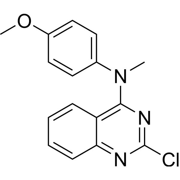
MPI-0441138 is an inducer of apoptosis and an inhibitor of mitosis. MPI-0441138 can induce DNA breaks and induce cytochrome c translocation, leading to tumor cell death.
Nur für Forschungszwecke. Wir verkaufen nicht an Patienten.
- CAS. Nr.: 827030-33-1
- Formel: C16H14ClN3O
- Molecular Weight:299.75
-
Speicherung:
Please store the product under the recommended conditions in the Certificate of Analysis.

To place orders, for customer services and technical support, please contact: MedChemExpress USA
Tel: 609-228-6898 E-mail: [email protected] [email protected]


-
 Biologische Aktivität
-
Chemical Information
- Protokoll
- Reinheit & Dokumentation
- Verweise
-
 Help & FAQs


-
HY-14590
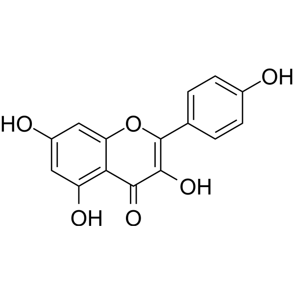
KaempferolEstrogen Receptor α Inhibitor -
HY-14590R
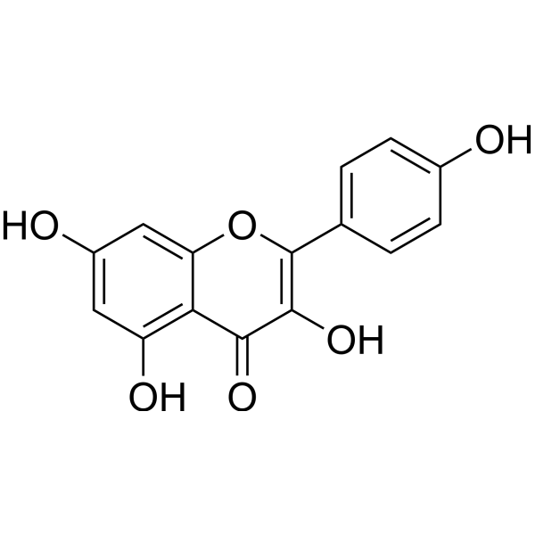
Kaempferol (Standard) -
HY-N1462
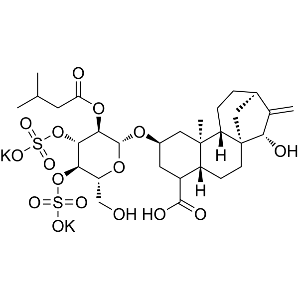
Atractyloside potassium saltADP/ATP Transport Inhibitor -
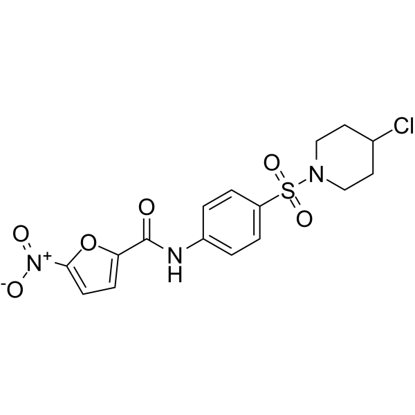
HY-101991
ML291UPR Signaling Activator -
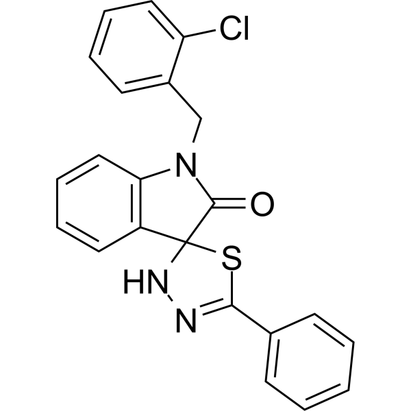
HY-103255
CFM-4CARP-1/APC-2 Binding Antagonist -
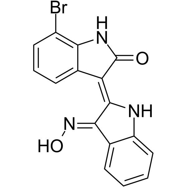
HY-121035
7BIOIndirubin Derivate -
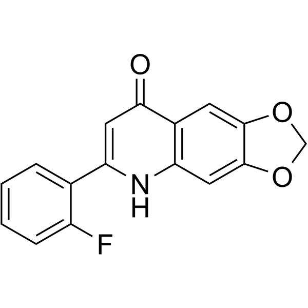
HY-103257
CHM-1Tubulin Polymerization Inhibitor -

HY-N1462R
Atractyloside potassium salt (Standard)ADP/ATP Transport Inhibitor -

HY-101991R
ML291 (Standard)Reference Standard -

HY-103257R
CHM-1 (Standard)Reference Standard -
HY-103256
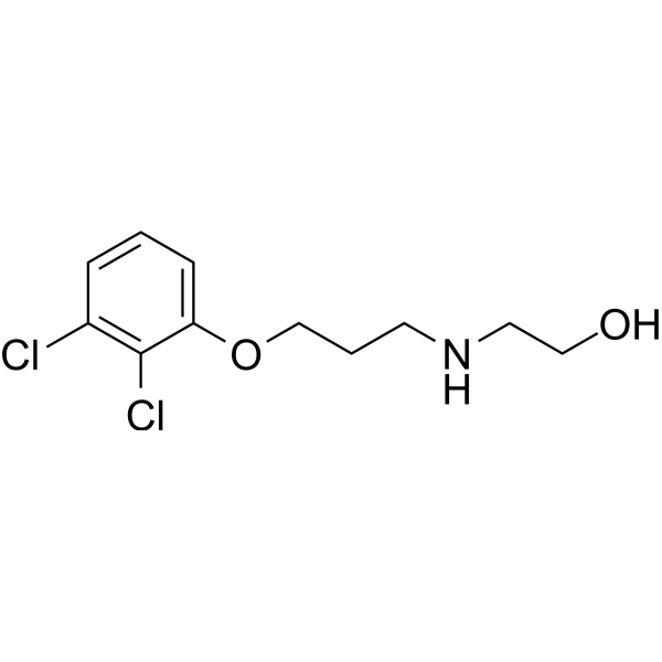
2,3-DCPEApoptosis Inducer


Biologische Aktivität
Beschreibung
Cellular Effect
|
Cell Line

|
Type | Value | Description | References |
|---|---|---|---|---|
| A2780 | IC50 |
1.085 nM
Compound: 25g
|
Antiproliferative activity against human A2780 cells incubated for 48 hrs by CCK8 assay
Antiproliferative activity against human A2780 cells incubated for 48 hrs by CCK8 assay
|
[PMID: 38972078] |
| A-431 | IC50 |
1.2 nM
Compound: 8
|
Cytotoxicity against human A-431 cells assessed as reduction in cell viability by Sulforhodamine B assay
Cytotoxicity against human A-431 cells assessed as reduction in cell viability by Sulforhodamine B assay
|
[PMID: 33647840] |
| A549 | IC50 |
1.28 nM
Compound: 25g
|
Antiproliferative activity against human A549 cells incubated for 48 hrs by CCK8 assay
Antiproliferative activity against human A549 cells incubated for 48 hrs by CCK8 assay
|
[PMID: 38972078] |
| HCC827 | IC50 |
1.25 nM
Compound: 25g
|
Antiproliferative activity against human HCC827 cells incubated for 48 hrs by CCK8 assay
Antiproliferative activity against human HCC827 cells incubated for 48 hrs by CCK8 assay
|
[PMID: 38972078] |
| HCT-116 | EC50 |
0.002 μM
Compound: 2b, EPI28265
|
Induction of apoptosis in human HCT116 cells assessed as caspase 3 activation after 24 hrs
Induction of apoptosis in human HCT116 cells assessed as caspase 3 activation after 24 hrs
|
[PMID: 18651728] |
| HCT-116 | EC50 |
0.002 μM
Compound: 6b, EP128265, MPI-0441138
|
Induction of apoptosis in human HCT116 cells assessed as caspase activation after 24 hrs by HTS assay
Induction of apoptosis in human HCT116 cells assessed as caspase activation after 24 hrs by HTS assay
|
[PMID: 19296653] |
| HCT-116 | GI50 |
0.002 μM
Compound: 2b, EPI28265
|
Growth inhibition of human HCT116 cells after 48 hrs by CellTiter-Glo assay
Growth inhibition of human HCT116 cells after 48 hrs by CellTiter-Glo assay
|
[PMID: 18651728] |
| HeLa | IC50 |
0.6 nM
Compound: 8
|
Cytotoxicity against human HeLa cells assessed as reduction in cell viability by Sulforhodamine B assay
Cytotoxicity against human HeLa cells assessed as reduction in cell viability by Sulforhodamine B assay
|
[PMID: 33647840] |
| HeLa | IC50 |
0.6 nM
Compound: 8
|
Cytotoxicity against human HeLa cells over expressing WT-beta3 assessed as reduction in cell viability by Sulforhodamine B assay
Cytotoxicity against human HeLa cells over expressing WT-beta3 assessed as reduction in cell viability by Sulforhodamine B assay
|
[PMID: 33647840] |
| MDA-MB-435 | IC50 |
0.6 nM
Compound: 8
|
Antiproliferative activity against human MDA-MB-435 cells assessed as reduction in cell viability by Sulforhodamine B assay
Antiproliferative activity against human MDA-MB-435 cells assessed as reduction in cell viability by Sulforhodamine B assay
|
[PMID: 33647840] |
| P388 | IC50 |
2.1 nM
Compound: 2b, EPI28265
|
Cytotoxicity against mouse P388 cells
Cytotoxicity against mouse P388 cells
|
[PMID: 18651728] |
| P388 | IC50 |
2.2 nM
Compound: 2b, EPI28265
|
Cytotoxicity against adriamycin-resistant mouse P388 cells expressing p-glycoprotein
Cytotoxicity against adriamycin-resistant mouse P388 cells expressing p-glycoprotein
|
[PMID: 18651728] |
| SK-OV-3 | IC50 |
0.78 nM
Compound: 25g
|
Antiproliferative activity against human SK-OV-3 cells incubated for 48 hrs by CCK8 assay
Antiproliferative activity against human SK-OV-3 cells incubated for 48 hrs by CCK8 assay
|
[PMID: 38972078] |
| SK-OV-3 | IC50 |
0.8 nM
Compound: 8
|
Cytotoxicity against human SK-OV-3 cells assessed as reduction in cell viability by Sulforhodamine B assay
Cytotoxicity against human SK-OV-3 cells assessed as reduction in cell viability by Sulforhodamine B assay
|
[PMID: 33647840] |
| SK-OV-3 | IC50 |
1.1 nM
Compound: 8
|
Cytotoxicity against human SK-OV-3 cells over expressing MDR1-6/6 assessed as reduction in cell viability by Sulforhodamine B assay
Cytotoxicity against human SK-OV-3 cells over expressing MDR1-6/6 assessed as reduction in cell viability by Sulforhodamine B assay
|
[PMID: 33647840] |
| SNU-398 | EC50 |
0.002 μM
Compound: 6b, EP128265, MPI-0441138
|
Induction of apoptosis in human SNU398 cells assessed as caspase activation after 24 hrs by HTS assay
Induction of apoptosis in human SNU398 cells assessed as caspase activation after 24 hrs by HTS assay
|
[PMID: 19296653] |
| T47D | EC50 |
0.002 μM
Compound: 2b, EPI28265
|
Induction of apoptosis in human T47D cells assessed as caspase 3 activation after 24 hrs
Induction of apoptosis in human T47D cells assessed as caspase 3 activation after 24 hrs
|
[PMID: 18651728] |
| T47D | EC50 |
0.002 μM
Compound: 6b, EP128265, MPI-0441138
|
Induction of apoptosis in human T47D cells assessed as caspase activation after 24 hrs by HTS assay
Induction of apoptosis in human T47D cells assessed as caspase activation after 24 hrs by HTS assay
|
[PMID: 19296653] |
| T47D | GI50 |
0.001 μM
Compound: 2b, EPI28265
|
Growth inhibition of human T47D cells after 48 hrs by CellTiter-Glo assay
Growth inhibition of human T47D cells after 48 hrs by CellTiter-Glo assay
|
[PMID: 18651728] |
| U-251 | IC50 |
32.5 nM
Compound: 8
|
Cytotoxicity against human U-251 cells assessed as reduction in cell viability by Sulforhodamine B assay
Cytotoxicity against human U-251 cells assessed as reduction in cell viability by Sulforhodamine B assay
|
[PMID: 33647840] |
In Vitro
MPI-0441138 (5 nM; 72 h) idnuces apoptosis and DNA fragmentation in 22Rv1, MDA-MB-231, Caco-2, and Du145 cells[1].
MPI-0441138 (5 nM; 1 h) induces AIF and cytochrome c translocation from mitochondria to cytosol and nucleus[1].
MedChemExpress (MCE) has not independently confirmed the accuracy of these methods. They are for reference only.
-
Cell Line:22Rv1, MDA-MB-231, Caco-2, and Du145 cells
-
Concentration:5 nM
-
Incubation Time:12, 24, 48, or 72 h
-
Result:Resulted robust caspase-3 activation and poly(ADP-ribose) polymerase cleavage, induced DNA fragmentation.


Chemical Information
-
CAS. Nr. 827030-33-1
-
Molecular Weight 299.75
-
Formel C16H14ClN3O
-
SMILES
CN(C1=CC=C(OC)C=C1)C2=C3C=CC=CC3=NC(Cl)=N2
-
Versand
Room temperature in continental US; may vary elsewhere.
-
Speicherung
Please store the product under the recommended conditions in the Certificate of Analysis.


Reinheit & Dokumentation
Verweise

Calculators
Konzentration (Stammlösung) × Volumen (Stammlösung) = Konzentration (Ziellösung) × Volumen (Ziellösung)


MPI-0441138 Related Classifications
HY-129715 Related Classifications
 Powered by Bioz