



Alexidine
Based on 5 publication(s) in Google Scholar
Alexidine, a bis-biguanide, exhibits antifungal and antibiofilm activity against a diverse range of fungal pathogens. Alexidine is an anticancer agent that targets a mitochondrial tyrosine phosphatase, PTPMT1, in mammalian cells and causes mitochondrial apoptosis.
For research use only. We do not sell to patients.
- CAS No.: 22573-93-9
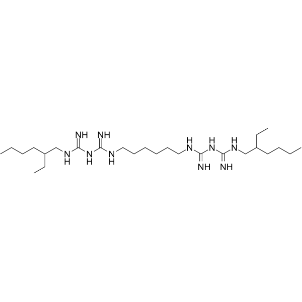
- Formula: C26H56N10
- Molecular Weight:508.79
-
Storage:
Please store the product under the recommended conditions in the Certificate of Analysis.

To place orders, for customer services and technical support, please contact: MedChemExpress USA
Tel: 609-228-6898 E-mail: [email protected] [email protected]


-
 Biological Activity
-
Chemical Information
- Protocols
- Purity & Documentation
- References
-
 Help & FAQs


Publications Citing Use of MedChemExpress (MCE) Alexidine
More

Biological Activity
Description
In Vitro
Alexidine (10 µM, 24 hours) treatment can decimate the biofilm community[1].
Alexidine (0-60 μg/ml, 24 hours) treatment can kill HUVECs and lung A549 cells[1].
MedChemExpress (MCE) has not independently confirmed the accuracy of these methods. They are for reference only.
-
Cell Line:C. albicans, C. auris, A. fumigatus
-
Concentration:10 µM
-
Incubation Time:24 hours
-
Result:Could significantly kill 80% of mature biofilm community.
-
Cell Line:HUVECs, lung A549 cells
-
Concentration:0-60 μg/ml
-
Incubation Time:24 hours
-
Result:resulted in 50% killing of HUVECs and lung A549 cells ( CC50 > 7.37 μg/ml ).
In Vivo
MedChemExpress (MCE) has not independently confirmed the accuracy of these methods. They are for reference only.
-
Animal Model:8-week-old C57BL/6 male mice
-
Dosage:3 µg/ml
-
Administration:Jugular vein-catheterized, 48 hours, 3 µg/ml, once
-
Result:Inhibited 67% of fungal biofilm growth and viability, compared to the control untreated biofilms.


Chemical Information
-
CAS No. 22573-93-9
-
Molecular Weight 508.79
-
Formula C26H56N10
-
SMILES
N=C(NCC(CC)CCCC)NC(NCCCCCCNC(NC(NCC(CC)CCCC)=N)=N)=N
-
Shipping
Room temperature in continental US; may vary elsewhere.
-
Storage
Please store the product under the recommended conditions in the Certificate of Analysis.


Publications (5)
-
Journal Impact Factor
-
Most Recent
-
Cell Death Dis
Pharmacological targeting of the mitochondrial phosphatase PTPMT1 sensitizes hepatocellular carcinoma to ferroptosis. [Abstract]2025 Apr 6;16(1):257. PMID: 40189563 -
Free Radic Biol Med
Alexidine dihydrochloride enhances the sensitivity of human hepatocellular carcinoma to disulfidptosis via ATF4-DDIT3 activation. [Abstract]2025 Jun 14:237:585-599. PMID: 40518125 -
Cell Rep
A generalized strategy to kill leukemic cells by targeting the regulatory systems governing mitochondrial membrane potential. [Abstract]2025 Nov 25;44(11):116496. PMID: 41166305 -
Biochem Biophys Res Commun
2026 Jan 18:796:153158. PMID: 41418346 -


Purity & Documentation
References

Calculators
Concentration (start) × Volume (start) = Concentration (final) × Volume (final)


Alexidine Related Classifications
HY-B1474 Related Classifications
 Powered by Bioz