



Pranidipine

Based on 1 Customer Validation
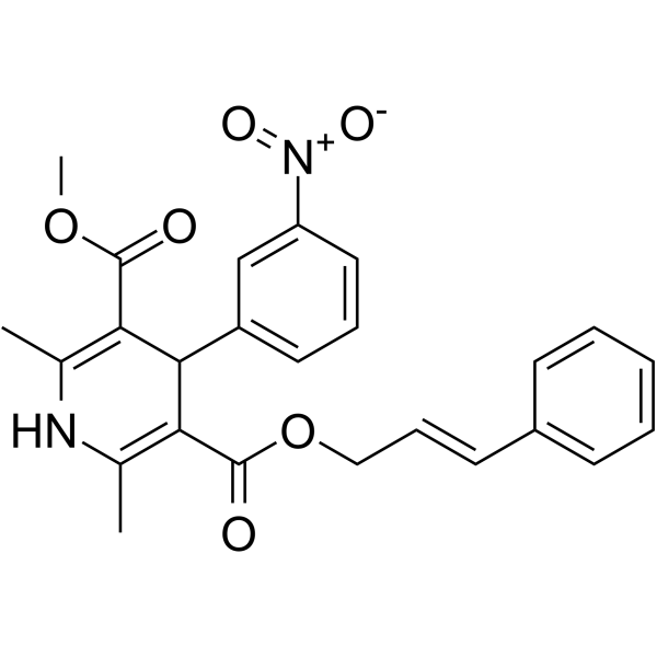
Pranidipine (OPC-13340) is an orally active L-type voltage-dependent calcium channel (L-VDCC) blocker with a Ki value of 0.16 nM. Pranidipine inhibits calcium-induced contraction, suppresses slow-response action potentials, shortens action potential duration, reduces systolic and diastolic blood pressure, and exerts vasodilatory effects. Pranidipine enhances its vasodilatory effect by blocking NO decomposition. Pranidipine can be used in research related to essential hypertension, angina pectoris, myocardial infarction, and dilated cardiomyopathy.
Nos produits utilisent uniquement pour la recherche. Nous ne vendons pas aux patients.
- Pureté : 99.52%
- CAS No.: 99522-79-9
- Formule: C25H24N2O6
- Masse moléculaire:448.47
-
Stockage:Powder -20°C, 3 years , 4°C, 2 years ; In solvent -80°C, 2 years , -20°C, 1 year

To place orders, for customer services and technical support, please contact: MedChemExpress USA
Tel: 609-228-6898 E-mail: [email protected] [email protected]


-
 Activité biologique
-
Chemical Information
-
Solvant et solubilité
- Protocole
- Pureté et documentation
- Références
-
 Help & FAQs


Voir tous les produits spécifiques à Isoform Calcium Channel
More

Activité biologique
Description
IC50 & Target
[1]|
L-type calcium channel 0.16 nM (Ki) |
Cellular Effect
|
Cell Line

|
Type | Value | Description | References |
|---|---|---|---|---|
| HEK293 | IC50 |
37 nM
Compound: 100
|
Antagonist activity at rabbit Cav1.2 expressed in HEK293 cells assessed as inhibition of voltage pulse-induced calcium current by FLIPR calcium 4 assay
Antagonist activity at rabbit Cav1.2 expressed in HEK293 cells assessed as inhibition of voltage pulse-induced calcium current by FLIPR calcium 4 assay
|
[PMID: 20382537] |
| HEK293 | IC50 |
37 nM
Compound: 100
|
Antagonist activity at rat Cav1.3 expressed in HEK293 cells assessed as inhibition of voltage pulse-induced calcium current by FLIPR calcium 4 assay
Antagonist activity at rat Cav1.3 expressed in HEK293 cells assessed as inhibition of voltage pulse-induced calcium current by FLIPR calcium 4 assay
|
[PMID: 20382537] |
In Vitro
Pranidipine (10-11-10-7 M) potently relaxes KCl-induced contractions of isolated rat thoracic aortic strips, with an ED50 of 5.9×10⁻¹⁰ M, exhibiting selective voltage-dependent calcium channel blocking activity[1].
Pranidipine (10-8-10-5 M) relaxes norepinephrine (HY-13715)-induced contractions of isolated rat thoracic aortic strips, with an ED50 of 8.7×10⁻⁶ M. It exhibits lower blocking potency for receptor-operated calcium channels compared with voltage-dependent calcium channels[1].
Pranidipine (3×10⁻⁶ M) shortens the action potential duration of isolated guinea pig right ventricular papillary muscles and inhibits slow-response action potentials, which is consistent with its calcium channel blocking activity[1].
Pranidipine (10-9-10-7 M; 20 min) does not alter the activity of constitutive nitric oxide synthase in cultured porcine aortic endothelial cells[2].
Pranidipine (10-6 M) enhances NO-mediated effects in an endothelial-smooth muscle cell co-culture system by inhibiting superoxide anion-induced NO decomposition, increasing cGMP accumulation, and reducing endothelin-1 production[3].
MedChemExpress (MCE) has not independently confirmed the accuracy of these methods. They are for reference only. Further protocols information, click here.
In Vivo
Pranidipine (3 mg/kg/day; chronic; 10 weeks) reduces blood pressure, urinary protein excretion, and glomerular/arteriolar sclerosis in subtotal nephrectomized spontaneously hypertensive rats[3].
Pranidipine protects the kidneys and brain from pathological damage and prolongs survival in salt-sensitive Dahl rats[3].
Pranidipine (3 mg/kg; 4 weeks) reduces cardiac pressures, prevents ventricular remodeling, and normalizes neurohormonal levels in Wistar rats with coronary artery ligation-induced myocardial infarction[3].
Pranidipine prevents brain, heart, and kidney injury and prolongs survival in stroke-prone spontaneously hypertensive rats[3].
MedChemExpress (MCE) has not independently confirmed the accuracy of these methods. They are for reference only.
-
Animal Model:Wistar rats (male, ~13 weeks or older, two kidney one-clip renal hypertensive model)[1]
-
Dosage:0.3-3 mg/kg (p.o.)
-
Administration:p.o.
-
Result:Caused dose-dependent decreases in SBP and DBP, with potency 3.5-10 times greater than Nicardipine.
Produced longer duration of hypotensive action than equipotent nicardipine at 1 mg/kg p.o., with a much lower HR-increasing effect.
-
Animal Model:Spontaneously Hypertensive Rats (subtotal nephrectomized)[3]
-
Dosage:3 mg/kg/day
-
Administration:chronic; 10 weeks
-
Result:Reduced systemic blood pressure, urinary protein excretion, and inhibited glomerular and arteriolar sclerosis.
-
Animal Model:Wistar rats (myocardial infarction-induced via coronary artery ligation)[3]
-
Dosage:3 mg/kg
-
Administration:4 weeks
-
Result:Reduced left ventricular end-diastolic pressure (LVEDP) and central venous pressure (CVP).
Prevented increases in left ventricular end-diastolic dimension (LVDd) and left/right ventricular weight.
Normalized plasma renin activity (PRA) and plasma catecholamine levels (epinephrine, norepinephrine, dopamine) that were elevated post-infarction.


Chemical Information
-
CAS No. 99522-79-9
-
Appearance Solid
-
Masse moléculaire 448.47
-
Formule C25H24N2O6
-
Color Light yellow to yellow
-
SMILES
O=C(C1=C(C)NC(C)=C(C(OC/C=C/C2=CC=CC=C2)=O)C1C3=CC=CC([N+]([O-])=O)=C3)OC
-
Synonyms
OPC-13340
-
Livraison
Room temperature in continental US; may vary elsewhere.
-
Stockage
Powder -20°C 3 years 4°C 2 years In solvent -80°C 2 years -20°C 1 year


Solvant et solubilité
In Vitro:
DMSO : 270 mg/mL (602.05 mM; Need ultrasonic; Hygroscopic DMSO has a significant impact on the solubility of product, please use newly opened DMSO)
Please refer to the solubility information to select the appropriate solvent. Once prepared, please aliquot and store the solution to prevent product inactivation from repeated freeze-thaw cycles.
Storage method and period of stock solution: -80°C, 2 years; -20°C, 1 year. When stored at -80°C, please use it within 2 years. When stored at -20°C, please use it within 1 year.
Please refer to the solubility information to select the appropriate solvent. Once prepared, please aliquot and store the solution to prevent product inactivation from repeated freeze-thaw cycles.
Storage method and period of stock solution: -80°C, 2 years; -20°C, 1 year. When stored at -80°C, please use it within 2 years. When stored at -20°C, please use it within 1 year.
Concentration (start) × Volume (start) = Concentration (final) × Volume (final)
In Vivo:
Select the appropriate dissolution method based on your experimental animal and administration route.
- For the following dissolution methods, please ensure to first prepare a clear stock solution using an In Vitro approach and then sequentially add co-solvents:
- To ensure reliable experimental results, the clarified stock solution can be appropriately stored based on storage conditions. As for the working solution for In Vivo experiments, it is recommended to prepare freshly and use it on the same day.
- The percentages shown for the solvents indicate their volumetric ratio in the final prepared solution. If precipitation or phase separation occurs during preparation, heat and/or sonication can be used to aid dissolution.
Add each solvent one by one: 10% DMSO 40% PEG300 5% Tween-80 45% Saline
Solubility: ≥ 2.25 mg/mL (5.02 mM); Clear solution
This protocol yields a clear solution of ≥ 2.25 mg/mL (saturation unknown).
Taking 1 mL working solution as an example, add 100 μL DMSO stock solution (22.5 mg/mL) to 400 μL PEG300, and mix evenly; then add 50 μL Tween-80 and mix evenly; then add 450 μL Saline to adjust the volume to 1 mL.
Preparation of Saline: Dissolve 0.9 g sodium chloride in ddH₂O and dilute to 100 mL to obtain a clear Saline solution.
In Vivo Dissolution Calculator
Please enter the basic information of animal experiments:
-
-
-
-
Recommended: Prepare an additional quantity of animals to account for potential losses during experiments.
Please enter your animal formula composition:
-
%DMSO +
Recommended: Keep the proportion of DMSO in working solution below 2% if your animal is weak.
-
%+
-
+%Tween-80 + +
-
%Saline +
The co-solvents required include: DMSO, . All of co-solvents are available by MedChemExpress (MCE). , Tween 80. All of co-solvents are available by MedChemExpress (MCE).
Working solution concentration: 0.22 mg/mL
Method for preparing stock solution: mg drug dissolved in μL DMSO. Stock solution concentration: mg/mL.
1. Take μL DMSO stock solution;
2. Add μL .
μL , mix evenly;
3. Then add μL Tween 80, mix evenly;
4. Then add μL
Please ensure that the stock solution in the first step is dissolved to a clear state, and add co-solvents in sequence. You can use ultrasonic heating (ultrasonic cleaner, recommended frequency 20-40 kHz), vortexing, etc. to assist dissolution.

Protocole
-
Cardiac voltage-sensitive optical mapping
Cardiac voltage-sensitive optical mapping records changes in transmembrane potential from cardiac tissue by staining the preparation with a voltage-sensitive dye and imaging fluorescence changes during electrical activation; the resulting optical action potentials can be used to map activation time, action potential duration, conduction velocity, wavefront propagation, and arrhythmia dynamics. The optical signal represents a relative fluorescence change from a tissue volume rather than a single-cell intracellular recording, so spatial resolution, sampling rate, voltage resolution, optical magnification, light penetration, and motion control must be considered together when interpreting optical action potentials.
-
Neuronal voltage-sensitive dye imaging
Neuronal voltage-sensitive dye imaging detects membrane-potential-dependent optical changes from dyes associated with neuronal membranes, enabling optical recording of electrical activity from single neurons, dendrites, axons, spines, or neuronal populations in brain slices and cultured neurons. VSD signals are typically reported as fractional fluorescence or absorbance changes over baseline, such as ΔF/F or ΔI/I, and published protocols use high-speed cameras or photodiode arrays because neuronal voltage signals occur on millisecond time scales. Fast VSD imaging can be applied at two common scales: bulk staining of brain slices to measure circuit-level spatiotemporal activity, and single-cell loading or biolistic delivery to record membrane-potential transients from individual neuronal compartments. Optical signals should be interpreted as membrane-potential-related readouts, and validation by simultaneous electrophysiology or pharmacological controls is recommended when the experimen
-
Acute brain-slice whole-cell patch-clamp recording
Acute brain-slice whole-cell patch-clamp recording measures membrane voltage or ionic current from visually targeted cells in living brain slices; after giga-seal formation, the membrane under the pipette is ruptured to provide low-resistance electrical access to the cell interior, enabling current-clamp analysis of excitability and voltage-clamp analysis of synaptic or membrane currents. Acute slices preserve local tissue architecture better than dissociated preparations and allow visually guided recording from defined brain regions or fluorescently labeled cells; however, whole-cell access also permits exchange between pipette solution and cytoplasm, so intracellular dialysis must be considered when interpreting signaling-dependent phenomena.
-
Research Protocol for Cardiovascular Diseases
Cardiovascular disease can be modeled as maladaptive cardiac remodeling, where ischemic injury or pressure overload activates inflammatory signaling, fibroblast activation, extracellular-matrix deposition, cardiomyocyte hypertrophy, vascular remodeling, and progressive ventricular dysfunction. The TGF-β/SMAD axis is a central profibrotic pathway after myocardial injury and pressure overload, while innate immune and cytokine pathways regulate leukocyte recruitment, scar formation, and adverse remodeling. Key unresolved questions include which inflammatory signals are reparative versus harmful, when fibrosis is protective versus maladaptive, and whether pathway inhibition improves function without weakening necessary infarct healing or compensatory remodeling.


Pureté et documentation
-
Fiche technique (282 KB)
-
SDS (252 KB)
- English - EN (252 KB)
- Français - FR (252 KB)
- Deutsch - DE (252 KB)
- Norwegian - NO (252 KB)
- Español - ES (252 KB)
- Swedish - SV (252 KB)
- Italian - IT (252 KB)
- Korean - KR (252 KB)
- Portuguese - PT (252 KB)
-
Instruction de manipulation (2659 KB)
Références
[1]. Nakayama N, et al. Antihypertensive activity of OPC-13340, a new potent and long-acting dihydropyridine calcium antagonist, in rats. J Cardiovasc Pharmacol. 1990;15(5):836-844. [Content Brief]
[2]. Mori T, et al. Pranidipine, a new 1,4-dihydropyridine calcium channel blocker, enhances cyclic GMP-independent nitric oxide-induced relaxation of the rat aorta. Mol Cell Biochem. 1998 Jan;178(1-2):335-43. [Content Brief]
[3]. Mori T, et al. Pranidipine, a 1,4-dihydropyridine calcium channel blocker that enhances nitric oxide-induced vascular relaxation. Cardiovasc Drug Rev. 2001;19(1):1-8. [Content Brief]


Complete Stock Solution Preparation Table
Please refer to the solubility information to select the appropriate solvent. Once prepared, please aliquot and store the solution to prevent product inactivation from repeated freeze-thaw cycles.
Storage method and period of stock solution: -80°C, 2 years; -20°C, 1 year. When stored at -80°C, please use it within 2 years. When stored at -20°C, please use it within 1 year.
| Optional Solvent | Concentration Solvent Mass | 1 mg | 5 mg | 10 mg | 25 mg |
|---|---|---|---|---|---|
| DMSO | 1 mM | 2.2298 mL | 11.1490 mL | 22.2980 mL | 55.7451 mL |
| 5 mM | 0.4460 mL | 2.2298 mL | 4.4596 mL | 11.1490 mL | |
| 10 mM | 0.2230 mL | 1.1149 mL | 2.2298 mL | 5.5745 mL | |
| 15 mM | 0.1487 mL | 0.7433 mL | 1.4865 mL | 3.7163 mL | |
| 20 mM | 0.1115 mL | 0.5575 mL | 1.1149 mL | 2.7873 mL | |
| 25 mM | 0.0892 mL | 0.4460 mL | 0.8919 mL | 2.2298 mL | |
| 30 mM | 0.0743 mL | 0.3716 mL | 0.7433 mL | 1.8582 mL | |
| 40 mM | 0.0557 mL | 0.2787 mL | 0.5575 mL | 1.3936 mL | |
| 50 mM | 0.0446 mL | 0.2230 mL | 0.4460 mL | 1.1149 mL | |
| 60 mM | 0.0372 mL | 0.1858 mL | 0.3716 mL | 0.9291 mL | |
| 80 mM | 0.0279 mL | 0.1394 mL | 0.2787 mL | 0.6968 mL | |
| 100 mM | 0.0223 mL | 0.1115 mL | 0.2230 mL | 0.5575 mL |


Pranidipine Related Classifications
HY-19664 Related Classifications
 Powered by Bioz
Keywords
- Pranidipine
- 99522-79-9
- OPC-13340
- OPC13340
- OPC 13340
- Calcium Channel
- NO Synthase
- porcine aortic endothelial cells
- spontaneously hypertensive rats
- human endothelial-smooth muscle cell co-cultures
- vascular smooth muscle cells
- dilated cardiomyopathy
- guinea pig right ventricular papillary muscles
- angina pectoris
- myocardial infarction
- essential hypertension
- L-type voltage-dependent calcium channel
- Inhibitor
- inhibitor
- inhibit