



Clove oil

Based on 1 Customer Validation
Clove oil (Clove leaf oil,rectified) is a volatile aromatic essential oil with antibacterial, anti-inflammatory and antioxidant activities. Clove oil disrupts the integrity of bacterial cell membranes, releases intracellular nucleic acids and proteins, and inhibits bacterial growth and reproduction. Clove oil scavenges DPPH, ABTS, superoxide anions, hydrogen peroxide and hydroxyl radicals. Clove oil reduces Fe3+ to Fe2+, chelates Fe2+ and iron ions, and inhibits lipid peroxidation in linoleic acid emulsions. Clove oil acts as a natural preservative to reduce the loss of oxidation products. Clove oil can be isolated from the flowers, stems and leaves of Eugenia aromatica and Eugenia caryophyllus, which belong to the Myrtaceae family. Clove oil can be used in studies related to bacterial infection, antioxidation and preservation.
For research use only. We do not sell to patients.
- CAS No.: 8000-34-8
-
Storage:
Please store the product under the recommended conditions in the Certificate of Analysis.

To place orders, for customer services and technical support, please contact: MedChemExpress USA
Tel: 609-228-6898 E-mail: [email protected] [email protected]


-
 Biological Activity
-
Chemical Information
-
Solvent & Solubility
- Protocols
- Purity & Documentation
- References
-
 Help & FAQs


-

HY-Y1888
Corn oil -
HY-117215
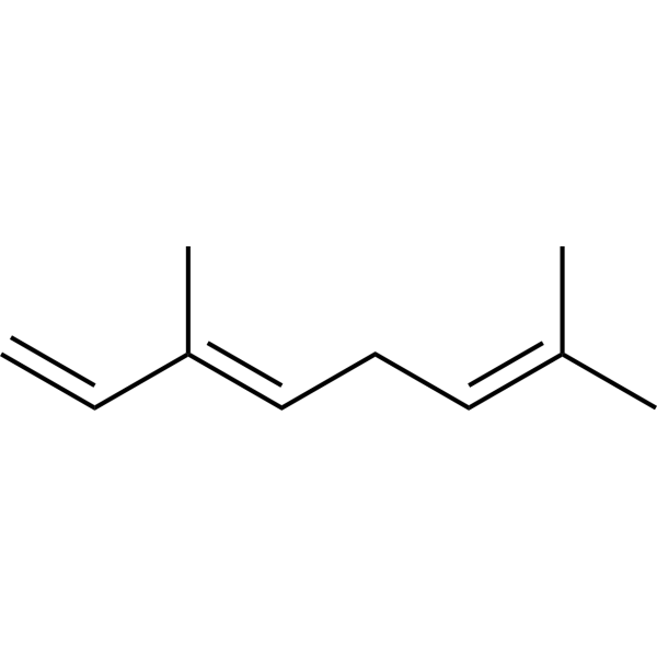
β-OcimeneBiochemical Assay Reagent -
HY-W003972
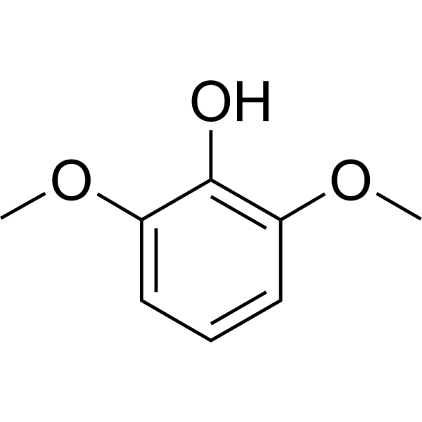
2,6-DimethoxyphenolLaccase Substrate -

HY-W145661
Gum storaxBiochemical Assay Reagent -

HY-W003972R
2,6-Dimethoxyphenol (Standard)Standard of 2,6-Dimethoxyphenol -

HY-159104
Cinnamon bark oil, from Cinnamonum zeylanicum BlumeAntifungal Agent -
HY-W015307
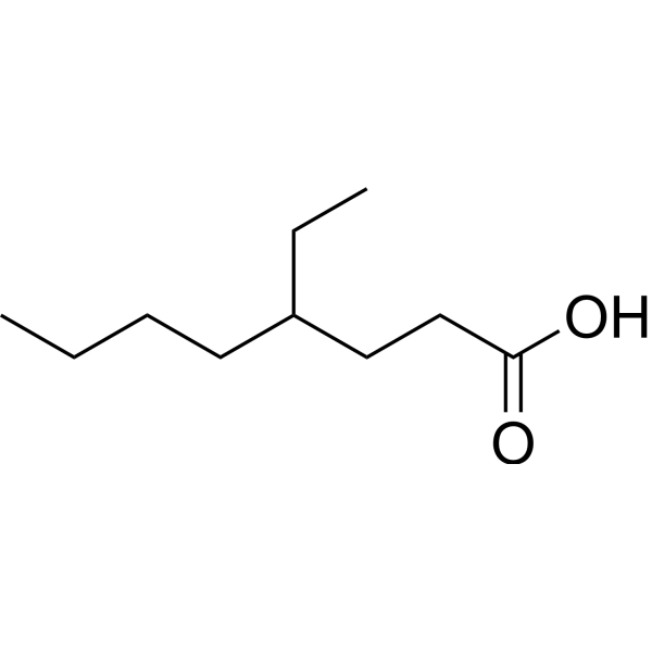
4-Ethyloctanoic acidFragrance Ingredient -
HY-W009443
Diethyl decanedioateBiochemical Assay Reagent -
HY-W010119
4-Methyl-2-phenyl-1,3-dioxolaneBiochemical Assay Reagent -
HY-N9502
Linalool oxideMonoterpene


Biological Activity
Description
In Vitro
Clove oil nanoemulsion (2 wt% clove oil content; 24 h) inhibits growth of Escherichia coli and Staphylococcus aureus via the filter paper diffusion method, with a larger inhibition zone (13.47 mm) observed for Staphylococcus aureus than for Escherichia coli (9.32 mm)[1].
Clove oil nanoemulsion (0.125-4.0 μL/mL; 24 h (MIC), 48 h (MBC)) inhibits growth of Escherichia coli with an MIC of 0.5 mg/mL and kills Escherichia coli with an MBC of 1 mg/mL; it inhibits growth of Staphylococcus aureus with an MIC of 0.25 mg/mL and kills Staphylococcus aureus with an MBC of 2 mg/mL[1].
Clove oil nanoemulsion (0.5 mg/mL (MIC), 1 mg/mL (MBC) for Escherichia coli; 0.25 mg/mL (MIC), 2 mg/mL (MBC) for Staphylococcus aureus; 3 h) disrupts the cell membrane integrity of Escherichia coli and Staphylococcus aureus, causing dose-dependent release of nucleic acids and proteins, with more pronounced nucleic acid release at MBC concentrations[1].
Clove oil (15-45 μg/mL; 30 h) potently inhibits lipid peroxidation of linoleic acid emulsion, with 97.9% inhibition at 15 μg/mL, 99.4% inhibition at 30 μg/mL, and 99.7% inhibition at 45 μg/mL[2].
Clove oil (0.01-0.5 μg/mL; 30 min) potently scavenges DPPH radicals in a cell-free assay with an IC50 of 0.08 μg/mL, achieving 91.2% inhibition at 0.5 μg/mL[3].
Clove oil (0.01-0.2 μg/mL; 1 h) potently scavenges hydroxyl radicals in a cell-free deoxyribose degradation assay with an IC50 of 0.02 μg/mL, achieving 93.7% inhibition at 0.2 μg/mL[3].
Clove oil (0.02-0.2 μg/mL; 1 h) acts as an iron chelator in a cell-free deoxyribose degradation assay, achieving 93% inhibition of metal-dependent hydroxyl radical generation at 0.2 μg/mL[3].
Clove oil (0.0025-0.01 wt%/vol; 10 days) inhibits both early and late stages of lipid peroxidation in a cell-free linoleic acid emulsion system, achieving up to 80.6% inhibition of TBARS formation at 0.01 wt%/vol and 73.91% inhibition of CD formation at 0.005 wt%/vol[3].
MedChemExpress (MCE) has not independently confirmed the accuracy of these methods. They are for reference only. Further protocols information, click here.
Clinical Trial
| NCT Number | Sponsor | Condition | Start Date |
Phase

|
|---|---|---|---|---|
| NCT01329991 | Plexxikon| | 2011-05 | PHASE1 |


Chemical Information
-
CAS No. 8000-34-8
-
Appearance Liquid (Density: 1.05 g/cm3)
-
Color Colorless to light yellow
-
SMILES
[Clove oil]
-
Synonyms
Clove leaf oil,rectified
-
Shipping
Room temperature in continental US; may vary elsewhere.
-
Storage
Please store the product under the recommended conditions in the Certificate of Analysis.


Solvent & Solubility
In Vitro:
DMSO : 100 mg/mL (Need ultrasonic; Hygroscopic DMSO has a significant impact on the solubility of product, please use newly opened DMSO)

Protocols
-
Research Protocol for Infectious Diseases
Infectious-disease experiments test how pathogens interact with host barriers, innate immune receptors, inflammatory signaling, pathogen replication, and tissue injury; pattern-recognition receptors such as TLRs, RIG-I-like receptors, NOD-like receptors, and inflammasomes detect microbial molecules and activate NF-κB, interferon, and cytokine responses. The central hypothesis is that infection severity reflects the balance between pathogen burden and host response: protective inflammation restricts pathogen growth, whereas excessive or mislocalized inflammation contributes to tissue damage and disease phenotype. Unresolved questions include which host pathways are protective versus pathogenic, why some infection models fail to translate to human disease, and which combined readouts best predict clinically relevant infection outcomes.
-
Aerobic Bacterial Batch Culture on Broth/Agar
Aerobic bacterial batch culture grows a closed inoculated population in broth or on agar without continuous medium replacement; growth readouts include turbidity/OD for total suspended biomass and colony-forming units for viable cells able to form colonies on agar. OD-based growth curves reflect light scattering by cells, but OD is instrument-, pathlength-, species-, cell-size-, and density-dependent, so OD should be calibrated or interpreted alongside viable counts when quantitative cell density is required.
-
Research Protocol for Inflammation-related Diseases
The NLRP3 inflammasome is a cytosolic innate immune signaling platform that integrates priming signals and danger-signal activation to promote caspase-1 activation, maturation of IL-1β and IL-18, and gasdermin D-mediated pyroptotic cell death. The core experimental logic is to determine whether inflammatory disease phenotypes are driven by increased NLRP3 expression, ASC-containing inflammasome assembly, caspase-1 cleavage, GSDMD cleavage, and extracellular release of IL-1β/IL-18 rather than by nonspecific cell injury alone. The pathway is strongly linked to inflammation-related disease phenotypes because monosodium urate crystals activate NALP3/NLRP3 inflammasome signaling in gout-like crystal inflammation, cholesterol crystals activate NLRP3 inflammasomes in atherogenesis models, and DSS-induced intestinal inflammation has been reported to involve NLRP3 inflammasome activity. However, experimental colitis studies also show context-dependent protective effects of NLRP3 inflammasome co
-
Ferroptosis Solutions
Ferroptosis is an iron-dependent, non-apoptotic form of regulated cell death characterized by lethal lipid peroxidation and sensitivity to suppression by iron chelators or lipophilic radical-trapping antioxidants. The core pathway links cystine uptake through system Xc−, glutathione availability, GPX4-dependent detoxification of phospholipid hydroperoxides, iron-dependent oxidative reactions, and polyunsaturated-phospholipid metabolism into a cell-death program that is biochemically and morphologically distinct from apoptosis, necrosis, and autophagy. The ferroptosis pathway is experimentally linked to phenotype through chemical and genetic perturbation. Erastin induces ferroptosis by inhibiting cystine uptake through system Xc− and weakening antioxidant defenses, while GPX4 inhibition or depletion causes lipid peroxide accumulation and ferroptotic cancer-cell death. ACSL4 and oxidizable arachidonoyl- or adrenoyl-containing phosphatidylethanolamines shape ferroptosis sensitivity by con


Purity & Documentation
-
Data Sheet (279 KB)
-
SDS (393 KB)
- English - EN (393 KB)
- Français - FR (393 KB)
- Deutsch - DE (393 KB)
- Norwegian - NO (393 KB)
- Español - ES (393 KB)
- Swedish - SV (393 KB)
- Italian - IT (393 KB)
- Korean - KR (393 KB)
- Portuguese - PT (393 KB)
-
Handling Instructions (2659 KB)
References

Calculators
Concentration (start) × Volume (start) = Concentration (final) × Volume (final)


Clove oil Related Classifications
HY-N1953 Related Classifications
 Powered by Bioz