



Cytochalasin J
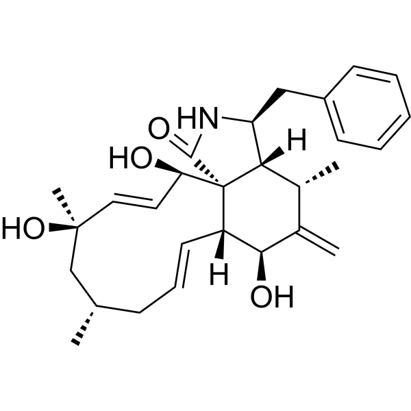
Cytochalasin J is a cytochalasin detivative. Cytochalasin J affects mitotic spindel microtubule organization and kinetochore structure, and exhibits cytotoxicity in human leukemia K562 cell with IC50 of 1.5 μM.
Nos produits utilisent uniquement pour la recherche. Nous ne vendons pas aux patients.
- CAS No.: 53760-20-6
- Formule: C28H37NO4
- Masse moléculaire:451.60
-
Stockage:
Please store the product under the recommended conditions in the Certificate of Analysis.

To place orders, for customer services and technical support, please contact: MedChemExpress USA
Tel: 609-228-6898 E-mail: [email protected] [email protected]


-
 Activité biologique
-
Chemical Information
- Protocole
- Pureté et documentation
- Références
-
 Help & FAQs


-
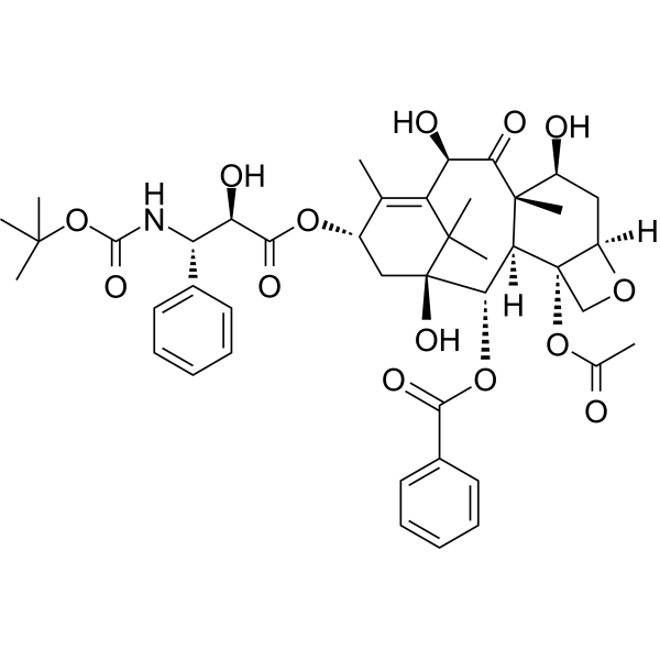
HY-B0011
DocetaxelMicrotubule Depolymerization Inhibitor -
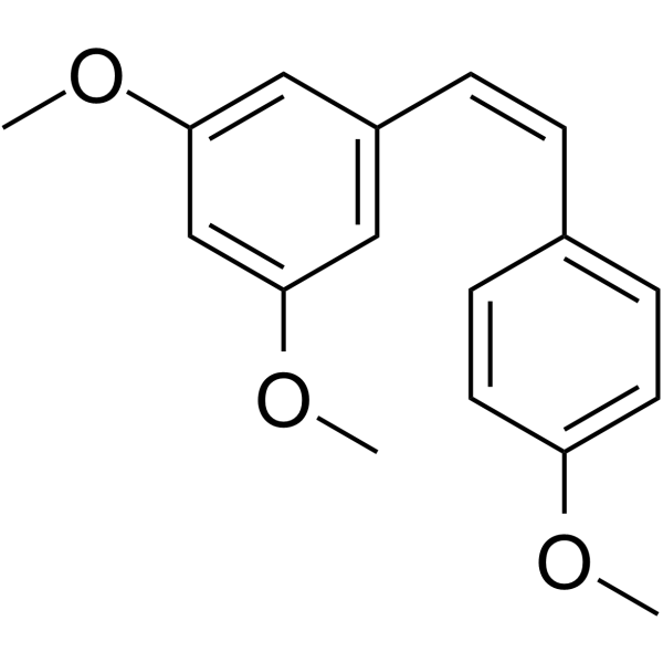
HY-121989
cis-Trismethoxy resveratrolTubulin Inhibitor -
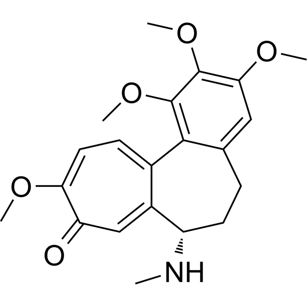
HY-N0282
DemecolcineTubulin Inhibitor -
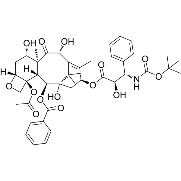
HY-B0011R
Docetaxel (Standard)Microtubule Depolymerization Inhibitor -
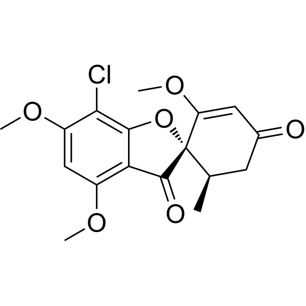
HY-17583
GriseofulvinAntifungal Antibiotic -

HY-N0282R
Demecolcine (Standard)Analytical Standard -
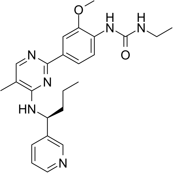
HY-10498
LexibulinTubulin Polymerisation Inhibitor -
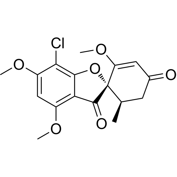
HY-17583R
Griseofulvin (Standard) -
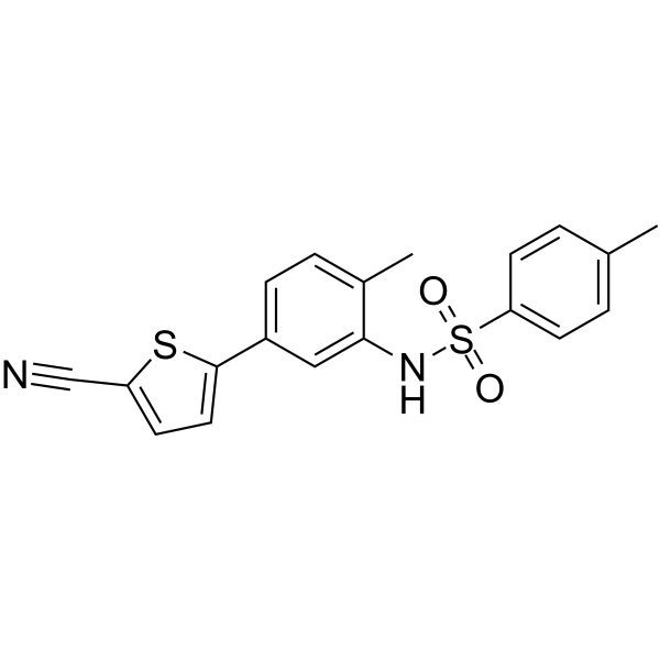
HY-16191
ELR510444Tubulin Inhibitor -

HY-10498R
Lexibulin (Standard)Tubulin Polymerisation Inhibitor -
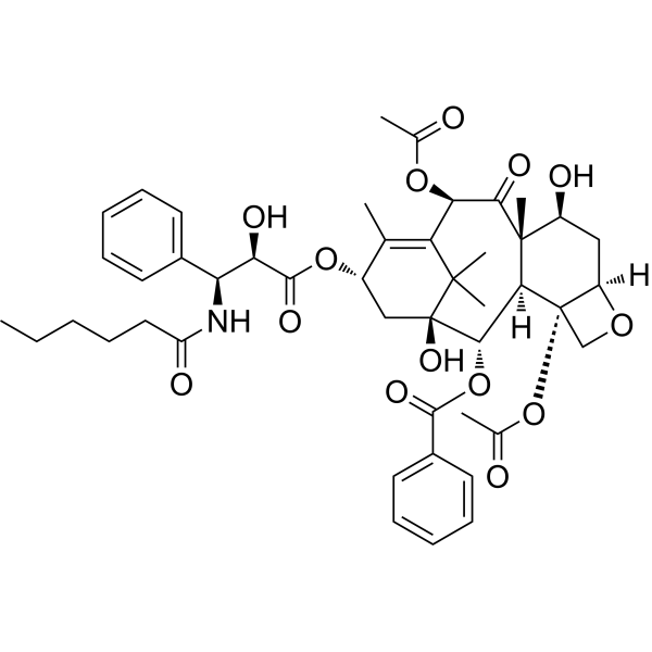
HY-137776
Paclitaxel C -

HY-137776R
Paclitaxel C (Standard)


Activité biologique
Description
Cellular Effect
|
Cell Line

|
Type | Value | Description | References |
|---|---|---|---|---|
| A549 | IC50 |
>50 μM
Compound: 17
|
Cytotoxicity against human A549 cells measured after 72 hrs by MTT assay
Cytotoxicity against human A549 cells measured after 72 hrs by MTT assay
|
[PMID: 35473314] |
| HepG2 | IC50 |
>50 μM
Compound: 17
|
Cytotoxicity against human HepG2 cells measured after 72 hrs by MTT assay
Cytotoxicity against human HepG2 cells measured after 72 hrs by MTT assay
|
[PMID: 35473314] |
| MDA-MB-231 | IC50 |
31 μM
Compound: 17
|
Cytotoxicity against human MDA-MB-231 cells measured after 72 hrs by MTT assay
Cytotoxicity against human MDA-MB-231 cells measured after 72 hrs by MTT assay
|
[PMID: 35473314] |
| MDA-MB-435 | IC50 |
4.9 μM
Compound: 17
|
Cytotoxicity against human MDA-MB-435 cells measured after 72 hrs by MTT assay
Cytotoxicity against human MDA-MB-435 cells measured after 72 hrs by MTT assay
|
[PMID: 35473314] |
| SNB-19 | IC50 |
6.8 μM
Compound: 17
|
Cytotoxicity against human SNB-19 cells measured after 72 hrs by MTT assay
Cytotoxicity against human SNB-19 cells measured after 72 hrs by MTT assay
|
[PMID: 35473314] |
| Vero | IC50 |
>50 μM
Compound: 17
|
Cytotoxicity against African green monkey Vero cells measured after 72 hrs by MTT assay
Cytotoxicity against African green monkey Vero cells measured after 72 hrs by MTT assay
|
[PMID: 35473314] |


Chemical Information
-
CAS No. 53760-20-6
-
Masse moléculaire 451.60
-
Formule C28H37NO4
-
SMILES
O=C([C@@]12[C@](/C=C/C[C@H](C)C[C@@](C)(O)/C=C/[C@H]2O)([H])[C@@H]3O)N[C@@H](CC4=CC=CC=C4)[C@]1([H])[C@H](C)C3=C
-
Structure Classification
-
Initial Source
Endothia gyrosa IFB-E023
-
Livraison
Room temperature in continental US; may vary elsewhere.
-
Stockage
Please store the product under the recommended conditions in the Certificate of Analysis.

Protocole
-
Cell Cytotoxicity Assay
Cytotoxicity assays are usually based on the assessment of cell membrane damage, which can also be indirectly detected by measuring cell viability. Detection methods include MTT assay, CKK-8 assay, LDH assay and ATP assay, etc.
-
Mammalian live/dead viability and cytotoxicity staining
Live/dead viability and cytotoxicity staining assays are based on the simultaneous detection of intracellular esterase activity in metabolically active (viable) cells and membrane integrity loss in non-viable cells. In commonly used dual-staining approaches, membrane-permeant fluorogenic substrates are converted by intracellular esterases into fluorescent products in live cells, while impermeant DNA-binding dyes selectively enter cells with compromised plasma membranes and label nucleic acids in dead or dying cells, enabling discrimination between viable and non-viable populations by fluorescence microscopy or flow cytometry.
-
Cell Viability Determination by MTT Colorimetric Assay
The following protocol uses the MTT colorimetric assay as a classic literature-established method for assessing cell viability/metabolic activity in cultured mammalian cells. MTT[3-(4,5-dimethylthiazol-2-yl)-2,5-diphenyltetrazolium bromide] is reduced by metabolically active cells to a colored formazan product; the amount of formazan is quantified spectrophotometrically and provides an indirect measure of metabolically active viable cells. Importantly, MTT reduction reflects cellular oxidoreductase/metabolic activity rather than an absolute direct count of living cells, so changes in cellular metabolism can alter the signal independently of cell number.


Pureté et documentation
Références
[1]. Xu S, et al., Cytotoxic cytochalasin metabolites of endophytic Endothia gyrosa. Chem Biodivers. 2009 May;6(5):739-45. [Content Brief]
[2]. Wrench GA, et al., Cytochalasin J treatment significantly alters mitotic spindle microtubule organization and kinetochore structure in PtK1 cells. Cell Motil Cytoskeleton. 1997;36(2):112-24. [Content Brief]

Calculators
Concentration (start) × Volume (start) = Concentration (final) × Volume (final)


Cytochalasin J Related Classifications
HY-126556 Related Classifications
 Powered by Bioz