



Wnt3a Surrogate Protein, Human (HEK293, Fc)
Based on 9 publication(s) in Google Scholar
Wnt3a Surrogate protein is a Wnt recombination protein that plays an important role in embryonic development, adult tissue homeostasis and injury repair. Wnt3a Surrogate protein, consisting of the domain and Fc portion of Wnt3a, helps organoids expand and maintain a source of Wnt. Organoids are a rapidly evolving model system for tissue development, and Wnt3a is considered as the essential protein for organoid growth. Wnt3a or the small molecule glycogen synthase kinase 3 (GSK3) inhibitor CHIR are commonly used as Wnt sources in experiments. However, the application of Wnt3a is hampered by Wnt lipidation, and cross-reactivity with Wnt frizzled (Fzd). Therefore, Wnt3a Surrogate protein was used instead of Wnt3a, as a water-soluble and Fzd-specific alternative Wnt agonist. And the Wnt3a Surrogate protein exhibits tissue selectivity consistent with Fzd isoform specificity. Wnt3a Surrogate Protein, Human (HEK293, Fc) is a recombinant Wnt3a protein of human origin, expressed by HEK293 cells with an Fc tag.
- Species: Human
- Source: HEK293
-
Storage:Stored at -80°C for 1 year from date of receipt. It is stable at -20°C for 3 months after opening. It is recommended to freeze aliquots at -80°C for extended storage. Avoid repeated freeze-thaw cycles.


To place orders, for customer services and technical support, please contact: MedChemExpress USA
Tel: 609-228-6898 E-mail: [email protected] [email protected]


-
 Biological Activity
-
Technical Parameters
-
Product Properties
- Documentation
- References
- Help & FAQs


-
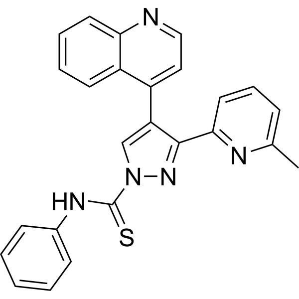
HY-10432
A 83-01 -

HY-15371
Forskolin -

HY-P70558
Noggin Protein, Human (HEK293) -

HY-10071
Y-27632 -

HY-B0215
Acetylcysteine -
HY-P7114
RSPO1/R-spondin-1 Protein, Human (CHO, His) -
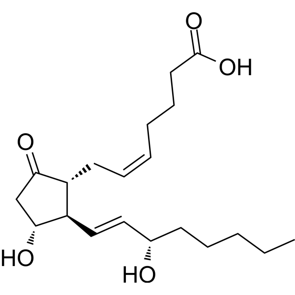
HY-101952
Prostaglandin E2 -
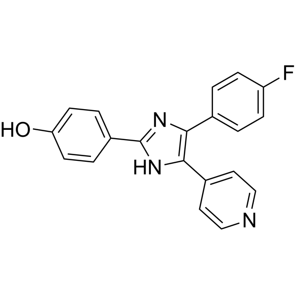
HY-10295
SB 202190 -
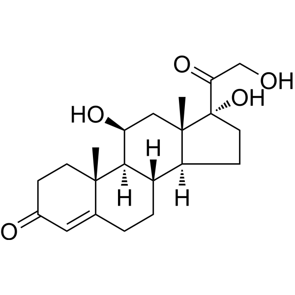
HY-N0583
Hydrocortisone -
HY-P7048
KGF-2/FGF-10 Protein, Human (169a.a) -

HY-10182
Laduviglusib -
HY-B0150
Nicotinamide


Biological Activity
Description
Wnt3a Surrogate protein is a Wnt recombination protein that plays an important role in embryonic development, adult tissue homeostasis and injury repair. Wnt3a Surrogate protein, consisting of the domain and Fc portion of Wnt3a, helps organoids expand and maintain a source of Wnt. Organoids are a rapidly evolving model system for tissue development, and Wnt3a is considered as the essential protein for organoid growth. Wnt3a or the small molecule glycogen synthase kinase 3 (GSK3) inhibitor CHIR are commonly used as Wnt sources in experiments. However, the application of Wnt3a is hampered by Wnt lipidation, and cross-reactivity with Wnt frizzled (Fzd). Therefore, Wnt3a Surrogate protein was used instead of Wnt3a, as a water-soluble and Fzd-specific alternative Wnt agonist. And the Wnt3a Surrogate protein exhibits tissue selectivity consistent with Fzd isoform specificity[1][2][3]. Wnt3a Surrogate Protein, Human (HEK293, Fc) is a recombinant Wnt3a protein of human origin, expressed by HEK293 cells with an Fc tag.
Background
Wnt is a secreted glycoprotein comprising 19 human proteins that signal through both canonical and non-canonical pathways. Wnts are involved in developmental morphology, stem cell maintenance, and wound healing, and can travel long distances or attach to the surface of exosomes. Among them, Wnt3a is an important molecule that can activate the Wnt/β-catenin signaling pathway and participate in various physiological and pathological processes, such as embryonic development, tissue repair, and tumor progression. Wnt3a also plays a role in cardiomyogenesis, skeletal muscle development, nervous system formation, and migration and invasion of trophoblast cells[4][5].
1. The Wnt family includes: Wnt1, Wnt2, Wnt2b (Wnt13), Wnt3, Wnt3a, Wnt4, Wnt5a, Wnt5b, Wnt6, Wnt7a, Wnt7b, Wnt8a, Wnt8b, Wnt9a (Wnt14), Wnt9b (Wnt14b), Wnt10a, Wnt10b, Wnt11 and Wnt16.
2. Wnt canonical pathway: also known as Wnt/β-catenin pathway.
3. Wnt non-canonical pathway: including Wnt-planar cell polarity pathway (Wnt-PCP pathway) and Wnt-calcium pathway (Wnt-Ca2+ pathway).
4. Introduction to Wnt3a: It is an important molecule in the process of cell signal transduction and development, and is highly conserved. Wnt3a interacts with Frizzled receptors, induces the accumulation of β-catenin, and activates the Wnt/β-catenin signaling pathway. Wnt3a also stimulates migration and invasion of trophoblasts.
Wnt3a function: In embryonic development, Wnt3a is involved in the formation of embryonic axis and directed cell differentiation; in adult tissues, Wnt3a is related to the regulation of stem cell self-renewal and cell fate determination, and is also related to the occurrence and development of certain tumors. For example, Wnt3a promotes or inhibits tumor progression through the canonical Wnt signaling pathway. Wnt3a is also associated with post-injury remodeling and cell expansion, and upregulation of WNT3a improves repair. Wnt3a also plays an important role in organ formation. It can promote cardiomyogenesis by increasing T cell-specific transcription factor (TCF)-dependent transcription and inducing cardiac-specific markers, bone morphogenetic proteins (BMPs) and myosin heavy chain (MHC). Studies have shown that Wnt3a induces myogenesis in differentiated body muscle groups, activates myogenic differentiation antigen (MyoD) expression, and promotes skeletal muscle development. Wnt3a activates the atypical protein kinase Cloys (pKCiota) and induces a punctate distribution of pKCiota in neurites and cytoplasm, with a particularly strong signal at the centrosome. Wnt3a plays a crucial role in the maintenance and differentiation of neuromesoderm stem cells. Thus, Wnt3a is involved in the formation of the nervous system.
Verified Bioactivity
Measured by its ability to induce Topflash reporter activity in HEK293T human embryonic kidney cells. The ED50 for this effect is <62 ng/mL.
MCE Validation Data
-
Purity - SDS-PAGE

Purity - SDS-PAGE


Publications (9)
-
Journal Impact Factor
-
Most Recent
-
Cell
2025 Sep 18;188(19):5175-5193.e21. PMID: 40818454 -
Cell Death Differ
2024 Nov;31(11):1452-1466. PMID: 38987382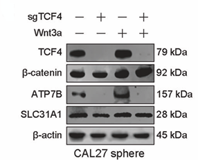
Wnt3a Surrogate Protein, Human (HEK293, Fc) purchased from MedChemExpress. Usage Cited in: Cell Death Differ. 2024 Nov;31(11):1452-1466. [Abstract]
WB analysis of the indicated markers in TCF4 knockout CAL27 and A375 sphere cells by Wnt3a treatment (50 ng/ml, 24 h) was performed.
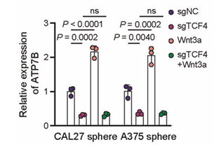
Wnt3a Surrogate Protein, Human (HEK293, Fc) purchased from MedChemExpress. Usage Cited in: Cell Death Differ. 2024 Nov;31(11):1452-1466. [Abstract]
Q-PCR analysis of the indicated markers in TCF4 knockout CAL27 and A375 sphere cells was performed following Wnt3a treatment (50 ng/ml, 24 h).
-
Cell Rep
Targeting EP2/EP4-driven expansion of suppressive VSIG4high macrophages overcomes immunotherapy resistance in colorectal cancer. [Abstract]2026 May 28;45(6):117450. PMID: 42213779 -
Curr Res Microb Sci
The lncRNA GAS5-encoded micropeptide facilitates influenza virus replication through modulation of the Wnt/β-catenin signaling pathway. [Abstract]2026 Jan 27:10:100559. PMID: 41657547 -
iScience
2025 Apr 3;28(5):112344. PMID: 40276762 -
FASEB J
ESCRT-III Subunit CHMP4C Regulates Angiogenesis by Targeting Endocytic Trafficking of GSK3β. [Abstract]2025 Nov 15;39(21):e71174. PMID: 41195682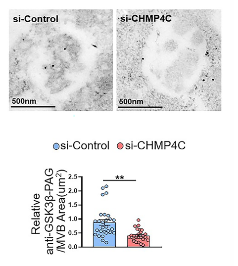
Wnt3a Surrogate Protein, Human (HEK293, Fc) purchased from MedChemExpress. Usage Cited in: FASEB J. 2025 Nov 15;39(21):e71174. [Abstract]
HUVECs were transfected with si-CHMP4C or si-Control. After 48 h of incubation, the cells were starved in serum-free ECM for 20 h, followed by stimulation with Wnt3a for another 0.5 h.
-
-
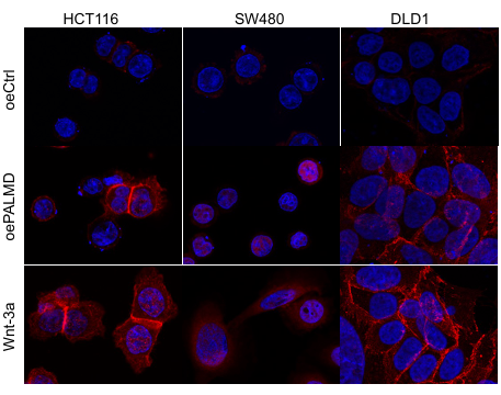
Wnt3a Surrogate Protein, Human (HEK293, Fc) purchased from MedChemExpress. Usage Cited in: bioRxiv. 2025 April 10.
Representative confocal microscopy of HCT116, SW480, and DLD1 cells after oeCtrl transfection, oePALMD transfection, or exogenous Wnt-3a stimulation (100 ng/mL; 2 h) was performed.
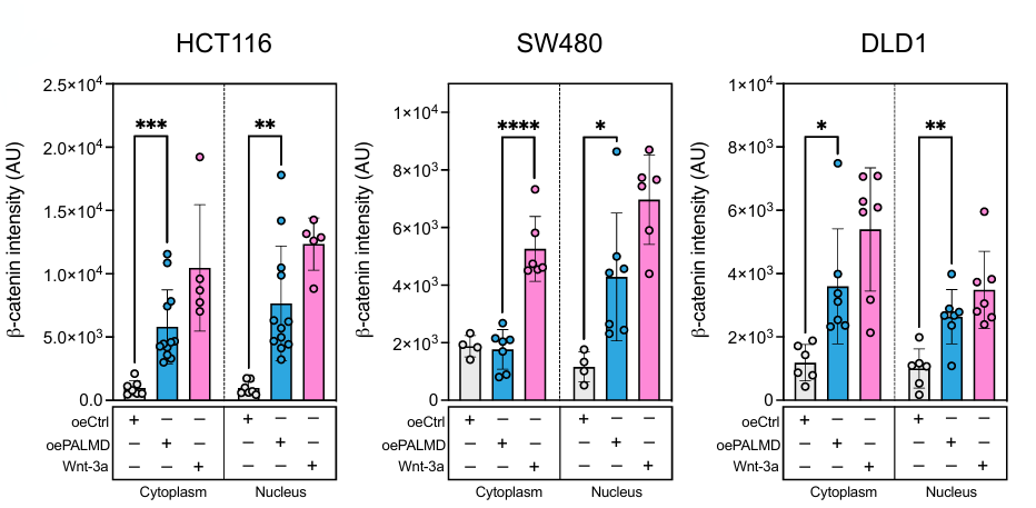
Wnt3a Surrogate Protein, Human (HEK293, Fc) purchased from MedChemExpress. Usage Cited in: bioRxiv. 2025 April 10.
Confocal immunofluorescence microscopy quantification demonstrated significant increases in nuclear β-catenin in HCT116, SW480, and DLD1 cells in oePALMD compared to oeCtrl (100 ng/ml exogenous Wnt-3a (2 h) stimulation as positive control), and confirmatory cell fractionation immunoblots were performed.
-


Technical Parameters
-
Species Human
-
Source HEK293
-
Tag Fc
-
Accession
Sequence information is not available or disclosed for this product.
-
Gene ID/
-
Synonyms
WNT3A; Wnt Family Member 3A; Wingless-Type MMTV Integration Site Family, Member 3A; Protein Wnt-3a
-
Predicted Molecular Mass
58.5 kDa
-
Molecular Weight
Approximately 66-73 kDa, based on SDS-PAGE under reducing conditions.
-
Glycosylation
Yes
-
Purity
≥ 95%, as determined by reducing SDS-PAGE.


Product Properties
Solution
Supplied as a 0.22 μm filtered solution of 25 mM Tris-HCl, 500 mM NaCl, pH 8.2.
Note: For SPR assay, please replace the buffer. Primary amine components (e.g., Tris, imidazole) can affect protein-coupled chips.
< 0.01 EU/µg as determined by LAL test.
Stored at -80°C for 1 year from date of receipt. It is stable at -20°C for 3 months after opening. It is recommended to freeze aliquots at -80°C for extended storage. Avoid repeated freeze-thaw cycles.
Shipping with dry ice.


Documentation
-
Data Sheet (264 KB)
-
SDS (251 KB)
- English - EN (251 KB)
- Français - FR (251 KB)
- Deutsch - DE (251 KB)
- Norwegian - NO (251 KB)
- Español - ES (251 KB)
- Swedish - SV (251 KB)
- Italian - IT (251 KB)
- Korean - KR (251 KB)
- Portuguese - PT (251 KB)
-
Handling Instructions (2659 KB)


References
[1]. Janda CY, et al. Surrogate Wnt agonists that phenocopy canonical Wnt and β-catenin signalling. Nature. 2017 May 11;545(7653):234-237. [Content Brief]
[2]. Chen H, et al. Development of Potent, Selective Surrogate WNT Molecules and Their Application in Defining Frizzled Requirements. Cell Chem Biol. 2020 May 21;27(5):598-609.e4. [Content Brief]
[3]. Miao Y, et al. Next-Generation Surrogate Wnts Support Organoid Growth and Deconvolute Frizzled Pleiotropy In Vivo. Cell Stem Cell. 2020 Nov 5;27(5):840-851.e6. [Content Brief]
[4]. He S, et al. Wnt3a: functions and implications in cancer. Chin J Cancer. 2015 Sep 14;34(12):554-62. [Content Brief]
[5]. Thomas BL, et al. WNT3A-loaded exosomes enable cartilage repair. J Extracell Vesicles. 2021 May;10(7):e12088. [Content Brief]

Calculators
Concentration (start) × Volume (start) = Concentration (final) × Volume (final)


 Powered by Bioz

