



Pexelizumab

Based on 1 Customer Validation
Pexelizumab (h5G1. 1-SC) is a humanized scFv monoclonal antibody directed against the C5 complement component. Pexelizumab inhibits apoptosis and leukocyte infiltration. Pexelizumab can be used for the research of cerebral IR injury and myocardial infarction.
For research use only. We do not sell to patients.
- Purity : 99.0%
- CAS No.: 219685-93-5
- Molecular Weight:26.271 kDa
-
Storage:
Please store the product under the recommended conditions in the Certificate of Analysis.


To place orders, for customer services and technical support, please contact: MedChemExpress USA
Tel: 609-228-6898 E-mail: [email protected] [email protected]


-
 Biological Activity
-
Chemical Information
- Protocols
- Purity & Documentation
- References
-
 Help & FAQs


-
HY-110136
PMX 205Complement System Antagonist -
HY-P1036
CompstatinPrimates’ C3 Inhibitor -

HY-P99050
SutimlimabC1s Inhibitor -

HY-P99365
RavulizumabAnti-C5 Antibody -
HY-112151
EG01377NRP1 Inhibitor -
HY-P3502
ZilucoplanC5 Inhibitor -

HY-P99786
PozelimabAnti-C5 Antibody -

HY-P99298
LampalizumabFactor D Inhibitor -
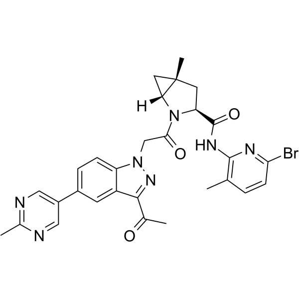
HY-139588
VemircopanComplement Factor D Inhibitor -
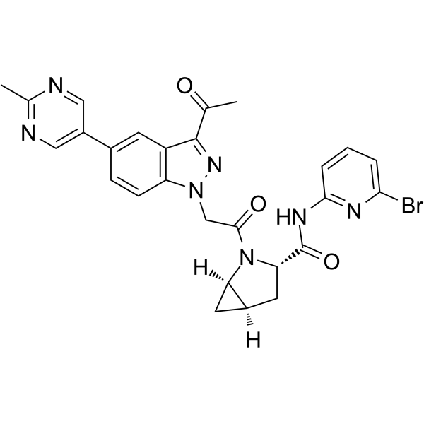
HY-138281
Complement factor D-IN-2Complement Factor D Inhibitor


Biological Activity
Description
Isotype
scFv-kappa-heavy
Species Reactivity
Human
IC50 & Target
Complement C5/C5a
In Vitro
Pexelizumab (h5G1. 1-SC) inhibits 5G1.1 mAb binding with an IC50 of 1.5 nM[2].
MedChemExpress (MCE) has not independently confirmed the accuracy of these methods. They are for reference only.
Clinical Trial
| NCT Number | Sponsor | Condition | Start Date |
Phase

|
|---|---|---|---|---|
| NCT01329991 | Plexxikon| | 2011-05 | PHASE1 |
Gene ID
Accession
Conjugated
Unconjugated
Reconsititution
The product can be reconstituted/diluted with sterile PBS or saline.
Format
-
scFv-kappa-heavy

Application
ELISA, FACS, Functional assay
Verified Bioactivity
-
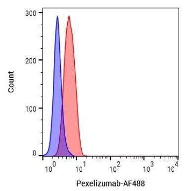 Flow Cytometry analysis of HepG2 cells labelling Complement C5 (red) with Pexelizumab (HY-P99886). Cells were fixed with 4% paraformaldehyde and permeabilised with 90% methanol. Then cells were stained with the primary antibody at 1/200 dilution for an hour at 4℃. Goat Anti-Human IgG H&L (AF488) (HY-P83776) was used as the secondary antibody at 1/1,000 dilution for 30 minutes at 4℃. Human IgG1 kappa (HY-P99001, blue) was used as the isotype control.


Chemical Information
-
CAS No. 219685-93-5
-
Appearance Liquid
-
Molecular Weight 26.271 kDa
-
Color Colorless to light yellow
-
SMILES
[Pexelizumab]
-
Synonyms
h5G1. 1-SC
-
Shipping
Shipping with dry ice.
-
Formulation
Please refer to the lot-specific COA for specific buffer information.
-
Storage
Please store the product under the recommended conditions in the Certificate of Analysis.


Purity & Documentation
-
Data Sheet (256 KB)
-
SDS (251 KB)
- English - EN (251 KB)
- Français - FR (251 KB)
- Deutsch - DE (251 KB)
- Norwegian - NO (251 KB)
- Español - ES (251 KB)
- Swedish - SV (251 KB)
- Italian - IT (251 KB)
- Korean - KR (251 KB)
- Portuguese - PT (251 KB)
-
Inhibitory Antibodies User Guide (603 KB)
References
[1]. APEX AMI Investigators, et al. Pexelizumab for acute ST-elevation myocardial infarction in patients undergoing primary percutaneous coronary intervention: a randomized controlled trial. JAMA. 2007 Jan 3;297(1):43-51. [Content Brief]
[2]. Thomas TC, et al. Inhibition of complement activity by humanized anti-C5 antibody and single-chain Fv. Mol Immunol. 1996 Dec;33(17-18):1389-401. [Content Brief]

Calculators
Concentration (start) × Volume (start) = Concentration (final) × Volume (final)


Pexelizumab Related Classifications
HY-P99886 Related Classifications
 Powered by Bioz