



Envafolimab

Based on 1 Customer Validation
Envafolimab (ASC 22; KN 035) is a recombinant protein of a humanized single-domain anti- PD-L1 antibody. Envafolimab is created by a fusion of the of anti-PD-L1 domain with Fc fragment of human IgG1 antibody. Envafolimab blocks interaction between PD-L1 and PD-1 with an IC50 value of 5.25 nM. Envafolimab has the potential for the research of solid tumors.
For research use only. We do not sell to patients.
- Purity : 99.77%
- CAS No.: 2102192-68-5
- Molecular Weight:80 kDa
-
Storage:
Please store the product under the recommended conditions in the Certificate of Analysis.


To place orders, for customer services and technical support, please contact: MedChemExpress USA
Tel: 609-228-6898 E-mail: [email protected] [email protected]


-
 Biological Activity
-
Chemical Information
- Protocols
- Purity & Documentation
- References
-
 Help & FAQs


-

HY-P9904
AtezolizumabAnti-PD-L1 Antibody -

HY-P9919
DurvalumabPD-L1 Blocking Antibody -

HY-P9902
PembrolizumabPD-1 Inhibitor -
HY-P9903
NivolumabPD-1 Inhibitor -

HY-P99048
SintilimabPD-1/PD-L1 Inhibitor -

HY-P99796
OzoralizumabAnti-TNFα Antibody -

HY-P99052
TislelizumabPD-1 Inhibitor -

HY-108730
AvelumabAnti-PD-L1 Antibody -
HY-P9905
CetuximabEGFR Inhibitor -

HY-P99114
SugemalimabAnti-PD-L1 Antibody -

HY-P99118
SerplulimabAnti-PD-1 Antibody -
HY-101093
CA-170VISTA/PD-L1 Inhibitor


Biological Activity
Description
Isotype
[G1VH-h-CH2-CH3]-dimer
Recommend Isotype Controls
Species Reactivity
Human
IC50 & Target
B7-H1/PD-L1/CD274
In Vitro
In Vivo
MedChemExpress (MCE) has not independently confirmed the accuracy of these methods. They are for reference only.
Clinical Trial
| NCT Number | Sponsor | Condition | Start Date |
Phase

|
|---|---|---|---|---|
| NCT01329991 | Plexxikon| | 2011-05 | PHASE1 |
Gene ID
Accession
Conjugated
Unconjugated
Reconsititution
The product can be reconstituted/diluted with sterile PBS or saline.
Format
-
[G1 VH-h-CH2-CH3]-dimer
2_S.png)
Application
ELISA, FACS, Functional assay
Verified Bioactivity
-
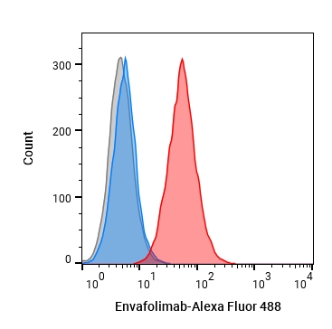 Flow cytometric analysis of 1.5X10⁶ MDA-MB-231 cells with Envafolimab (HY-P99115, red). Cells were fixed with 4% paraformaldehyde. Then stained with the primary antibody at 1/200 dilution for an hour at 4℃. Goat Anti-Human IgG H&L (AF488) (HY-P83776) was used as the secondary antibody at 1/1,000 dilution for 30 minutes at 4℃. Human IgG1 kappa (HY-P99001, blue) was used as the isotype control, cells without incubation with primary antibody were used as the unlabeled control (black).


Chemical Information
-
CAS No. 2102192-68-5
-
Appearance Liquid
-
Molecular Weight 80 kDa
-
Color Colorless to light yellow
-
SMILES
[Envafolimab]
-
Synonyms
ASC 22; KN 035
-
Shipping
Shipping with dry ice.
-
Formulation
Please refer to the lot-specific COA for specific buffer information.
-
Storage
Please store the product under the recommended conditions in the Certificate of Analysis.

Protocols
-
Baculovirus-insect cell protein expression and purification
Baculovirus-insect cell expression uses recombinant baculovirus to deliver a target gene into insect cells, where late or very-late viral transcription drives recombinant protein production; the method was classically demonstrated by expression of human β-interferon in baculovirus-infected insect cells. The readout is target protein accumulation, assessed by activity, fluorescence if a fluorescent reporter is used, SDS-PAGE, Western blot, or purified protein yield. The system can express soluble, secreted, membrane-associated, and multiprotein targets, but expression outcome depends on the construct, baculovirus vector, insect cell line, multiplicity of infection, infection cell density, harvest time, and target-specific stability.
-
Cell Cytotoxicity Assay
Cytotoxicity assays are usually based on the assessment of cell membrane damage, which can also be indirectly detected by measuring cell viability. Detection methods include MTT assay, CKK-8 assay, LDH assay and ATP assay, etc.
-
E. coli fusion-tag soluble protein purification
The purification of soluble recombinant proteins in Escherichia coli is achieved by fusing the target protein with a solubility-enhancing affinity tag (e. g. , His-tag, GST, MBP, Fh8, CSQ-tag, or thioredoxin) to improve expression yield, prevent aggregation, and enable efficient purification via affinity chromatography. The fusion protein is expressed under inducible promoters (e. g. , IPTG-induced T7 promoter), lysed from bacterial cells, and purified using resin-based affinity chromatography (e. g. , Ni-NTA for His-tag, amylose resin for MBP, chitin resin for intein tags, or HIC for Fh8). Tags can be removed post-purification using site-specific proteases (e. g. , TEV, enterokinase) or through intracellular cleavage systems. Solubility screening using multiple fusion partners (e. g. , Expresso® system) allows optimization of expression conditions for difficult-to-express proteins.
-
Inclusion-body expression, solubilization, refolding and purification
Inclusion-body recovery uses insoluble recombinant protein aggregates from E. coli as a starting material; the workflow is cell disruption, inclusion-body isolation/washing, denaturant or mild solubilization, refolding into soluble protein, and final chromatographic purification. The readouts are soluble protein recovery, purity by SDS-PAGE/chromatography, structural recovery by methods such as circular dichroism when used, and biological activity when an assay is available.
-
Protocol For Protein Expression And Purification
Recombinant protein expression in Escherichia coli followed by purification of a His-tagged soluble protein by immobilized metal affinity chromatography (IMAC), with optional MBP fusion and TEV tag removal when the construct includes these elements. The biological readout is production of the encoded target protein, detected as an inducible band at the expected molecular mass by SDS-PAGE and quantified by total protein assay or chromatographic absorbance; the purification readout is enrichment of the target protein in elution fractions after selective binding of polyhistidine residues to immobilized Ni2+/metal-chelate resin and elution by imidazole-containing buffer. Expression is driven by an inducible bacterial expression system, commonly T7/lac-based, in which IPTG or lactose/auto-induction activates transcription and translation of the cloned gene; lower induction temperature, lower inducer concentration, induction timing, and solubility-enhancing fusion tags can influence the frac
-
Mammalian transient protein expression and purification
Mammalian transient protein expression introduces plasmid DNA into HEK293 or CHO cells for short-term recombinant protein production, allowing secreted, glycosylated, Fc-tagged, His-tagged, or membrane proteins to be produced without stable clone generation.


Purity & Documentation
-
Data Sheet (257 KB)
-
SDS (251 KB)
- English - EN (251 KB)
- Français - FR (251 KB)
- Deutsch - DE (251 KB)
- Norwegian - NO (251 KB)
- Español - ES (251 KB)
- Swedish - SV (251 KB)
- Italian - IT (251 KB)
- Korean - KR (251 KB)
- Portuguese - PT (251 KB)
-
Inhibitory Antibodies User Guide (603 KB)
References
[1]. Papadopoulos KP, et al. First-in-Human Phase I Study of Envafolimab, a Novel Subcutaneous Single-Domain Anti-PD-L1 Antibody, in Patients with Advanced Solid Tumors. Oncologist. 2021 Sep;26(9):e1514-e1525. [Content Brief]
[2]. Akinleye A, et al. Immune checkpoint inhibitors of PD-L1 as cancer therapeutics. J Hematol Oncol. 2019 Sep 5;12(1):92. [Content Brief]
[3]. Zhang F, Wei H, Wang X, Bai Y, Wang P, Wu J, Jiang X, Wang Y, Cai H, Xu T, Zhou A. Structural basis of a novel PD-L1 nanobody for immune checkpoint blockade. Cell Discov. 2017 Mar 7;3:17004. [Content Brief]

Calculators
Concentration (start) × Volume (start) = Concentration (final) × Volume (final)


Envafolimab Related Classifications
HY-P99115 Related Classifications
 Powered by Bioz