



CAY10471

Based on 1 Customer Validation
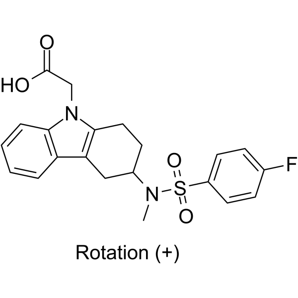
CAY10471 (TM30089) is a potent, selective, and orally active prostaglandin D2 receptor CRTH2 antagonist. CAY10471 attenuates the progression of tubulointerstitial fibrosis and chronic contact hypersensitivity (CHS) in animal model.
For research use only. We do not sell to patients.
- Purity : 99.9%
- CAS No.: 627865-18-3
- Formula: C21H21FN2O4S
- Molecular Weight:416.47
-
Storage:
-20°C, sealed storage, away from moisture
* In solvent : -80°C, 6 months; -20°C, 1 month (sealed storage, away from moisture)

To place orders, for customer services and technical support, please contact: MedChemExpress USA
Tel: 609-228-6898 E-mail: [email protected] [email protected]


-
 Biological Activity
-
Chemical Information
-
Solvent & Solubility
- Protocols
- Purity & Documentation
- References
-
 Help & FAQs




Biological Activity
Description
IC50 & Target
[1]|
CRTH2 |
In Vitro
CAY10471 (1 μM; 1-24 hours) decreases 15dPGJ2-induced phosphorylation of p38 MAP kinase significantly in PC12 cells[1].
MedChemExpress (MCE) has not independently confirmed the accuracy of these methods. They are for reference only. Further protocols information, click here.
-
Cell Line:PC12 cells
-
Concentration:1 μM
-
Incubation Time:1 or 24 hours
-
Result:Blocked 15d-PGJ2-induced p38 MAP kinase activation.
In Vivo
MedChemExpress (MCE) has not independently confirmed the accuracy of these methods. They are for reference only.
-
Animal Model:Balb/c mice, DP−/− mice, CRTH2−/− mice[2]
-
Dosage:2 mg/kg
-
Administration:Oral treatment; once daily; challenged on day 22 or over 10 consecutive days
-
Result:Significantly suppressed both CHS and IgE-CAI inflammatory responses.
-
Animal Model:C57BL/6 mice[3]
-
Dosage:20 mg/kg
-
Administration:Oral treatment; twice daily; beginning 3/4/5 days before UUO
-
Result:Slowed the progression of renal fibrosis in the obstructed kidneys.


Chemical Information
-
CAS No. 627865-18-3
-
Appearance Solid
-
Molecular Weight 416.47
-
Formula C21H21FN2O4S
-
Color White to off-white
-
SMILES
O=C(CN1C2=C(CC(CC2)N(C)S(=O)(C3=CC=C(C=C3)F)=O)C4=C1C=CC=C4)O.[Rotation (+)]
-
Synonyms
TM30089
-
Shipping
Room temperature in continental US; may vary elsewhere.
-
Storage
-20°C, sealed storage, away from moisture
* In solvent : -80°C, 6 months; -20°C, 1 month (sealed storage, away from moisture)


Solvent & Solubility
In Vitro:
DMSO : 30 mg/mL (72.03 mM; Need ultrasonic and warming; Hygroscopic DMSO has a significant impact on the solubility of product, please use newly opened DMSO)
Please refer to the solubility information to select the appropriate solvent. Once prepared, please aliquot and store the solution to prevent product inactivation from repeated freeze-thaw cycles.
Storage method and period of stock solution: -80°C, 6 months; -20°C, 1 month (sealed storage, away from moisture). When stored at -80°C, please use it within 6 months. When stored at -20°C, please use it within 1 month.
Please refer to the solubility information to select the appropriate solvent. Once prepared, please aliquot and store the solution to prevent product inactivation from repeated freeze-thaw cycles.
Storage method and period of stock solution: -80°C, 6 months; -20°C, 1 month (sealed storage, away from moisture). When stored at -80°C, please use it within 6 months. When stored at -20°C, please use it within 1 month.
Concentration (start) × Volume (start) = Concentration (final) × Volume (final)

Protocols
-
Fibrosis/Collagen Morphometry
Fibrosis and collagen morphometry is based on the quantitative visualization of fibrillar collagen deposition in tissue sections using histochemical stains such as Sirius Red (Picrosirius Red) or Masson's trichrome, followed by image-based or polarization-enhanced analysis to estimate collagen proportional area as a surrogate of extracellular matrix accumulation during fibrotic remodeling. Sirius Red combined with polarized light microscopy enhances detection of collagen fibers due to birefringence properties, enabling more specific visualization of collagen type I and III fibrils compared to conventional bright-field histology, while whole-section or region-restricted digital morphometry reduces field-selection bias in fibrosis assessment. Alternative quantitative approaches include second harmonic generation (SHG) and two-photon excited fluorescence microscopy, which enable label-free detection of fibrillar collagen and have been validated against histological staining and biochemica
-
Collagen: Sirius Red Staining
Sirius Red or picrosirius red staining is a histochemical method for visualizing collagen-rich extracellular matrix in tissue sections, and collagen fibers are detected as red-stained structures under bright-field microscopy with enhanced birefringence under polarized light. Picrosirius red is useful for assessing total collagen organization, distribution, and fibrosis burden, but polarized color should not be interpreted as a definitive collagen type I versus type III readout because color is affected by fiber orientation, thickness, and packing.
-
Connective Tissue: Masson's Trichrome/Collagen Trichrome Staining
Masson’s Trichrome (collagen/trichrome staining) is a histological technique that differentially stains tissue compartments using sequential acidic dyes to distinguish collagen from muscle and cytoplasmic components based on dye affinity and tissue permeability differences, enabling visualization of fibrosis and connective tissue architecture in histological sections. The classical formulation typically uses Weigert's iron hematoxylin for nuclear staining, Biebrich scarlet-acid fuchsin for cytoplasm and muscle, and aniline blue (or light green variants) for collagen, producing a characteristic blue/green collagen signal contrasted against red cytoplasm and dark nuclei. The staining principle relies on selective displacement of smaller dye molecules by larger anionic dyes in collagen-rich regions under controlled acidified conditions, which enhances collagen-specific dye retention. This property makes the method widely used for fibrosis assessment in organs such as heart, liver, lung, a
-
Contact Hypersensitivity Dermatitis
Contact hypersensitivity (CHS) dermatitis is a T cell-mediated delayed-type (Type IV) immune reaction in which low-molecular-weight haptens applied to the skin bind host proteins to form complete antigens, triggering sensitization followed by a secondary inflammatory response upon re-exposure (elicitation phase), which is commonly quantified by ear swelling as a readout of skin inflammation in murine models. This model is widely used to study allergic contact dermatitis because it is antigen-specific, reproducible, and reflects key immunological events including dendritic cell activation, T cell priming in draining lymph nodes, and effector T cell-driven tissue inflammation. DNFB- and oxazolone-induced CHS models are standard systems for evaluating both acute and chronic T cell-dependent skin inflammation and for testing immunomodulatory interventions.
-
How to Choose the Right Model Animal
Choosing the right model animal is a validity-driven decision in which the species, strain, sex, age, genetic background, disease-induction method, outcome measures, and welfare burden must match the scientific question rather than laboratory tradition or convenience. A model should be selected by judging face validity, construct validity, and predictive validity: whether it resembles the human phenotype, whether it reproduces relevant mechanisms, and whether results are likely to predict human biology or treatment response. Animal studies often fail to translate because of species differences, weak disease resemblance, poor experimental design, inadequate reporting, publication bias, and underuse of randomization, blinding, and sample-size justification. Unresolved questions include how to rank competing models objectively, how much human-disease complexity must be reproduced for a given objective, and when non-animal systems such as organoids, ex vivo tissue, or computational models


Purity & Documentation
-
Data Sheet (279 KB)
-
SDS (394 KB)
- English - EN (394 KB)
- Français - FR (394 KB)
- Deutsch - DE (394 KB)
- Norwegian - NO (394 KB)
- Español - ES (394 KB)
- Swedish - SV (394 KB)
- Italian - IT (394 KB)
- Korean - KR (394 KB)
- Portuguese - PT (394 KB)
-
Handling Instructions (2659 KB)
References
[1]. Hatanaka M, et al. 15d-prostaglandin J2 enhancement of nerve growth factor-induced neurite outgrowth is blocked by the chemoattractant receptor- homologous molecule expressed on T-helper type 2 cells (CRTH2) antagonist CAY10471 in PC12 cells. J Pharmacol Sci. 2010;113(1):89-93. Epub 2010 Apr 16. [Content Brief]
[2]. Matsushima Y, et al. Distinct roles of prostaglandin D2 receptors in chronic skin inflammation.Mol Immunol. 2011 Oct;49(1-2):304-10. [Content Brief]
[3]. Ito H, et al. PGD2-CRTH2 pathway promotes tubulointerstitial fibrosis.J Am Soc Nephrol. 2012 Nov;23(11):1797-809. [Content Brief]


Complete Stock Solution Preparation Table
Please refer to the solubility information to select the appropriate solvent. Once prepared, please aliquot and store the solution to prevent product inactivation from repeated freeze-thaw cycles.
Storage method and period of stock solution: -80°C, 6 months; -20°C, 1 month (sealed storage, away from moisture). When stored at -80°C, please use it within 6 months. When stored at -20°C, please use it within 1 month.
| Optional Solvent | Concentration Solvent Mass | 1 mg | 5 mg | 10 mg | 25 mg |
|---|---|---|---|---|---|
| DMSO | 1 mM | 2.4011 mL | 12.0057 mL | 24.0113 mL | 60.0283 mL |
| 5 mM | 0.4802 mL | 2.4011 mL | 4.8023 mL | 12.0057 mL | |
| 10 mM | 0.2401 mL | 1.2006 mL | 2.4011 mL | 6.0028 mL | |
| 15 mM | 0.1601 mL | 0.8004 mL | 1.6008 mL | 4.0019 mL | |
| 20 mM | 0.1201 mL | 0.6003 mL | 1.2006 mL | 3.0014 mL | |
| 25 mM | 0.0960 mL | 0.4802 mL | 0.9605 mL | 2.4011 mL | |
| 30 mM | 0.0800 mL | 0.4002 mL | 0.8004 mL | 2.0009 mL | |
| 40 mM | 0.0600 mL | 0.3001 mL | 0.6003 mL | 1.5007 mL | |
| 50 mM | 0.0480 mL | 0.2401 mL | 0.4802 mL | 1.2006 mL | |
| 60 mM | 0.0400 mL | 0.2001 mL | 0.4002 mL | 1.0005 mL |


CAY10471 Related Classifications
HY-13706A Related Classifications
 Powered by Bioz