



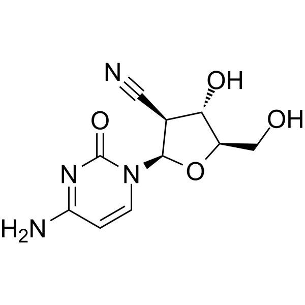
CNDAC
CNDAC is a metabolite of the orally active agent Sapacitabine (HY-16445), and a nucleoside analog. CNDAC induces DNA damage and apoptosis.
Nos produits utilisent uniquement pour la recherche. Nous ne vendons pas aux patients.
- CAS No.: 135598-68-4
- Formule: C10H12N4O4
- Masse moléculaire:252.23
-
Stockage:
Please store the product under the recommended conditions in the Certificate of Analysis.

To place orders, for customer services and technical support, please contact: MedChemExpress USA
Tel: 609-228-6898 E-mail: [email protected] [email protected]


-
 Activité biologique
-
Chemical Information
- Protocole
- Pureté et documentation
- Références
-
 Help & FAQs


-
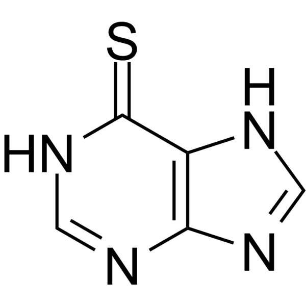
HY-13677
6-Mercaptopurine -
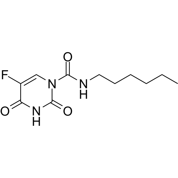
HY-B0182
CarmofurAcid Ceramidase/SARS-CoV-2 Mpro/FAAH/NAAA Inhibitor -
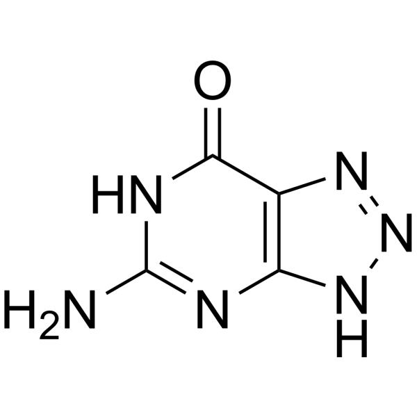
HY-B1468
8-AzaguaninePurine Analogue -
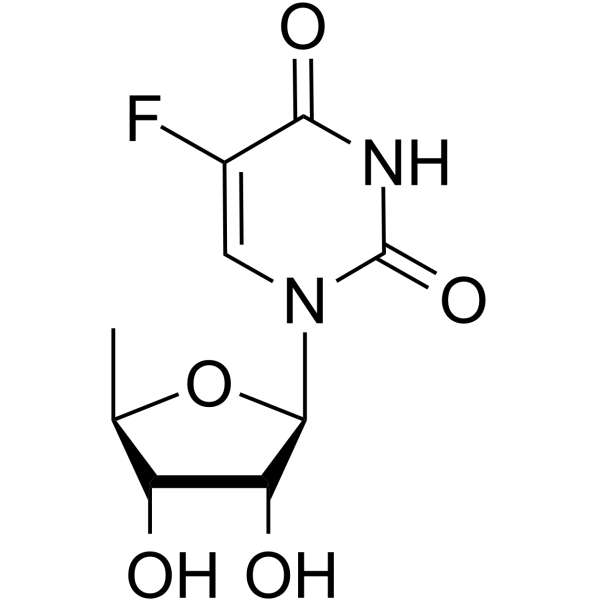
HY-B0021
DoxifluridineNucleoside Antimetabolite/Analog Inhibitor -

HY-13677R
6-Mercaptopurine (Standard) -

HY-B1468R
8-Azaguanine (Standard)Purine Analogue -

HY-B0182R
Carmofur (Standard)Acid Ceramidase/SARS-CoV-2 Mpro/FAAH/NAAA Inhibitor -

HY-B0021R
Doxifluridine (Standard)Nucleoside Antimetabolite/Analog Inhibitor
Voir tous les produits spécifiques à Isoform DNA/RNA Synthesis
More

Activité biologique
Description
Cellular Effect
|
Cell Line

|
Type | Value | Description | References |
|---|---|---|---|---|
| BC-3 | IC50 |
0.23 μM
Compound: 1, CNDAC
|
Cytotoxicity against Kaposi's sarcoma-associated herpesvirus infected BC3 cells after 120 hrs by MTT assay
Cytotoxicity against Kaposi's sarcoma-associated herpesvirus infected BC3 cells after 120 hrs by MTT assay
|
[PMID: 17402726] |
| DG-75 | IC50 |
0.21 μM
Compound: 1, CNDAC
|
Cytotoxicity against DG75 cells after 120 hrs by MTT assay
Cytotoxicity against DG75 cells after 120 hrs by MTT assay
|
[PMID: 17402726] |
| HOS-TE85 | IC50 |
6.8 μg/mL
Compound: CNDAC
|
Inhibitory effect tested in vitro for the growth of Osteosarcoma MNNG-HOS cell line
Inhibitory effect tested in vitro for the growth of Osteosarcoma MNNG-HOS cell line
|
[PMID: 1895307] |
| KHOS-321H | IC50 |
4.5 μg/mL
Compound: CNDAC
|
Inhibitory effect tested in vitro for the growth of Osteosarcoma KHOS-321H cell line
Inhibitory effect tested in vitro for the growth of Osteosarcoma KHOS-321H cell line
|
[PMID: 1895307] |
| MKN-1 | IC50 |
2.1 μg/mL
Compound: CNDAC
|
Inhibitory effect tested in vitro for the growth of Stomach adenocarcinoma MKN-1 cell line
Inhibitory effect tested in vitro for the growth of Stomach adenocarcinoma MKN-1 cell line
|
[PMID: 1895307] |
| MKN-28 | IC50 |
1.7 μg/mL
Compound: CNDAC
|
Inhibitory effect tested in vitro for the growth of Stomach adenocarcinoma MKN- 28 cell line
Inhibitory effect tested in vitro for the growth of Stomach adenocarcinoma MKN- 28 cell line
|
[PMID: 1895307] |
| MKN-7 | IC50 |
1.4 μg/mL
Compound: CNDAC
|
Inhibitory effect tested in vitro for the growth of Stomach adenocarcinoma MKN-7 cell line
Inhibitory effect tested in vitro for the growth of Stomach adenocarcinoma MKN-7 cell line
|
[PMID: 1895307] |
| MKN-74 | IC50 |
0.04 μg/mL
Compound: CNDAC
|
Inhibitory effect tested in vitro for the growth of Stomach adenocarcinoma MKN -24 cell line
Inhibitory effect tested in vitro for the growth of Stomach adenocarcinoma MKN -24 cell line
|
[PMID: 1895307] |
| NUGC-4 | IC50 |
5.2 μg/mL
Compound: CNDAC
|
Inhibitory effect tested in vitro for the growth of Stomach adenocarcinoma NUGC-4 cell line
Inhibitory effect tested in vitro for the growth of Stomach adenocarcinoma NUGC-4 cell line
|
[PMID: 1895307] |
| OST | IC50 |
6.4 μg/mL
Compound: CNDAC
|
Inhibitory effect tested in vitro for the growth of Osteosarcoma OST cell line
Inhibitory effect tested in vitro for the growth of Osteosarcoma OST cell line
|
[PMID: 1895307] |
| PC-13 | IC50 |
>100 μg/mL
Compound: CNDAC
|
Inhibitory effect tested in vitro for the growth of Lung adenocarcinoma PC-13 cell line
Inhibitory effect tested in vitro for the growth of Lung adenocarcinoma PC-13 cell line
|
[PMID: 1895307] |
| PC-3 | IC50 |
5.6 μg/mL
Compound: CNDAC
|
Inhibitory effect tested in vitro for the growth of Lung adenocarcinoma PC-3 cell line
Inhibitory effect tested in vitro for the growth of Lung adenocarcinoma PC-3 cell line
|
[PMID: 1895307] |
| PC-8 | IC50 |
4.6 μg/mL
Compound: CNDAC
|
Inhibitory effect tested in vitro for the growth of Lung adenocarcinoma PC-8 cell line
Inhibitory effect tested in vitro for the growth of Lung adenocarcinoma PC-8 cell line
|
[PMID: 1895307] |
| PC-9 | IC50 |
4.4 μg/mL
Compound: CNDAC
|
Inhibitory effect tested in vitro for the growth of Lung adenocarcinoma PC-9 cell line
Inhibitory effect tested in vitro for the growth of Lung adenocarcinoma PC-9 cell line
|
[PMID: 1895307] |
| QG-56 | IC50 |
40 μg/mL
Compound: CNDAC
|
Inhibitory effect tested in vitro for the growth of Lung squamous-cell carcinoma QG-56 cell line
Inhibitory effect tested in vitro for the growth of Lung squamous-cell carcinoma QG-56 cell line
|
[PMID: 1895307] |
| SK-ES1 | IC50 |
2.2 μg/mL
Compound: CNDAC
|
Inhibitory effect tested in vitro for the growth of Osteosarcoma SK-ES-1 cell line
Inhibitory effect tested in vitro for the growth of Osteosarcoma SK-ES-1 cell line
|
[PMID: 1895307] |
| ST-KM-1 | IC50 |
2.8 μg/mL
Compound: CNDAC
|
Inhibitory effect tested in vitro for the growth of Stomach adenocarcinoma ST-KM cell line
Inhibitory effect tested in vitro for the growth of Stomach adenocarcinoma ST-KM cell line
|
[PMID: 1895307] |
| SW480 | IC50 |
0.27 μg/mL
Compound: CNDAC
|
Inhibitory effect tested in vitro for the growth of colon adenocarcinoma SW-480 cell line
Inhibitory effect tested in vitro for the growth of colon adenocarcinoma SW-480 cell line
|
[PMID: 1895307] |
In Vitro
CNDAC has a unique mechanism of action: after incorporation into DNA, it induces single-strand breaks (SSBs) that are converted into double-strand breaks (DSBs) when cells go through a second S phase[1].
Lack of Rad51D and XRCC3 sensitizes cells to CNDAC (0-1 μM; 24 h)[1].
CNDAC (0-100 μM; 3 days) inhibits proliferation of HL-60 and THP-1 cells[2].
CNDAC (0-10 μM; 3-6 days) induces apoptosis in HL-60 and THP-1 cells[2].
CNDAC (6 μM; 48 h) induces cell cycle arrest in the G2 phase following a delayed S phase in HCT116 cells[3].
MedChemExpress (MCE) has not independently confirmed the accuracy of these methods. They are for reference only.
-
Cell Line:Rad51D-deficient 51D1, Rad51D-complemented 51D1.3, wild-type AA8 and XRCC3-deficient irs1SF CHO cells
-
Concentration:0-1 μM
-
Incubation Time:24 h
-
Result:Inhibited cell survival with IC50s of 0.006, 0.32, 0.48 and 0.0053 μM against Rad51D-deficient 51D1, Rad51D-complemented 51D1.3, wild-type AA8 and XRCC3-deficient irs1SF cell lines, respectively.
-
Cell Line:HL-60 and THP-1 cells
-
Concentration:0-100 μM
-
Incubation Time:3 days
-
Result:Inhibited proliferation with IC50s of 1.5832 μM and 0.84 μM against HL-60 and THP-1 cells, respectively.
-
Cell Line:HL-60 and THP-1 cells
-
Concentration:0, 0.5, 1, 2, 3, 4, 5 and 10 μM
-
Incubation Time:3, 4, 5, and 6 days
-
Result:Induced apoptosis in both cells.
-
Cell Line:HCT116
-
Concentration:6 μM
-
Incubation Time:48 h
-
Result:36 and 36% of cells were arrested in late-S and G2/M phases, respectively.
In Vivo
MedChemExpress (MCE) has not independently confirmed the accuracy of these methods. They are for reference only.
-
Animal Model:CDF1 mice, P388 tumor model[4]
-
Dosage:20 mg/kg
-
Administration:Intraperitoneal injection, daily for 10 days
-
Result:Greatly increased the survival time and survival rate.


Chemical Information
-
CAS No. 135598-68-4
-
Masse moléculaire 252.23
-
Formule C10H12N4O4
-
SMILES
O=C(N=C(N)C=C1)N1[C@H]2[C@@H](C#N)[C@H](O)[C@@H](CO)O2
-
Livraison
Room temperature in continental US; may vary elsewhere.
-
Stockage
Please store the product under the recommended conditions in the Certificate of Analysis.


Pureté et documentation
Références
[1]. Liu XJ, et al. Sapacitabine, the prodrug of CNDAC, is a nucleoside analog with a unique action mechanism of inducing DNA strand breaks. hin J Cancer. 2012 Aug;31(8):373-80. [Content Brief]
[2]. Jagan S, et al. Bone Marrow and Peripheral Blood AML Cells Are Highly Sensitive to CNDAC, the Active Form of Sapacitabine. Adv Hematol. 2012;2012:727683. [Content Brief]
[3]. Serova M, et al. Antiproliferative effects of sapacitabine (CYC682), a novel 2'-deoxycytidine-derivative, in human cancer cells. Br J Cancer. 2007 Sep 3;97(5):628-36. [Content Brief]
[4]. Azuma A, et al. Nucleosides and nucleotides. 122. 2'-C-cyano-2'-deoxy-1-beta-D-arabinofuranosylcytosine and its derivatives. A new class of nucleoside with a broad antitumor spectrum. J Med Chem. 1993 Dec 24;36(26):4183-9. [Content Brief]

Calculators
Concentration (start) × Volume (start) = Concentration (final) × Volume (final)


CNDAC Related Classifications
HY-16445A Related Classifications
 Powered by Bioz