



Atrosimab

Based on 1 Customer Validation
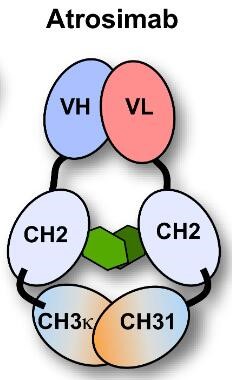
Atrosimab is an Fv-Fc1K fusion protein with an EC50 value of 0.37 nM against humans. Atrosimab inhibits TNF-induced TNFR1 activation, release of IL-6 and IL-8, and cell death, and alleviates neuroinflammation. Atrosimab is applicable to research related to inflammatory diseases, neurodegenerative diseases, acute and chronic inflammation, experimental arthritis, non-alcoholic steatohepatitis, and experimental autoimmune encephalomyelitis.
Para uso exclusivo en investigación. No vendemos a pacientes.
- Pureza : 99%
- Peso molecular:72.338 kDa
-
Almacenamiento:
Please store the product under the recommended conditions in the Certificate of Analysis.


To place orders, for customer services and technical support, please contact: MedChemExpress USA
Tel: 609-228-6898 E-mail: [email protected] [email protected]


-
 Actividad biológica
-
Chemical Information
- Protocolo
- Pureza y Documentación
- Referencias
-
 Help & FAQs




Ver todos los productos específicos de isoformas TNF Receptor
More

Actividad biológica
Descripciòn
Isotype
Fv-Fc
Species Reactivity
Human
In Vitro
Atrosimab (serial dilutions; 4-128 nM) binds purified human TNFR1-Fc with high affinity, and lacks binding to human complement protein C1q and human Fcγ receptors Ia, IIb, IIIa[1].
Atrosimab (100 nM; 1, 3, 7 days) exhibits good stability in human plasma for at least 7 days and has an aggregation temperature of 64°C[1].
Atrosimab (serial dilutions; 16-20 h, 24 h) potently inhibits TNF-mediated TNFR1 activation in HeLa, HT1080, and Kym-1 cells, with IC50 values of 54.5 nM, 24.2 nM, and 16.2 nM, respectively[1].
Atrosimab (serial dilutions; 16-20 h) does not exhibit TNFR1 agonistic activity in HT1080 cells even when cross-linked by three different goat anti-human IgG sera[1].
Atrosimab (24 h) does not affect spontaneous or glutamate-induced altered firing rates of huTNFR1-k/i mouse embryonic day 14 primary cortical neurons after 24 h incubation[2].
Atrosimab (100 μg; heated from 35°C to 80°C with 2 min equilibration per 1°C interval; 2-500 nM; up to 9 months) is highly thermally and long-term stable, binds selectively and with high affinity to human, rhesus, and cynomolgus TNFR1 (but not mouse/rat TNFR1 or any TNFR2), and recognizes an epitope on human TNFR1 that includes residues P23, L67, R68, H69, and Q24[3].
Atrosimab (up to 1000 nM; 16-20 h at 37°C, 5% CO2; 1-7 days at 37°C in 50% human serum) potently and dose-dependently inhibits TNF-induced IL-8 release in HT1080 cells with IC50 values of 21.9-34.9 nM, does not exhibit agonistic activity in these cells, and maintains functional stability after incubation in human serum at 37°C for up to 7 days[3].
Atrosimab (100 μg/mL; 24 h) inhibits TNF-induced VCAM-1 and ICAM-1 expression in hCMEC/D3 human brain microvascular endothelial cells[3].
MedChemExpress (MCE) has not independently confirmed the accuracy of these methods. They are for reference only. Further protocols information, click here.
-
Cell Line:HT1080 cells
-
Concentration:serial dilutions of Atrosimab; ~15.8 nM (constant anti-human IgG serum)
-
Incubation Time:16-20 h
-
Result:Showed no induction of IL-8 release across all tested concentrations, indicating no cross-linking-mediated TNFR1 agonistic activity.
Parmacokinetics
| Species | Dose | Route | T1/2 (Distribution) | T1/2β | AUC0-t | C0 | Vss | CL |
|---|---|---|---|---|---|---|---|---|
| Mice[1] | 400 μg | i.v. | 2.2 h | 41.7 h | 5856.0 μg·h/mL | 324.7 μg/mL | 3.4 μg/mL | 0.29 μg/mL |
| Mice[3] | 30.0 mg/kg | i.v. | 2.26 h | 32.77 h | 6989.7 μg·h/mL | / | / | / |
| Mice[3] | 1.0 mg/kg | i.v. | 1.05 h | 31.37 h | 365.78 μg·h/mL | / | / | / |
| Mice[3] | 30.0 mg/kg | s.c. | / | 33.7 h | 6190.7 μg·h/mL | / | / | / |
| Mice[3] | 1.0 mg/kg | s.c. | / | 54.56 h | 321.83 μg·h/mL | / | / | / |
| Mice[3] | 30.0 mg/kg | i.v. | 0.66 h | 31.52 h | 7983.8 μg·h/mL | / | / | / |
| Mice[3] | 1.0 mg/kg | i.v. | 5.41 h | 9.52 h | 177.8 μg·h/mL | / | / | / |
| Mice[3] | 30.0 mg/kg | s.c. | / | 41 h | 6816.7 μg·h/mL | / | / | / |
| Mice[3] | 1.0 mg/kg | s.c. | / | 16.75 h | 214.07 μg·h/mL | / | / | / |
In Vivo
Atrosimab (30 mg/kg; i.v.; single dose) potently blocks acute TNF-induced inflammatory responses in huTNFR1-k/i mice[3].
Atrosimab (20-80 mg/kg; s.c.; twice weekly; 11 weeks) prevents arthritis development in a prophylactic setting and Atrosimab (5-45 mg/kg; s.c.; twice weekly; 6 weeks) alleviates established arthritis with dose-dependent efficacy in Tg197hTNFR1KI mice[3].
Atrosimab (45 mg/kg; i.p.; twice weekly; 8 weeks) alleviates liver fibrosis, steatosis, and apoptotic injury in a huTNFR1-k/i mouse model of NASH[3].
Atrosimab (30-60 mg/kg; i.p.; single doses on days 1, 4, 8, 12 of manifest disease) ameliorates motor symptoms, demyelination, and axonal injury in a huTNFR1-k/i mouse model of EAE, with 30 mg/kg being sufficient for maximal therapeutic activity[3].
MedChemExpress (MCE) has not independently confirmed the accuracy of these methods. They are for reference only.
-
Animal Model:huTNFR1-k/i (7-month-old male; C57Bl/6J background; NMDA-induced acute cholinergic neurodegeneration via stereotactic injection into NBM)[2]
-
Dosage:3 μg
-
Administration:stereotactic co-injection into NBM; infused at 0.1 μl/min at two dorsoventral depths: 0.3 μl at -0.44 mm and 0.3 μl at -0.46 mm relative to Bregma; total volume 0.6 μl
-
Result:Increased post-shock latency to 274.8 s in passive avoidance paradigm, indicating improved long-term associative memory.\nPrevented significant body weight loss, with mean body weight change of -2.43 g from surgery to perfusion.\nIncreased surface area of ChAT-positive cholinergic fibers in layer V of somatosensory cortex to 11.04%.\nIncreased ratio of cholinergic neurons in lesioned vs. non-lesioned NBM hemisphere to 0.14.\nIncreased ratio of NeuN-positive neuronal surface area in lesioned vs. non-lesioned somatosensory cortex layer V to 0.48.\nReduced volume of IBA-1-positive activated microglia/lesion size to 0.373 mm3.\nReduced ratio of CD68 mean intensity (lesioned vs. non-lesioned NBM hemisphere) to 1.22.\nReduced ratio of GFAP mean intensity (lesioned vs. non-lesioned NBM hemisphere) to 2.51. Showed no significant effect on short-term spatial memory, anxiety-like behavior, locomotor activity, or hippocampal synapsin-1 levels.
-
Animal Model:huTNFR1-k/i (C57BL/6 background)[3]
-
Dosage:30 mg/kg
-
Administration:i.v.; single dose
-
Result:Completely blocked TNF-induced body weight loss.\nSignificantly reduced serum IL-6 levels at 1 hour and 6 hours post-TNF injection to almost baseline levels.\nSignificantly reduced TNF-induced systemic CRP levels at 6 hours post-TNF injection compared to control-treated mice.
-
Animal Model:Tg197hTNFR1KI[3]
-
Dosage:20-80 mg/kg (prophylactic setting); 5-45 mg/kg (therapeutic setting)
-
Administration:s.c.; twice weekly; 11 weeks (prophylactic setting); 6 weeks (therapeutic setting)
-
Result:Prevented development of arthritic disease, prevented disease-associated inhibition of body weight gain, and resulted in no histopathological changes beyond the level of 4-week-old control mice (all doses, prophylactic setting).\nHalted disease progression (5 mg/kg, therapeutic setting).\nShowed improved therapeutic activity comparable to approved anti-TNF therapeutics (15 mg/kg, 45 mg/kg, therapeutic setting).\nImproved joint histopathology, with histopathological scores below the level of 9-week-old control mice (all doses, therapeutic setting).
-
Animal Model:huTNFR1-k/i (male)[3]
-
Dosage:45 mg/kg
-
Administration:i.p.; twice weekly; 8 weeks
-
Result:Significantly reduced liver fibrosis (assessed by Sirius Red staining).\nReduced liver steatosis (assessed by Oil Red O staining).\nDecreased activated caspase-3-positive cells (a marker of apoptotic liver injury) compared to saline-treated mice.
-
Animal Model:huTNFR1-k/i (female, 6-8 weeks old)[3]
-
Dosage:30-60 mg/kg
-
Administration:i.p.; single doses on days 1, 4, 8, 12 of manifest disease
-
Result:Ameliorated EAE motor symptoms.\nReduced cumulative EAE score.\nPrevented disease-associated weight loss.\nReduced cumulative weight loss.\nSignificantly reduced spinal cord demyelination (assessed by Luxol fast blue staining).\nSignificantly reduced axonal injury (assessed by amyloid precursor protein accumulation) compared to control-treated mice.\nShowed no efficacy difference between the 30 mg/kg and 60 mg/kg doses.
Ensayo clínico
| NCT Number | Sponsor | Condition | Start Date |
Phase

|
|---|---|---|---|---|
| NCT01329991 | Plexxikon| | 2011-05 | PHASE1 |
Gene ID
Accession
Conjugated
Unconjugated
Reconsititution
The product can be reconstituted/diluted with sterile PBS or saline.
Format
-
Fv-Fc1κ
Aplicación
ELISA, FACS, Functional assay
Verified Bioactivity
-
 Loaded Atrosimab on ProA biosensor, can bind TNFR-1/CD120a Protein, Human (HEK293, His, HY-P700263) with an affinity constant of 2.076E-09 M as determined in BLI assay.


Chemical Information
-
Appearance Liquid
-
Peso molecular 72.338 kDa
-
Color Colorless to light yellow
-
SMILES
[Atrosimab]
-
Synonyms
ATM-001
-
Envío
Shipping with dry ice.
-
Formulation
Please refer to the lot-specific COA for specific buffer information.
-
Almacenamiento
Please store the product under the recommended conditions in the Certificate of Analysis.

Protocolo
-
Collagen-Induced Arthritis
Collagen-induced arthritis (CIA) is an autoimmune murine model of rheumatoid arthritis in which immunization with type II collagen (CII) emulsified in an adjuvant induces a T cell- and autoantibody-driven inflammatory arthritis characterized by synovial hyperplasia, immune cell infiltration, and joint destruction. The model typically relies on genetically susceptible mouse strains (e. g. , DBA/1) and reproduces key features of human rheumatoid arthritis, including anti-collagen immune responses and progressive joint inflammation. Disease onset generally occurs within ~3-4 weeks after immunization, depending on antigen/adjuvant combinations and protocol variation. The immunopathology is driven by adaptive immune activation against CII, leading to systemic and local joint inflammation mediated by pro-inflammatory cytokines and effector immune cells, making CIA a standard preclinical platform for evaluating immunomodulatory and anti-arthritic interventions.
-
Cell Cytotoxicity Assay
Cytotoxicity assays are usually based on the assessment of cell membrane damage, which can also be indirectly detected by measuring cell viability. Detection methods include MTT assay, CKK-8 assay, LDH assay and ATP assay, etc.
-
Mammalian live/dead viability and cytotoxicity staining
Live/dead viability and cytotoxicity staining assays are based on the simultaneous detection of intracellular esterase activity in metabolically active (viable) cells and membrane integrity loss in non-viable cells. In commonly used dual-staining approaches, membrane-permeant fluorogenic substrates are converted by intracellular esterases into fluorescent products in live cells, while impermeant DNA-binding dyes selectively enter cells with compromised plasma membranes and label nucleic acids in dead or dying cells, enabling discrimination between viable and non-viable populations by fluorescence microscopy or flow cytometry.
-
Cotton Pellet Granuloma
Cotton pellet granuloma is a classical in vivo chronic inflammation model used to evaluate the anti-inflammatory potential of test substances by measuring their ability to inhibit granuloma tissue formation around an implanted foreign body (cotton pellet) in rodents. The method is based on the biological response to a sterile implanted material, which induces proliferative phase inflammation characterized by fibroblast proliferation and collagen-rich granuloma formation, and the final readout reflects the extent of chronic inflammatory tissue growth surrounding the pellet. In multiple preclinical pharmacological evaluations, inhibition of cotton pellet-induced granuloma formation has been used as an indicator of anti-inflammatory activity in both synthetic and natural product screening contexts.
-
Apoptosis Solutions
Apoptosis is a regulated, generally non-lytic cell-death pathway that removes unwanted, damaged, infected, or abnormal cells through coordinated morphological changes, caspase activation, DNA fragmentation, and membrane remodeling. The intrinsic apoptosis pathway is controlled mainly by mitochondrial outer membrane permeabilization, BCL-2 family proteins, cytochrome c release, apoptosome formation, caspase-9 activation, and downstream executioner caspase-3/7 activation. The extrinsic apoptosis pathway is initiated by death receptors such as Fas, TNFR, and TRAIL receptors, which recruit adaptor proteins and activate caspase-8 before engaging executioner caspases or mitochondrial amplification through BID cleavage. Apoptosis is linked to many phenotypes, including cancer cell killing, tissue homeostasis, immune regulation, neurodegeneration, infection response, and treatment-induced cytotoxicity; unresolved questions include how apoptosis interacts with necroptosis, pyroptosis, ferroptos
-
Research Protocol for Inflammation-related Diseases
The NLRP3 inflammasome is a cytosolic innate immune signaling platform that integrates priming signals and danger-signal activation to promote caspase-1 activation, maturation of IL-1β and IL-18, and gasdermin D-mediated pyroptotic cell death. The core experimental logic is to determine whether inflammatory disease phenotypes are driven by increased NLRP3 expression, ASC-containing inflammasome assembly, caspase-1 cleavage, GSDMD cleavage, and extracellular release of IL-1β/IL-18 rather than by nonspecific cell injury alone. The pathway is strongly linked to inflammation-related disease phenotypes because monosodium urate crystals activate NALP3/NLRP3 inflammasome signaling in gout-like crystal inflammation, cholesterol crystals activate NLRP3 inflammasomes in atherogenesis models, and DSS-induced intestinal inflammation has been reported to involve NLRP3 inflammasome activity. However, experimental colitis studies also show context-dependent protective effects of NLRP3 inflammasome co
-
Research Protocol for Neurological Diseases
PINK1/Parkin-mediated mitophagy pathway is a mitochondrial quality-control signaling axis in which mitochondrial depolarization stabilizes PINK1 on damaged mitochondria, activates Parkin recruitment and E3 ubiquitin ligase activity, promotes ubiquitination of outer mitochondrial membrane proteins, recruits selective autophagy adaptors, and drives lysosomal degradation of damaged mitochondria. In neurological disease research, this pathway is experimentally important because neurons, especially dopaminergic neurons, are highly dependent on mitochondrial integrity, and defective mitochondrial turnover can lead to mitochondrial dysfunction, oxidative stress, impaired neuronal survival, α-synuclein accumulation, and neuroinflammatory damage-associated signals. The genetic disease link is strongest in Parkinson’s disease because mutations in PRKN/parkin cause autosomal recessive juvenile parkinsonism, mutations in PINK1 cause hereditary early-onset Parkinson’s disease, and Drosophila studie


Pureza y Documentación
-
Ficha de datos (268 KB)
-
SDS (251 KB)
- English - EN (251 KB)
- Français - FR (251 KB)
- Deutsch - DE (251 KB)
- Norwegian - NO (251 KB)
- Español - ES (251 KB)
- Swedish - SV (251 KB)
- Italian - IT (251 KB)
- Korean - KR (251 KB)
- Portuguese - PT (251 KB)
-
Inhibitory Antibodies User Guide (603 KB)
Referencias
[1]. Richter F, et al. Improved monovalent TNF receptor 1-selective inhibitor with novel heterodimerizing Fc. MAbs. 2019;11(4):653-665. [Content Brief]
[2]. Ortí-Casañ N, et al. The TNFR1 antagonist Atrosimab reduces neuronal loss, glial activation and memory deficits in an acute mouse model of neurodegeneration. Sci Rep. 2023;13(1):10622. Published 2023 Jun 30. [Content Brief]
[3]. Richter F, et al. The TNFR1 Antagonist Atrosimab Is Therapeutic in Mouse Models of Acute and Chronic Inflammation. Front Immunol. 2021;12:705485. Published 2021 Jul 7. [Content Brief]

Calculators
Concentration (start) × Volume (start) = Concentration (final) × Volume (final)


Atrosimab Related Classifications
HY-P991049 Related Classifications
 Powered by Bioz