
PAA4
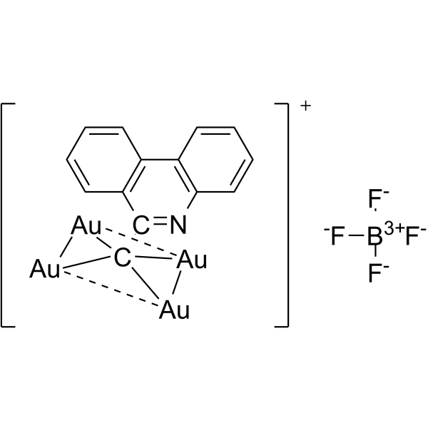
PAA4 is a hypercoordinate carbon-centered tetranuclear gold (I) cluster prodrug. PAA4 releases active Au (I) ions upon triggering by GSH (HY-D0187), and this process is accelerated in the acidic microenvironment of bladder cancer cells with high GSH expression. PAA4 inhibits the activities of cytosolic TrxR1 and mitochondrial TrxR2, inducing ROS accumulation, lipid peroxidation, ferroptosis, loss of mitochondrial membrane potential and DNA damage. PAA4 can be used in bladder cancer-related research.
Para uso exclusivo en investigación. No vendemos a pacientes.
- Fòrmula: C14H8Au4BF4N
- Peso molecular:1064.89
-
Almacenamiento:
Please store the product under the recommended conditions in the Certificate of Analysis.
Ver todos los productos específicos de isoformas DNA/RNA Synthesis
More
Actividad biológica
Descripciòn
In Vitro
PAA4 (0-4.0 μM; 24 h) exerts selective cytotoxicity against human bladder cancer EJ cells (IC50 = 0.8 μM), compared with normal HUVEC cells (IC50 = 2.1 μM) and SV-HUC-1 cells (IC50 = 2.3 μM)[1].
PAA4 (1-4 μM; 6-8 h) induces ferroptosis in human bladder cancer EJ cells, as evidenced by increased PTGS2 expression, elevated lipid peroxidation levels, and other changes[1].
PAA4 (1.5 μM; 4-24 h) is efficiently internalized by human bladder cancer EJ cells, with an uptake rate of 30%, and approximately 10% of the gold localizes to mitochondria; at the same or lower concentrations, its cellular uptake rate is higher than that of the mononuclear gold (I) complex PA1[1].
PAA4 (1.5 μM; 4 h) releases active AuI ions in human bladder cancer EJ cells[1].
PAA4 (1.5 μM; 4 h) potently inhibits the activities of cytosolic TrxR1 and mitochondrial TrxR2 in human bladder cancer EJ cells, reducing their activities to 21% and 22% of that in the control group, respectively[1].
PAA4 (1.5-2.0 μM; 4 h) increases the intracellular ROS level in human bladder cancer EJ cells by 3.4-fold[1].
PAA4 (1-4 μM; 1-6 h) upregulates the pro-oxidative genes CYBA and CYBB, and induces DNA damage in human bladder cancer EJ cells[1].
PAA4 (1.5 μM; 4 h) induces loss of mitochondrial membrane potential in human bladder cancer EJ cells[1].
MedChemExpress (MCE) has not independently confirmed the accuracy of these methods. They are for reference only. Further protocols information, click here.
-
Cell Line:human bladder cancer EJ cells
-
Concentration:1, 2 and 4 μM
-
Incubation Time:6 h with or without pre-treatment for 2 hours with Deferoxamine (DFO) (HY-B1625) (100 μM) or N-Acetyl-L-cysteine (NAC) (HY-B0215) (3.0 mM).
-
Result:Increased PTGS2 expression, and reduced cytotoxicity upon pretreatment with ferroptosis inhibitors DFO or NAC.
In Vivo
PAA4 (1.5 μM; intravesical; once every other day; 5 total doses, 60 min incubation per dose) demonstrates potent in vivo anti-bladder cancer activity in the orthotopic model, drastically reducing tumor bioluminescence signal at 21 days, extending median survival beyond 50 days, inducing tumor ferroptosis, and exhibiting good biocompatibility with no detectable liver or kidney toxicity[1].
MedChemExpress (MCE) has not independently confirmed the accuracy of these methods. They are for reference only.
-
Animal Model:BALB/c nude mice (female, 4-6 weeks, ~18 g, air-pouch bladder cancer model)[1]
-
Dosage:1.5 μM
-
Administration:intra-pouch; single incubation; 30 min
-
Result:Reduced mean tumor volume to 564 mm3 after 22 days compared to 1067 mm3 in PBS control.
Extended median survival time to 36 days compared to 25 days in PBS control.
Showed no significant body weight loss.
-
Animal Model:BALB/c nude mice (female, 6-8 weeks, 16-18 g, orthotopic bladder cancer model)[1]
-
Dosage:1.5 μM
-
Administration:intravesical; once every other day; 5 total doses, 60 min incubation per dose
-
Result:Drastically reduced bioluminescence intensity (luciferase signal) at 21 days post-first treatment compared to PBS control.
Extended median survival time beyond 50 days, with over 50% survival at 50 days compared to 28 days in PBS control.
Showed no significant differences in blood levels of ALT, AST, ALP, BUN, and CRE from saline-treated controls.
Detected significant PTGS2 immunofluorescence in tumor sections, confirming ferroptosis induction.
Chemical Information
-
Peso molecular 1064.89
-
Fòrmula C14H8Au4BF4N
-
SMILES
[F-][B+3]([F-])([F-])[F-].C1(C(C234[Au]5[Au]2[Au]3[Au]45)=NC6=C7C=CC=C6)=C7C=CC=C1
-
Envío
Room temperature in continental US; may vary elsewhere.
-
Almacenamiento
Please store the product under the recommended conditions in the Certificate of Analysis.
Protocolo
-
RNA extraction experimental
By lysing cells, releasing RNA, and removing impurities such as proteins and DNA, high-purity RNA products are finally obtained. The commonly used traditional method is the guanidine isothiocyanate/phenol/chloroform method (Trizol), which is suitable for a variety of animal materials including animal tissues, microorganisms, cultured cells, etc., and most plant materials.
-
Mitochondrial membrane-potential fluorescent assay
Mitochondrial membrane potential fluorescent assays estimate ΔΨm in living cells using lipophilic cationic dyes such as TMRM, TMRE, rhodamine 123, and JC-1, which accumulate in mitochondria according to membrane polarization; loss of signal after FCCP or CCCP treatment is interpreted as mitochondrial depolarization. TMRM/TMRE and rhodamine 123 are commonly used for semi-quantitative live-cell microscopy or flow cytometry, while JC-1 can report a shift from red aggregate fluorescence to green monomer fluorescence during depolarization; interpretation requires controls because dye concentration, quenching mode, cell type, dye efflux, and mitochondrial mass can affect fluorescence independently of ΔΨm.
-
ROS/oxidative-stress fluorescent staining
ROS/oxidative-stress fluorescent staining uses cell-permeant fluorogenic probes that become fluorescent after oxidation inside cells or tissues; commonly used examples include DCFH-DA/DCFDA for broad cellular oxidant detection, DHE for superoxide-related signal detection, MitoSOX for mitochondrial superoxide-related signal detection, and CellROX probes for oxidative-stress-associated fluorescence readouts. The assay detects probe oxidation rather than a single ROS species unless the probe and analysis method have been chemically validated for that species. DCFH-DA enters cells, is deacetylated by intracellular esterases to DCFH, and produces fluorescent DCF after oxidation, so the readout is used as an operational measure of total cellular oxidative stress rather than a species-specific ROS measurement. DHE and MitoSOX can report superoxide-related oxidation, but red fluorescence alone can include non-specific ethidium-like oxidation products; HPLC or optimized spectral approaches are
-
Fluorescent plasma-membrane potential dye assay
Fluorescent plasma-membrane potential dye assays measure changes in cell membrane potential using voltage-sensitive dyes whose fluorescence changes when cells depolarize or hyperpolarize. Anionic bis-oxonol dyes such as DiBAC4(3) enter depolarized cells more readily and show increased fluorescence after intracellular binding, while hyperpolarization reduces dye accumulation and fluorescence. FMP/FLIPR membrane-potential dyes are used for faster, homogeneous microplate assays of ion-channel or receptor-mediated membrane-potential changes.
-
Mitochondrial membrane-potential and mitochondrial mass staining
Mitochondrial membrane potential staining measures the electrochemical polarization across the mitochondrial inner membrane in live cells using lipophilic cationic fluorescent probes; early rhodamine-based work showed that selective mitochondrial dye accumulation is lost when the mitochondrial transmembrane potential is dissipated. JC-1 reports mitochondrial polarization by shifting from green monomer fluorescence to red J-aggregate fluorescence as dye concentration increases within energized mitochondria; therefore, the red/green fluorescence ratio is used as a relative readout of mitochondrial membrane potential. TMRE or TMRM staining provides a single-channel relative readout because these cationic rhodamine esters accumulate in polarized mitochondria, and lower fluorescence indicates reduced mitochondrial polarization when acquisition and dye-loading conditions are controlled. Mitochondrial mass staining is commonly performed with MitoTracker Green FM or related MitoTracker dyes as
-
Ferroptosis Solutions
Ferroptosis is an iron-dependent, non-apoptotic form of regulated cell death characterized by lethal lipid peroxidation and sensitivity to suppression by iron chelators or lipophilic radical-trapping antioxidants. The core pathway links cystine uptake through system Xc−, glutathione availability, GPX4-dependent detoxification of phospholipid hydroperoxides, iron-dependent oxidative reactions, and polyunsaturated-phospholipid metabolism into a cell-death program that is biochemically and morphologically distinct from apoptosis, necrosis, and autophagy. The ferroptosis pathway is experimentally linked to phenotype through chemical and genetic perturbation. Erastin induces ferroptosis by inhibiting cystine uptake through system Xc− and weakening antioxidant defenses, while GPX4 inhibition or depletion causes lipid peroxide accumulation and ferroptotic cancer-cell death. ACSL4 and oxidizable arachidonoyl- or adrenoyl-containing phosphatidylethanolamines shape ferroptosis sensitivity by con
-
Genotoxicity/Mutagenicity Study
The bacterial reverse mutation assay detects point mutations that restore amino-acid prototrophy in auxotrophic Salmonella typhimurium or Escherichia coli tester strains; after exposure to a test article, mutagenic activity is read out as an increased number of revertant colonies on minimal agar compared with the vehicle control. The assay uses tester strains with different mutation targets so that base-substitution and frameshift mutagens can be detected, and testing is performed with and without exogenous mammalian metabolic activation because some chemicals require biotransformation to become mutagenic.
Pureza y Documentación
Referencias
Calculators
Concentration (start) × Volume (start) = Concentration (final) × Volume (final)

