



Hydroxyphenyl Fluorescein (solution)

Based on 1 Customer Validation
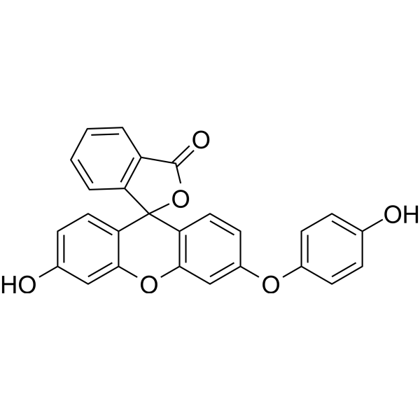
Hydroxyphenyl Fluorescein (HPF) (solution) is a stable ROS fluorescent probe dye. Hydroxyphenyl Fluorescein has stronger specificity and stability than H2DCFDA (HY-D0940). Hydroxyphenyl Fluorescein can produce strong green fluorescence through hydroxyl radical reaction with intracellular peroxynitroso. Hydroxyphenyl Fluorescein can be applied for fluorescence microscopy, high-throughput imager, luciferase microplate reader or flow cytometry. Ex/Em=490/515 nm.
Solvent and concentration: DMSO: 10 mM
For research use only. We do not sell to patients.
- CAS No.: 359010-69-8
- Formula: C26H16O6
- Molecular Weight:424.41
-
Storage:
Please store the product under the recommended conditions in the Certificate of Analysis.


To place orders, for customer services and technical support, please contact: MedChemExpress USA
Tel: 609-228-6898 E-mail: [email protected] [email protected]


-
 Biological Activity
-
Chemical Information
- Protocols
- Purity & Documentation
- References
-
 Help & FAQs


-

HY-DY1004
BODIPY 493/503 (solution)Neutral Lipid Dye -
HY-111330
Hydroxyphenyl FluoresceinROS Probe -

HY-DY1015
Dihydroethidium (solution)Fluorescent -
HY-15902
ABTS diammonium saltHRP Substrate -

HY-B0215
AcetylcysteineMucolytic Agent -
HY-D0819
CY5-SEReactive Dye -
HY-W090090
BODIPY 493/503Neutral Lipid Dye -
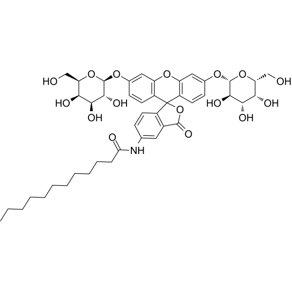
HY-126839
C12FDGβ-Galactosidase Fluorescent Substrate -
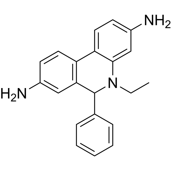
HY-D0079
DihydroethidiumFluorescent -
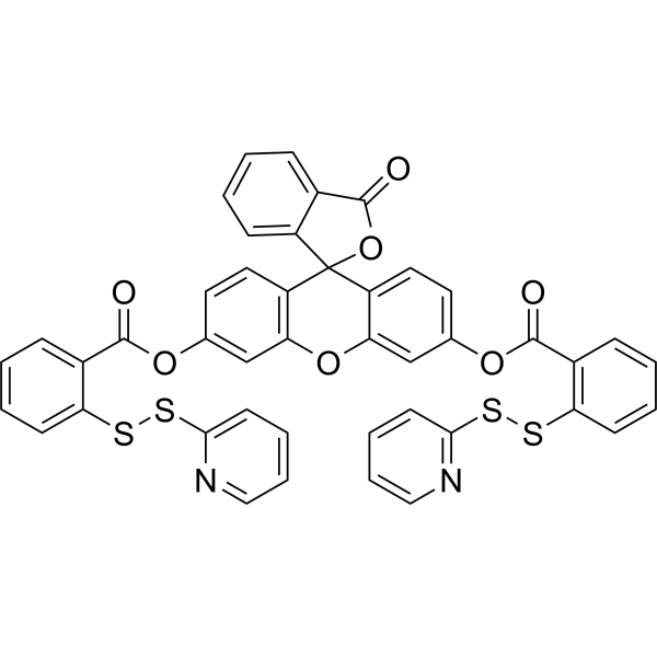
HY-129420
WSP-5Fluorescent Probe -

HY-B0215R
Acetylcysteine (Standard)Mucolytic Agent -
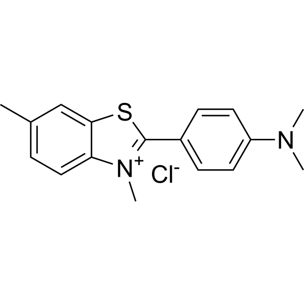
HY-D0218
Thioflavin Tβ-amyloid Dye


Biological Activity
Description
Solvent and concentration: DMSO: 10 mM
In Vitro
Guidelines (Following is our recommended protocol. This protocol only provides a guideline, and should be modified according to your specific needs).
1. Preparation of HPF working solution
1.1 Stock solution
It is recommended to store the stock solution at -20°C -80°C away from light and avoid repetitive freeze-thaw cycles.
1.2 Preparation of HPF working solution
Dilute the stock solution in serum-free cell culture medium or PBS.The corresponding stock solution can be diluted according to the actual situation. Note that if the solvent is DMSO, the cytotoxicity of DMSO must be considered, and a solvent control should be prepared; if the solvent is pure water, the working solution needs to be filtered and sterilized before adding cells.
Note: Please adjust the concentration of HPF working solution according to the actual situation.
2. Cell staining
2.1 Cell preparation
For suspension cells: Centrifuge at 1000 g at 4°C for 3-5 minutes and then discard the supernatant. Wash twice with PBS, 5 minutes each time.
For adherent cells: Discard the cell culture medium, and add trypsin to dissociate cells to make a single-cell suspension. Centrifuge at 1000 g at 4°C for 3-5 minutes and then discard the supernatant. Wash twice with PBS, 5 minutes each time.
2.2 Add 1 mL of HPF working solution, and then incubate at room temperature for 15-30 minutes.
2.3 Centrifuge at 400 g at 4°C for 3-4 minutes and then discard the supernatant.
2.4 Wash twice with PBS, 5 minutes each time.
2.5 Resuspend cells with serum-free cell culture medium or PBS, and then detect by fluorescence microscope or flow cytometer.
MedChemExpress (MCE) has not independently confirmed the accuracy of these methods. They are for reference only. Further protocols information, click here.


Chemical Information
-
CAS No. 359010-69-8
-
Appearance Liquid
-
Molecular Weight 424.41
-
Formula C26H16O6
-
Color Colorless to light yellow
-
SMILES
OC1=CC=C(OC2=CC3=C(C4(C5=CC=C(C=C5O3)O)C6=C(C(O4)=O)C=CC=C6)C=C2)C=C1
-
Shipping
Room temperature in continental US; may vary elsewhere.
-
Storage
Please store the product under the recommended conditions in the Certificate of Analysis.

Protocols
-
Flow Cytometry
Flow cytometry (FC) is a technique for high-speed, step-by-step quantitative analysis and sorting of single cells or other biological particles in a suspension by detecting labeled fluorescent signals.
-
Flow cytometric DNA-content cell-cycle staining
Flow cytometric DNA-content cell-cycle staining measures the fluorescence intensity of DNA-bound fluorochromes in single cells or nuclei to estimate DNA content distributions, allowing assignment of populations to G0/G1, S, and G2/M phases by DNA histogram deconvolution. Propidium iodide (PI) intercalates into DNA, and PI fluorescence is proportional to cellular DNA content when staining is performed under conditions that make DNA accessible and minimize non-DNA signal. Cells with G2/M DNA content are expected to show approximately twice the fluorescence intensity of G0/G1 cells, while S-phase cells occupy intermediate fluorescence values. PI-based DNA-content analysis can also detect cells with fractional DNA content, often reported as sub-G1, when DNA fragmentation and extraction during staining reduce retained DNA signal in apoptotic cells. DAPI is an alternative DNA fluorochrome for univariate DNA-content analysis, while bivariate approaches combining DNA content with proliferation
-
ROS/oxidative-stress fluorescent staining
ROS/oxidative-stress fluorescent staining uses cell-permeant fluorogenic probes that become fluorescent after oxidation inside cells or tissues; commonly used examples include DCFH-DA/DCFDA for broad cellular oxidant detection, DHE for superoxide-related signal detection, MitoSOX for mitochondrial superoxide-related signal detection, and CellROX probes for oxidative-stress-associated fluorescence readouts. The assay detects probe oxidation rather than a single ROS species unless the probe and analysis method have been chemically validated for that species. DCFH-DA enters cells, is deacetylated by intracellular esterases to DCFH, and produces fluorescent DCF after oxidation, so the readout is used as an operational measure of total cellular oxidative stress rather than a species-specific ROS measurement. DHE and MitoSOX can report superoxide-related oxidation, but red fluorescence alone can include non-specific ethidium-like oxidation products; HPLC or optimized spectral approaches are
-
Protocol for Phospho-flow cytometry
Phospho-flow cytometry detects intracellular phosphorylated signaling proteins in single cells using phospho-specific antibodies after rapid fixation and permeabilization; the fluorescence intensity reflects phosphorylation state and therefore kinase-pathway activation, inhibition, or drug response in defined cell subsets. Unlike Western blot, phospho-flow preserves single-cell resolution and can measure signaling heterogeneity in cancer cells, primary immune cells, dissociated mouse tumors, macrophages, organoid-derived cells, and drug-screening samples when validated antibodies and fixation/permeabilization conditions are used.


Purity & Documentation
-
Data Sheet (273 KB)
-
SDS (252 KB)
- English - EN (252 KB)
- Français - FR (252 KB)
- Deutsch - DE (252 KB)
- Norwegian - NO (252 KB)
- Español - ES (252 KB)
- Swedish - SV (252 KB)
- Italian - IT (252 KB)
- Korean - KR (252 KB)
- Portuguese - PT (252 KB)
-
Handling Instructions (2659 KB)
References

Calculators
Concentration (start) × Volume (start) = Concentration (final) × Volume (final)


Hydroxyphenyl Fluorescein (solution) Related Classifications
HY-DY1035 Related Classifications
 Powered by Bioz