



Psammaplin A

Based on 1 Customer Validation
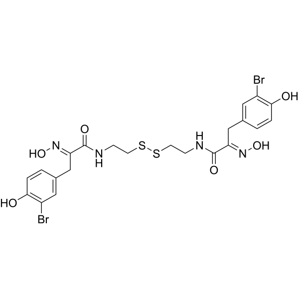
Psammaplin A is a marine metabolite. Psammaplin A is a selective HDAC1 (IC50: 45 nM), DNA methyltransferases (IC50: 18.6 nM) and aminopeptidase N (APN) (IC50: 18 μM) inhibitor. Psammaplin A also inhibits DNA topoisomerase and farnesyl protein transferase. Psammaplin A is a PPARγ activator and induces apoptosis. Psammaplin A has antitumor and anti-inflammatory activities. Psammaplin A has antibacterial activity against Gram-positive bacteria and inhibits DNA synthesis and DNA gyrase activity. Psammaplin A inhibits angiogenesis.
For research use only. We do not sell to patients.
- Purity : 96.0%
- CAS No.: 110659-91-1
- Formula: C22H24Br2N4O6S2
- Molecular Weight:664.39
-
Storage:
4°C, stored under nitrogen
* In solvent : -80°C, 6 months; -20°C, 1 month (stored under nitrogen)

To place orders, for customer services and technical support, please contact: MedChemExpress USA
Tel: 609-228-6898 E-mail: [email protected] [email protected]


-
 Biological Activity
-
Chemical Information
-
Solvent & Solubility
- Protocols
- Purity & Documentation
- References
-
 Help & FAQs


-
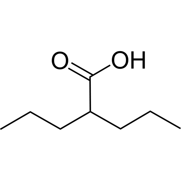
HY-10585
Valproic acidHDAC Inhibitor -
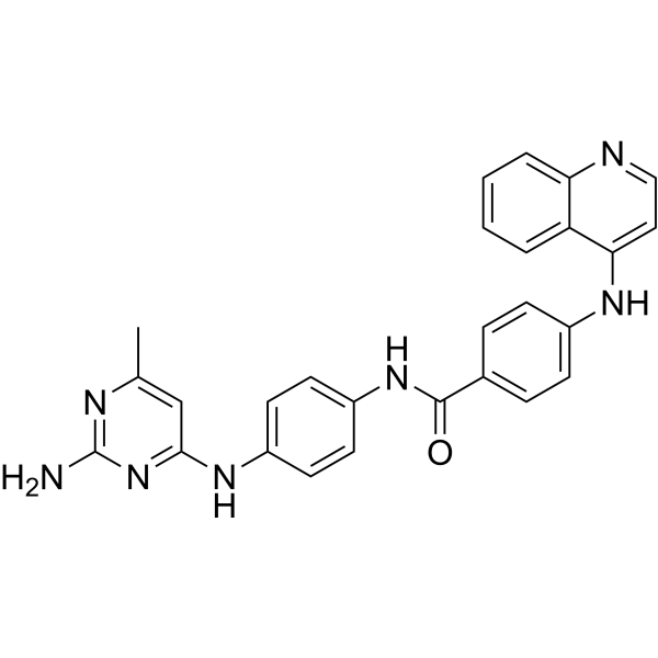
HY-13962
SGI-1027Apoptosis Inducer -
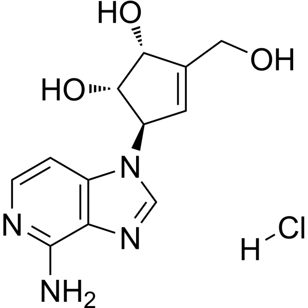
HY-12186
3-Deazaneplanocin A hydrochlorideEZH2 Inhibitor -
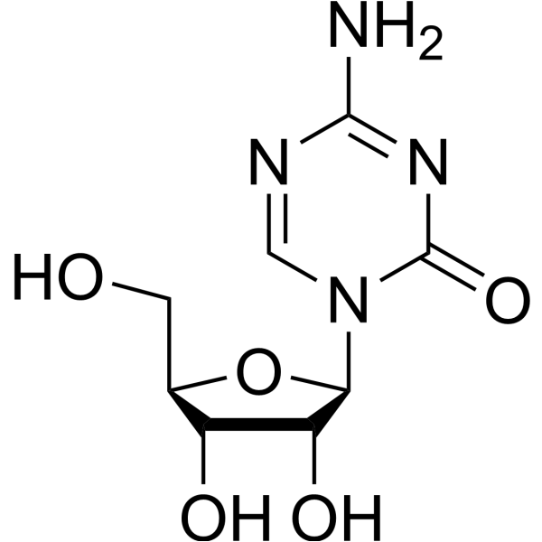
HY-10586
5-AzacytidineDNMT Inhibitor -

HY-10586R
5-Azacytidine (Standard)DNMT Inhibitor -
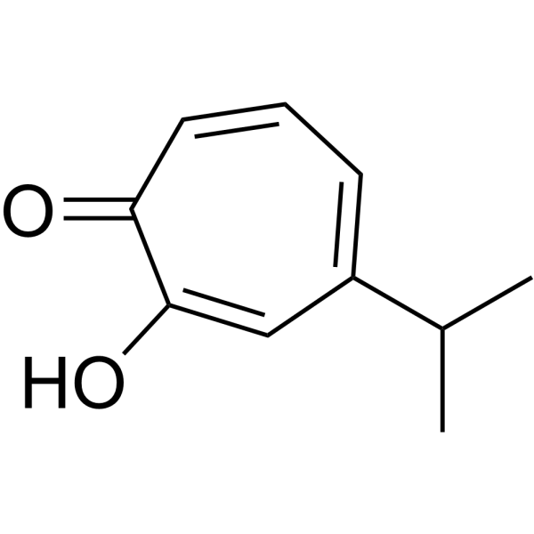
HY-B2230
HinokitiolVirus Protease Inhibitor -
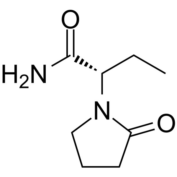
HY-B0106
LevetiracetamAntiepileptic Agent -
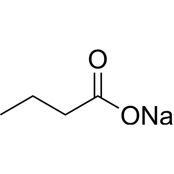
HY-B0350A
Sodium butyrateHDAC Inhibitor -
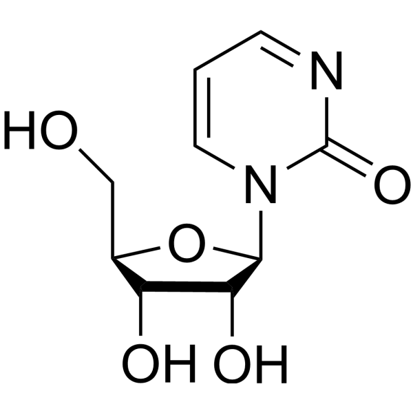
HY-13420
ZebularineDNA Methylation Inhibitor -
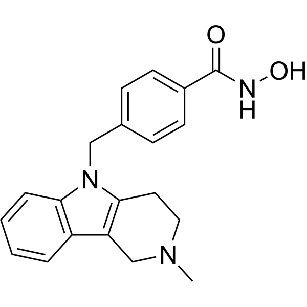
HY-13271A
Tubastatin AHDAC6 Inhibitor -
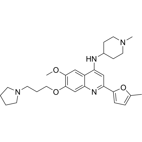
HY-101925
CM-272G9a/DNMTs Inhibitor -
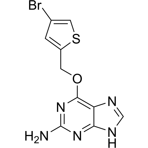
HY-13668
LomeguatribMGMT Inhibitor


Biological Activity
Description
|
HDAC1 45 nM (IC50) |
DNA Methyltransferase 18.6 nM (IC50) |
PPARγ |
APN 18 μM (IC50) |
Cellular Effect
|
Cell Line

|
Type | Value | Description | References |
|---|---|---|---|---|
| A549 | ED50 |
0.57 μg/mL
Compound: 1
|
Cytotoxicity against human A549 cells
Cytotoxicity against human A549 cells
|
[PMID: 14640526] |
| A549 | GI50 |
4.5 μM
Compound: Psammaplin A
|
Cytotoxicity against human A549 cells assessed as reduction in cell growth inhibition after 96 hrs by MTT assay
Cytotoxicity against human A549 cells assessed as reduction in cell growth inhibition after 96 hrs by MTT assay
|
[PMID: 27460171] |
| A549 | GI50 |
7.5 μM
Compound: 11c
|
Growth inhibition of human A549 cells after 96 hrs by sulphorhodamine B assay
Growth inhibition of human A549 cells after 96 hrs by sulphorhodamine B assay
|
[PMID: 22280363] |
| A549 | IC50 |
1.18 μM
Compound: 1, PsA
|
Cytotoxicity against human A549 cells by SRB assay
Cytotoxicity against human A549 cells by SRB assay
|
[PMID: 25884112] |
| A549 | IC50 |
1.76 μM
Compound: PsA
|
Cytotoxicity against human A549 cells assessed as reduction in cell viability by SRB assay
Cytotoxicity against human A549 cells assessed as reduction in cell viability by SRB assay
|
[PMID: 33636429] |
| CHO-K1 | EC50 |
0.9 μM
Compound: 4
|
Cytotoxicity against CHO-K1 cells by Alamar blue assay
Cytotoxicity against CHO-K1 cells by Alamar blue assay
|
[PMID: 16962325] |
| HCT-116 | IC50 |
0.61 μM
Compound: PsA
|
Cytotoxicity against human HCT-116 cells assessed as reduction in cell viability by SRB assay
Cytotoxicity against human HCT-116 cells assessed as reduction in cell viability by SRB assay
|
[PMID: 33636429] |
| HCT-116 | IC50 |
1.62 μM
Compound: 1, PsA
|
Cytotoxicity against human HCT116 cells by SRB assay
Cytotoxicity against human HCT116 cells by SRB assay
|
[PMID: 25884112] |
| HCT-116 | IC50 |
3.05 μM
Compound: PsA
|
Antiproliferative activity against human HCT-116 cells incubated for 48 hrs by MTT assay
Antiproliferative activity against human HCT-116 cells incubated for 48 hrs by MTT assay
|
[PMID: 38851056] |
| HCT-15 | ED50 |
0.68 μg/mL
Compound: 1
|
Cytotoxicity against human HCT15 cells
Cytotoxicity against human HCT15 cells
|
[PMID: 14640526] |
| HeLa | IC50 |
0.05 μM
Compound: Psammaplin A
|
Inhibition of HDAC in human HeLa cells using Fluor deLys as substrate assessed as deacetylation of substrate by fluorescence assay
Inhibition of HDAC in human HeLa cells using Fluor deLys as substrate assessed as deacetylation of substrate by fluorescence assay
|
[PMID: 27460171] |
| HL-60 | GI50 |
0.29 μM
Compound: Psammaplin A
|
Cytotoxicity against human HL60 cells assessed as reduction in cell growth inhibition after 72 hrs by trypan blue-staining based hemocytometric analysis
Cytotoxicity against human HL60 cells assessed as reduction in cell growth inhibition after 72 hrs by trypan blue-staining based hemocytometric analysis
|
[PMID: 27460171] |
| HL-60 | GI50 |
0.37 μM
Compound: 1; PsA
|
Growth inhibition of human HL-60 cells after 72 hrs by trypan blue dye based assay
Growth inhibition of human HL-60 cells after 72 hrs by trypan blue dye based assay
|
[PMID: 33488962] |
| MCF7 | EC50 |
5.7 μM
Compound: 1
|
Activation of PPARgamma transfected in human MCF7 cells by luciferase reporter gene assay
Activation of PPARgamma transfected in human MCF7 cells by luciferase reporter gene assay
|
[PMID: 16643023] |
| MCF7 | GI50 |
1.27 μM
Compound: 11c
|
Growth inhibition of human MCF7 cells after 96 hrs by sulphorhodamine B assay
Growth inhibition of human MCF7 cells after 96 hrs by sulphorhodamine B assay
|
[PMID: 22280363] |
| MCF7 | GI50 |
2.26 μM
Compound: Psammaplin A
|
Cytotoxicity against human MCF7 cells assessed as reduction in cell growth inhibition after 96 hrs by MTT assay
Cytotoxicity against human MCF7 cells assessed as reduction in cell growth inhibition after 96 hrs by MTT assay
|
[PMID: 27460171] |
| MCF7 | IC50 |
5 μM
Compound: 1
|
Antiproliferative activity against human MCF7 cells by neutral red method
Antiproliferative activity against human MCF7 cells by neutral red method
|
[PMID: 16643023] |
| MDA-MB-231 | IC50 |
1.31 μM
Compound: PsA
|
Cytotoxicity against human MDA-MB-231 cells assessed as reduction in cell viability by SRB assay
Cytotoxicity against human MDA-MB-231 cells assessed as reduction in cell viability by SRB assay
|
[PMID: 33636429] |
| P388 | IC50 |
0.3 μg/mL
Compound: 9
|
Cytotoxicity against mouse P388 cells assessed as cell growth inhibition
Cytotoxicity against mouse P388 cells assessed as cell growth inhibition
|
[PMID: 34826681] |
| PC-3 | GI50 |
3.52 μM
Compound: Psammaplin A
|
Cytotoxicity against human PC3 cells assessed as reduction in cell growth inhibition after 96 hrs by MTT assay
Cytotoxicity against human PC3 cells assessed as reduction in cell growth inhibition after 96 hrs by MTT assay
|
[PMID: 27460171] |
| SK-HEP1 | IC50 |
1.29 μM
Compound: PsA
|
Cytotoxicity against human SK-HEP1 cells assessed as reduction in cell viability by SRB assay
Cytotoxicity against human SK-HEP1 cells assessed as reduction in cell viability by SRB assay
|
[PMID: 33636429] |
| SK-MEL-2 | ED50 |
0.13 μg/mL
Compound: 1
|
Cytotoxicity against human SK-MEL-2 cells
Cytotoxicity against human SK-MEL-2 cells
|
[PMID: 14640526] |
| SK-OV-3 | ED50 |
0.14 μg/mL
Compound: 1
|
Cytotoxicity against human SKOV3 cells
Cytotoxicity against human SKOV3 cells
|
[PMID: 14640526] |
| SNU-638 | IC50 |
0.56 μM
Compound: PsA
|
Cytotoxicity against human SNU-638 cells assessed as reduction in cell viability by SRB assay
Cytotoxicity against human SNU-638 cells assessed as reduction in cell viability by SRB assay
|
[PMID: 33636429] |
| T-cell | EC50 |
0.2 μM
Compound: Psammaplin A
|
Reactivation of latent HIV1 NL4-3-Luc expression in human naive CD4+ T cells treated 7 days post-infection measured after 48 hrs by luminescence plate reader analysis
Reactivation of latent HIV1 NL4-3-Luc expression in human naive CD4+ T cells treated 7 days post-infection measured after 48 hrs by luminescence plate reader analysis
|
[PMID: 24495105] |
| WI-38 | GI50 |
3.44 μM
Compound: 11c
|
Growth inhibition of human WI38 cells after 96 hrs by sulphorhodamine B assay
Growth inhibition of human WI38 cells after 96 hrs by sulphorhodamine B assay
|
[PMID: 22280363] |
| XF498 | ED50 |
0.57 μg/mL
Compound: 1
|
Cytotoxicity against human XF498 cells
Cytotoxicity against human XF498 cells
|
[PMID: 14640526] |
In Vitro
Psammaplin A inhibits the viability of A549 (GI50: 7.5 μM), MCF7 (GI50: 1.27 μM), and WI38 (GI50: 3.44 μM) cells[1].
Psammaplin A (72 h) inhibits the viability of A549 (IC50: 1.18 μM) and HCT116 (IC50: 1.62 μM) cells[2].
Psammaplin A (0.5-5 μM) inhibits bFGF-induced angiogenesis in bovine aortic endothelial cells (BAEC) in a dose-dependent manner[3].
Psammaplin A (0-100 μg/mL, 1-4 h) inhibits the toxicity of INT-407 cells induced by V. vulnificus in a concentration- and time-dependent manner[4].
Psammaplin A (0-100 μg/mL, 0-13 h) inhibits the growth of V. vulnificus in a concentration-dependent manner[4].
Psammaplin A (0-40 μM, 12 h) inhibits the viability of the macrophage cell line RAW264.7 in a concentration-dependent manner[6].
Psammaplin A (0-500 μM, 2 h) inhibits SV40 DNA replication in HeLa cells in a concentration-dependent manner.
Psammaplin A (125-500 μM, 30 min) inhibits the catalytic activity of topoisomerase I[6].
Psammaplin A (125-500 μM, 15 min) inhibits the binding activity of RPA to single-stranded DNA[6].
Psammaplin A (1-40 μM, 30 min) inhibits the activity of polymerase α-primase in a concentration-dependent manner[6].
Psammaplin A (3-30 μM, 4-16 h) induces apoptosis in T47D and MCF-7 cells[8].
MedChemExpress (MCE) has not independently confirmed the accuracy of these methods. They are for reference only.
-
Cell Line:MCF7 cells
-
Concentration:5 μM
-
Incubation Time:24 h
-
Result:Up-regulated the expression of Ac-H3 protein.
-
Cell Line:INT-407 cells
-
Concentration:0, 12.5, 25, 50, 100 μg/mL
-
Incubation Time:1, 2, 3, 4 h
-
Result:Reduced cytotoxicity.
Reduced cell damage.
-
Cell Line:Macrophage cell line RAW 264.7
-
Concentration:0, 1, 5, 10, 20, 40 μM
-
Incubation Time:12 h
-
Result:Reduced the number of viable cells.
-
Cell Line:MCF-7 and T47D cells
-
Concentration:3, 10, 30 μM
-
Incubation Time:4, 8, 16 h
-
Result:Increased cells apoptosis.
In Vivo
Psammaplin A (5-50 μg, i.p., once) improves the survival rate of mice in the V. vulnificus-infected model[4].
MedChemExpress (MCE) has not independently confirmed the accuracy of these methods. They are for reference only.
-
Animal Model:A549 cell-implanted xenograft model (male athymic BALB/c mice, 6 weeks old, 18-20 g)[2]
-
Dosage:30 mg/kg
-
Administration:Intraperitoneal injection (i.p.), three times a week for 35 d
-
Result:Inhibited the tumor growth.
Inhibited the expression of the proliferation biomarker Ki-67 in both central and edge region of tumor tissues.
-
Animal Model:V. vulnificus-infected model (35 female ICR mice, 8 weeks old, 20-22 g)[4]
-
Dosage:5, 10, 25 and 50 μg
-
Administration:Intraperitoneal injection (i.p.), once
-
Result:Prolonged survival time of mice.
Inhibited damage to the intestines, liver and spleen of mice.
- Research Protocol for Infectious Diseases
- Human pluripotent stem cell endothelial-cell differentiation
- Vascular/Branching Fractal Analysis
- Annexin V plus membrane-impermeant dye apoptosis staining
- BrdU Incorporation Assay
- Research Protocol for Inflammation-related Diseases
- Gram Staining of Tissue Sections
- Bacterial live/dead nucleic-acid viability staining
- EdU Incorporation Assay (Click Chemistry-Based DNA Synthesis Measurement)
- Detection of Apoptosis
- TUNEL staining for apoptotic DNA fragmentation
- Endothelial Tube Formation Assay
- Apoptosis Solutions
- Endothelial Cell Migration/Angiogenic Sprouting Assay


Chemical Information
-
CAS No. 110659-91-1
-
Appearance Solid
-
Molecular Weight 664.39
-
Formula C22H24Br2N4O6S2
-
Color Off-white to yellow
-
SMILES
O=C(NCCSSCCNC(/C(CC1=CC(Br)=C(O)C=C1)=N/O)=O)/C(CC2=CC(Br)=C(O)C=C2)=N/O
-
Structure Classification
-
Initial Source
-
Shipping
Room temperature in continental US; may vary elsewhere.
-
Storage
4°C, stored under nitrogen
* In solvent : -80°C, 6 months; -20°C, 1 month (stored under nitrogen)


Solvent & Solubility
In Vitro:
DMSO : 33.3 mg/mL (50.12 mM; Need ultrasonic and warming; Hygroscopic DMSO has a significant impact on the solubility of product, please use newly opened DMSO)
Please refer to the solubility information to select the appropriate solvent. Once prepared, please aliquot and store the solution to prevent product inactivation from repeated freeze-thaw cycles.
Storage method and period of stock solution: -80°C, 6 months; -20°C, 1 month (stored under nitrogen). When stored at -80°C, please use it within 6 months. When stored at -20°C, please use it within 1 month.
Please refer to the solubility information to select the appropriate solvent. Once prepared, please aliquot and store the solution to prevent product inactivation from repeated freeze-thaw cycles.
Storage method and period of stock solution: -80°C, 6 months; -20°C, 1 month (stored under nitrogen). When stored at -80°C, please use it within 6 months. When stored at -20°C, please use it within 1 month.
Concentration (start) × Volume (start) = Concentration (final) × Volume (final)


Purity & Documentation
-
Data Sheet (277 KB)
-
SDS (393 KB)
- English - EN (393 KB)
- Français - FR (393 KB)
- Deutsch - DE (393 KB)
- Norwegian - NO (393 KB)
- Español - ES (393 KB)
- Swedish - SV (393 KB)
- Italian - IT (393 KB)
- Korean - KR (393 KB)
- Portuguese - PT (393 KB)
-
Handling Instructions (2659 KB)
References
[1]. Baud MG, et al. Defining the mechanism of action and enzymatic selectivity of psammaplin A against its epigenetic targets. J Med Chem. 2012 Feb 23;55(4):1731-50. [Content Brief]
[2]. Hong S, et al. Efficient synthesis and biological activity of Psammaplin A and its analogues as antitumor agents. Eur J Med Chem. 2015;96:218-30. [Content Brief]
[3]. Shim JS, et al. Psammaplin A, a marine natural product, inhibits aminopeptidase N and suppresses angiogenesis in vitro. Cancer Lett. 2004 Jan 20;203(2):163-9. [Content Brief]
[4]. Lee BC, et al. In vitro and in vivo anti-Vibrio vulnificus activity of psammaplin A, a natural marine compound. Mol Med Rep. 2016 Sep;14(3):2691-6. [Content Brief]
[5]. Piña IC, et al. Psammaplins from the sponge Pseudoceratina purpurea: inhibition of both histone deacetylase and DNA methyltransferase. J Org Chem. 2003 May 16;68(10):3866-73. [Content Brief]
[6]. Jiang Y, et al. Cytotoxicity of psammaplin A from a two-sponge association may correlate with the inhibition of DNA replication. BMC Cancer. 2004 Sep 30;4:70. [Content Brief]
[8]. Mora FD, et al. Bioassay for the identification of natural product-based activators of peroxisome proliferator-activated receptor-gamma (PPARgamma): the marine sponge metabolite psammaplin A activates PPARgamma and induces apoptosis in human breast tumor cells. J Nat Prod. 2006 Apr;69(4):547-52. [Content Brief]


Complete Stock Solution Preparation Table
Please refer to the solubility information to select the appropriate solvent. Once prepared, please aliquot and store the solution to prevent product inactivation from repeated freeze-thaw cycles.
Storage method and period of stock solution: -80°C, 6 months; -20°C, 1 month (stored under nitrogen). When stored at -80°C, please use it within 6 months. When stored at -20°C, please use it within 1 month.
| Optional Solvent | Concentration Solvent Mass | 1 mg | 5 mg | 10 mg | 25 mg |
|---|---|---|---|---|---|
| DMSO | 1 mM | 1.5051 mL | 7.5257 mL | 15.0514 mL | 37.6285 mL |
| 5 mM | 0.3010 mL | 1.5051 mL | 3.0103 mL | 7.5257 mL | |
| 10 mM | 0.1505 mL | 0.7526 mL | 1.5051 mL | 3.7629 mL | |
| 15 mM | 0.1003 mL | 0.5017 mL | 1.0034 mL | 2.5086 mL | |
| 20 mM | 0.0753 mL | 0.3763 mL | 0.7526 mL | 1.8814 mL | |
| 25 mM | 0.0602 mL | 0.3010 mL | 0.6021 mL | 1.5051 mL | |
| 30 mM | 0.0502 mL | 0.2509 mL | 0.5017 mL | 1.2543 mL | |
| 40 mM | 0.0376 mL | 0.1881 mL | 0.3763 mL | 0.9407 mL | |
| 50 mM | 0.0301 mL | 0.1505 mL | 0.3010 mL | 0.7526 mL |


Psammaplin A Related Classifications
HY-N2150 Related Classifications
 Powered by Bioz