



Pemrametostat
Based on 36 publication(s) in Google Scholar
GSK3326595 is a protein arginine methyltransferase 5 (PRMT5) inhibitor. GSK3326595 decreases SARS-CoV-2 infection, inhibits cancer cell proliferation and induces pro-inflammatory macrophage polarization and increases hepatic triglyceride levels without affecting atherosclerosis. GSK3326595 can be used for research of relapsed/refractory mantle cell lymphoma.
For research use only. We do not sell to patients.
- Purity : 99.82%
- CAS No.: 1616392-22-3
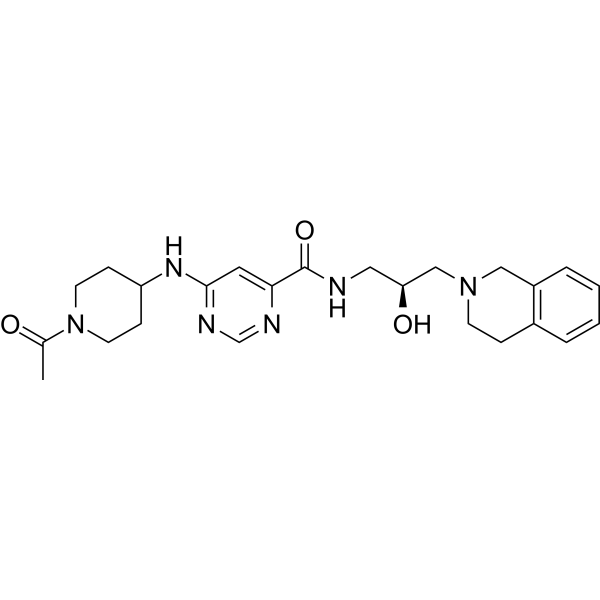
- Formula: C24H32N6O3
- Molecular Weight:452.55
-
Storage:Powder -20°C, 3 years , 4°C, 2 years ; In solvent -80°C, 2 years , -20°C, 1 year

To place orders, for customer services and technical support, please contact: MedChemExpress USA
Tel: 609-228-6898 E-mail: [email protected] [email protected]


-
 Biological Activity
-
Chemical Information
-
Solvent & Solubility
- Protocols
- Purity & Documentation
- References
-
 Help & FAQs


-
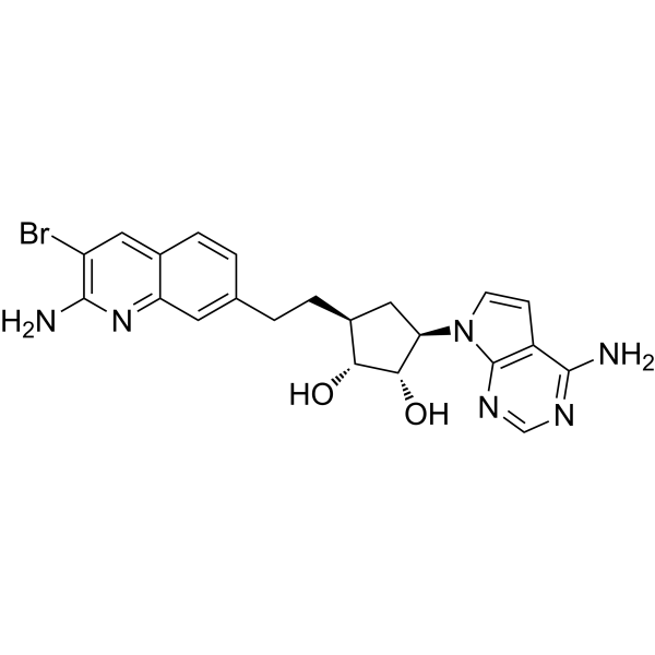
HY-101564
OnametostatPRMT5 Inhibitor -
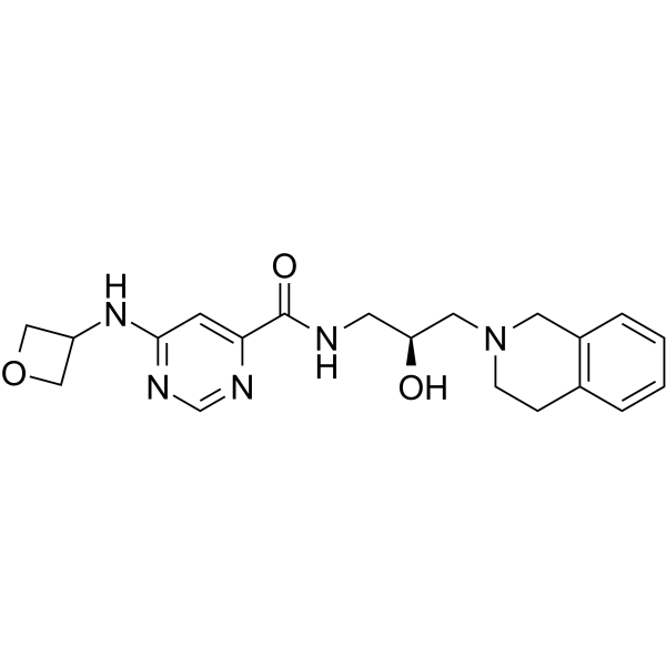
HY-12727
EPZ015666PRMT5 Inhibitor -
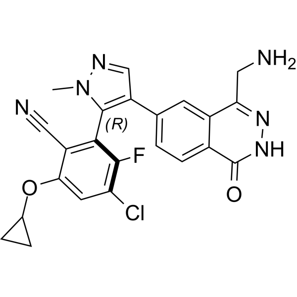
HY-139611
NavlimetostatPRMT5/MTA Complex Inhibitor -
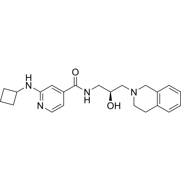
HY-100235
GSK591PRMT5 Inhibitor -
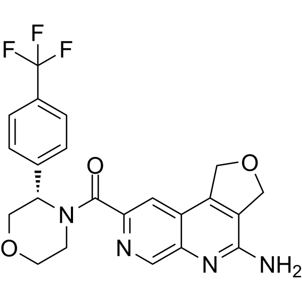
HY-153390
AMG-193PRMT5 Inhibitor -
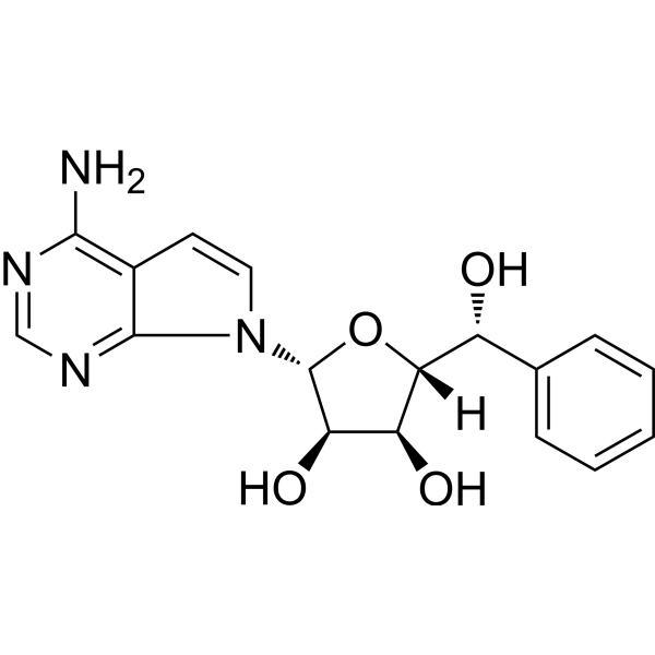
HY-107777
LLY-283PRMT5 Inhibitor -
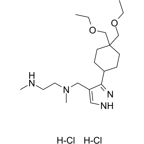
HY-128717A
GSK3368715 dihydrochloridePRMT Inhibitor -
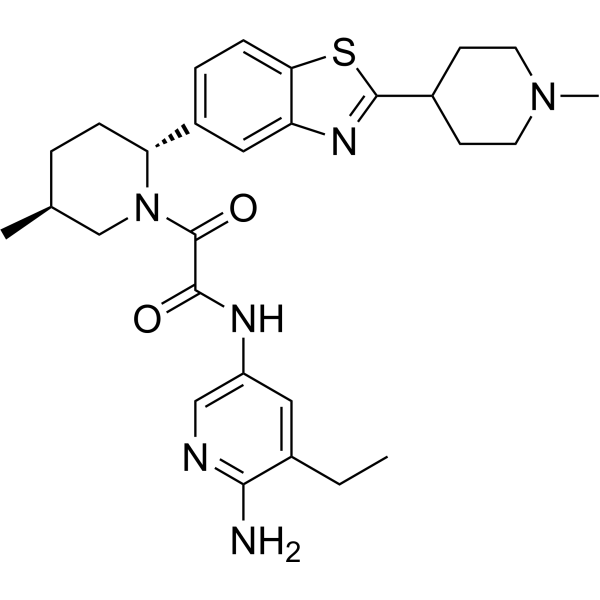
HY-156680
VopimetostatPRMT5 Inhibitor -
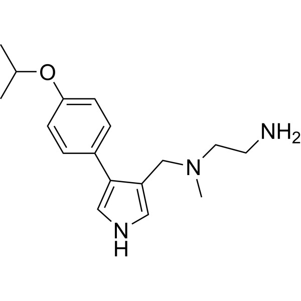
HY-19615
MS023Type I PRMT Inhibitor -
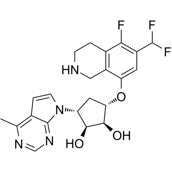
HY-131493
PF-06939999PRMT5 Inhibitor -
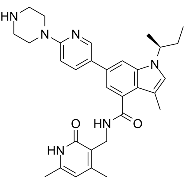
HY-13470
GSK126EZH2 Inhibitor -
HY-12970
EPZ020411PRMT6 Inhibitor
Publications Citing Use of MedChemExpress (MCE) Pemrametostat
More- Gut. 2024 Sep 11:gutjnl-2024-332998. [Abstract]
- Nat Commun. 2025 Dec 3;16(1):10129. [Abstract]
- Nat Commun. 2021 Jun 8;12(1):3444. [Abstract]
- J Adv Res. 2025 Nov 5:S2090-1232(25)00874-4. [Abstract]
- J Exp Clin Cancer Res. 2022 Oct 5;41(1):293. [Abstract]
- Adv Sci (Weinh). 2025 Sep;12(33):e08280. [Abstract]
- MedComm (2020). 2024 Sep 20;5(10):e705. [Abstract]
- Cell Death Dis. 2026 Feb 2;17(1):195. [Abstract]
- Cancer Lett. 2026 May 1:645:218395. [Abstract]
- Clin Cancer Res. 2025 Jun 24. [Abstract]
- Oncogene. 2021 Apr;40(15):2711-2724. [Abstract]
- EMBO Mol Med. 2023 Aug 7;15(8):e17248. [Abstract]
- Blood Adv. 2022 Apr 28;bloodadvances.2021006306. [Abstract]
- Cell Mol Life Sci. 2023 Jan 17;80(2):43. [Abstract]
- Biomed J. 2026 Jun 10:101002. [Abstract]
- EMBO Rep. 2025 Oct 6. [Abstract]
- Invest Ophthalmol Vis Sci. 2025 Jun 2;66(6):8. [Abstract]
- Transl Oncol. 2025 Feb:52:102264. [Abstract]
- Sci Rep. 2023 Jul 3;13(1):10752. [Abstract]
- Cancers (Basel). 2023 Apr 27;15(9):2501. [Abstract]
- J Cell Mol Med. 2024 Mar;28(5):e17856. [Abstract]
- J Biol Chem. 2022 Oct;298(10):102434. [Abstract]
- Cancer Res Commun. 2026 Sep 1;6(9):2039-2055.
- Breast Cancer (Dove Med Press). 2023 Nov 6:15:785-799. [Abstract]
- Mol Carcinog. 2023 Aug;62(8):1119-1135. [Abstract]
- Glycobiology. 2024 Dec 10;34(12):cwae081. [Abstract]
- Biochim Biophys Acta Gen Subj. 2026 Nov;1870(11):130993.
- Biomed Pharmacother. 2026 Sep:202:119803.
- bioRxiv. 2026 Apr 13.
- bioRxiv. 2026 Feb 18:2026.02.16.706212. [Abstract]
- SSRN. 2025 Dec 1.
- Res Sq. 2025 Jul 14.
- bioRxiv. 2024 Aug 16:2024.08.15.608201. [Abstract]
- Medical University of South Carolina. 2023 Aug 12.
- Research Square Print. November 11th, 2022.
- Springer. 2021 May 12.
 Customer Validation & Images

-
WB
-
Cell Proliferation/Viability Assay
-
WB
-
IHC
-
Cell Proliferation/Viability Assay
All Histone Methyltransferase Isoforms
More

Biological Activity
Description
IC50 & Target
|
PRMT5 |
CDK4 |
CDK6 |
Cellular Effect
|
Cell Line

|
Type | Value | Description | References |
|---|---|---|---|---|
| 5637 | GI50 |
0.069 μM
Compound: GSK3326595
|
Antiproliferative activity against human 5637 cells assessed as cell growth inhibition incubated for 6 days by celltiter-glo assay
Antiproliferative activity against human 5637 cells assessed as cell growth inhibition incubated for 6 days by celltiter-glo assay
|
[PMID: 29946150] |
| A 172 | GI50 |
1.167 μM
Compound: GSK3326595
|
Antiproliferative activity against human A-172 cells assessed as cell growth inhibition incubated for 6 days by celltiter-glo assay
Antiproliferative activity against human A-172 cells assessed as cell growth inhibition incubated for 6 days by celltiter-glo assay
|
[PMID: 29946150] |
| BJAB | GI50 |
1.9 μM
Compound: GSK3326595
|
Antiproliferative activity against human BJAB cells assessed as cell growth inhibition incubated for 6 days by celltiter-glo assay
Antiproliferative activity against human BJAB cells assessed as cell growth inhibition incubated for 6 days by celltiter-glo assay
|
[PMID: 29946150] |
| DOHH-2 | GI50 |
0.213 μM
Compound: GSK3326595
|
Antiproliferative activity against human DOHH-2 cells assessed as cell growth inhibition incubated for 6 days by celltiter-glo assay
Antiproliferative activity against human DOHH-2 cells assessed as cell growth inhibition incubated for 6 days by celltiter-glo assay
|
[PMID: 29946150] |
| HCC1143 | GI50 |
0.246 μM
Compound: GSK3326595
|
Antiproliferative activity against human HCC1143 cells assessed as cell growth inhibition viability incubated for 6 days by celltiter-glo assay
Antiproliferative activity against human HCC1143 cells assessed as cell growth inhibition viability incubated for 6 days by celltiter-glo assay
|
[PMID: 29946150] |
| HCC1806 | GI50 |
0.098 μM
Compound: GSK3326595
|
Antiproliferative activity against human HCC1806 cells assessed as cell growth inhibition incubated for 6 days by celltiter-glo assay
Antiproliferative activity against human HCC1806 cells assessed as cell growth inhibition incubated for 6 days by celltiter-glo assay
|
[PMID: 29946150] |
| HCC70 | GI50 |
0.13 μM
Compound: GSK3326595
|
Antiproliferative activity against human HCC70 cells assessed as cell growth inhibition incubated for 6 days by celltiter-glo assay
Antiproliferative activity against human HCC70 cells assessed as cell growth inhibition incubated for 6 days by celltiter-glo assay
|
[PMID: 29946150] |
| HCT-116 | IC50 |
189 nM
Compound: GSK3326595
|
Antiproliferative activity against MTAP knockout human HCT-116 cells assessed as reduction in cell viability incubated for 5 days followed by replacement with fresh medium containing compound and measured after 5 days by CTG assay
Antiproliferative activity against MTAP knockout human HCT-116 cells assessed as reduction in cell viability incubated for 5 days followed by replacement with fresh medium containing compound and measured after 5 days by CTG assay
|
[PMID: 35041419] |
| HCT-116 | IC50 |
237 nM
Compound: GSK3326595
|
Antiproliferative activity against human HCT-116 cells expressing wild type MTAP assessed as reduction in cell viability incubated for 5 days followed by replacement with fresh medium containing compound and measured after 5 days by CTG assay
Antiproliferative activity against human HCT-116 cells expressing wild type MTAP assessed as reduction in cell viability incubated for 5 days followed by replacement with fresh medium containing compound and measured after 5 days by CTG assay
|
[PMID: 35041419] |
| HT-1376 | GI50 |
17.599 μM
Compound: GSK3326595
|
Antiproliferative activity against human HT-1376 cells assessed as cell growth inhibition incubated for 6 days by celltiter-glo assay
Antiproliferative activity against human HT-1376 cells assessed as cell growth inhibition incubated for 6 days by celltiter-glo assay
|
[PMID: 29946150] |
| J82 | GI50 |
29.326 μM
Compound: GSK3326595
|
Antiproliferative activity against human J82 cells assessed as cell growth inhibition incubated for 6 days by celltiter-glo assay
Antiproliferative activity against human J82 cells assessed as cell growth inhibition incubated for 6 days by celltiter-glo assay
|
[PMID: 29946150] |
| JeKo-1 | GI50 |
5.402 μM
Compound: GSK3326595
|
Antiproliferative activity against human JeKo-1 cells assessed as cell growth inhibition incubated for 6 days by celltiter-glo assay
Antiproliferative activity against human JeKo-1 cells assessed as cell growth inhibition incubated for 6 days by celltiter-glo assay
|
[PMID: 29946150] |
| JM1 | GI50 |
0.079 μM
Compound: GSK3326595
|
Antiproliferative activity against human JM1 cells assessed as cell growth inhibition incubated for 6 days by celltiter-glo assay
Antiproliferative activity against human JM1 cells assessed as cell growth inhibition incubated for 6 days by celltiter-glo assay
|
[PMID: 29946150] |
| JVM-2 | GI50 |
0.009 μM
Compound: GSK3326595
|
Antiproliferative activity against human JVM-2 cells assessed as cell growth inhibition incubated for 6 days by celltiter-glo assay
Antiproliferative activity against human JVM-2 cells assessed as cell growth inhibition incubated for 6 days by celltiter-glo assay
|
[PMID: 29946150] |
| KMS-11 | GI50 |
0.066 μM
Compound: GSK3326595
|
Antiproliferative activity against human KMS-11 cells assessed as cell growth inhibition incubated for 6 days by celltiter-glo assay
Antiproliferative activity against human KMS-11 cells assessed as cell growth inhibition incubated for 6 days by celltiter-glo assay
|
[PMID: 29946150] |
| LN-229 | GI50 |
0.254 μM
Compound: GSK3326595
|
Antiproliferative activity against human LN-229 cells assessed as cell growth inhibition incubated for 6 days by celltiter-glo assay
Antiproliferative activity against human LN-229 cells assessed as cell growth inhibition incubated for 6 days by celltiter-glo assay
|
[PMID: 29946150] |
| LP-1 | GI50 |
1.945 μM
Compound: GSK3326595
|
Antiproliferative activity against human LP-1 cells assessed as cell growth inhibition incubated for 6 days by celltiter-glo assay
Antiproliferative activity against human LP-1 cells assessed as cell growth inhibition incubated for 6 days by celltiter-glo assay
|
[PMID: 29946150] |
| Maver1 | GI50 |
0.085 μM
Compound: GSK3326595
|
Antiproliferative activity against human MAVER-1 cells assessed as cell growth inhibition incubated for 6 days by celltiter-glo assay
Antiproliferative activity against human MAVER-1 cells assessed as cell growth inhibition incubated for 6 days by celltiter-glo assay
|
[PMID: 29946150] |
| MCF7 | GI50 |
0.062 μM
Compound: GSK3326595
|
Antiproliferative activity against human MCF7 cells assessed as cell growth inhibition incubated for 6 days by celltiter-glo assay
Antiproliferative activity against human MCF7 cells assessed as cell growth inhibition incubated for 6 days by celltiter-glo assay
|
[PMID: 29946150] |
| MDA-MB-231 | GI50 |
0.544 μM
Compound: GSK3326595
|
Antiproliferative activity against human MDA-MB-231 cells assessed as cell growth inhibition incubated for 6 days by celltiter-glo assay
Antiproliferative activity against human MDA-MB-231 cells assessed as cell growth inhibition incubated for 6 days by celltiter-glo assay
|
[PMID: 29946150] |
| MDA-MB-453 | GI50 |
0.029 μM
Compound: GSK3326595
|
Antiproliferative activity against human MDA-MB-453 cells assessed as cell growth inhibition incubated for 6 days by celltiter-glo assay
Antiproliferative activity against human MDA-MB-453 cells assessed as cell growth inhibition incubated for 6 days by celltiter-glo assay
|
[PMID: 29946150] |
| MDA-MB-468 | GI50 |
0.057 μM
Compound: GSK3326595
|
Antiproliferative activity against human MDA-MB-468 cells assessed as cell growth inhibition incubated for 6 days by celltiter-glo assay
Antiproliferative activity against human MDA-MB-468 cells assessed as cell growth inhibition incubated for 6 days by celltiter-glo assay
|
[PMID: 29946150] |
| NCI-H526 | GI50 |
0.275 μM
Compound: GSK3326595
|
Antiproliferative activity against human NCI-H526 cells assessed as cell growth inhibition incubated for 6 days by celltiter-glo assay
Antiproliferative activity against human NCI-H526 cells assessed as cell growth inhibition incubated for 6 days by celltiter-glo assay
|
[PMID: 29946150] |
| NU-DHL-1 | GI50 |
0.333 μM
Compound: GSK3326595
|
Antiproliferative activity against human NU-DHL-1 cells assessed as cell growth inhibition incubated for 6 days by celltiter-glo assay
Antiproliferative activity against human NU-DHL-1 cells assessed as cell growth inhibition incubated for 6 days by celltiter-glo assay
|
[PMID: 29946150] |
| Pfeiffer | GI50 |
0.421 μM
Compound: GSK3326595
|
Antiproliferative activity against human Pfeiffer cells assessed as cell growth inhibition incubated for 6 days by celltiter-glo assay
Antiproliferative activity against human Pfeiffer cells assessed as cell growth inhibition incubated for 6 days by celltiter-glo assay
|
[PMID: 29946150] |
| RCK8 | GI50 |
0.529 μM
Compound: GSK3326595
|
Antiproliferative activity against human RCK8 cells assessed as cell growth inhibition incubated for 6 days by celltiter-glo assay
Antiproliferative activity against human RCK8 cells assessed as cell growth inhibition incubated for 6 days by celltiter-glo assay
|
[PMID: 29946150] |
| Rec1 | GI50 |
2.108 μM
Compound: GSK3326595
|
Antiproliferative activity against human REC1 cells assessed as cell growth inhibition incubated for 6 days by celltiter-glo assay
Antiproliferative activity against human REC1 cells assessed as cell growth inhibition incubated for 6 days by celltiter-glo assay
|
[PMID: 29946150] |
| RL | GI50 |
0.014 μM
Compound: GSK3326595
|
Antiproliferative activity against human RL cells assessed as cell growth inhibition incubated for 6 days by celltiter-glo assay
Antiproliferative activity against human RL cells assessed as cell growth inhibition incubated for 6 days by celltiter-glo assay
|
[PMID: 29946150] |
| SCaBER | GI50 |
0.198 μM
Compound: GSK3326595
|
Antiproliferative activity against human SCaBER cells assessed as cell growth inhibition incubated for 6 days by celltiter-glo assay
Antiproliferative activity against human SCaBER cells assessed as cell growth inhibition incubated for 6 days by celltiter-glo assay
|
[PMID: 29946150] |
| SF-295 | GI50 |
24.722 μM
Compound: GSK3326595
|
Antiproliferative activity against human SF-295 cells assessed as cell growth inhibition incubated for 6 days by celltiter-glo assay
Antiproliferative activity against human SF-295 cells assessed as cell growth inhibition incubated for 6 days by celltiter-glo assay
|
[PMID: 29946150] |
| SF-539 | GI50 |
0.061 μM
Compound: GSK3326595
|
Antiproliferative activity against human SF-539 cells assessed as cell growth inhibition incubated for 6 days by celltiter-glo assay
Antiproliferative activity against human SF-539 cells assessed as cell growth inhibition incubated for 6 days by celltiter-glo assay
|
[PMID: 29946150] |
| SK-BR-3 | GI50 |
0.073 μM
Compound: GSK3326595
|
Antiproliferative activity against human SK-BR-3 cells assessed as cell growth inhibition incubated for 6 days by celltiter-glo assay
Antiproliferative activity against human SK-BR-3 cells assessed as cell growth inhibition incubated for 6 days by celltiter-glo assay
|
[PMID: 29946150] |
| SUD4 | GI50 |
0.492 μM
Compound: GSK3326595
|
Antiproliferative activity against human SU-DHL-4 cells assessed as cell growth inhibition incubated for 6 days by celltiter-glo assay
Antiproliferative activity against human SU-DHL-4 cells assessed as cell growth inhibition incubated for 6 days by celltiter-glo assay
|
[PMID: 29946150] |
| SU-DHL10 | GI50 |
0.054 μM
Compound: GSK3326595
|
Antiproliferative activity against human SU-DHL-10 cells assessed as cell growth inhibition incubated for 6 days by celltiter-glo assay
Antiproliferative activity against human SU-DHL-10 cells assessed as cell growth inhibition incubated for 6 days by celltiter-glo assay
|
[PMID: 29946150] |
| SU-DHL-16 | GI50 |
0.099 μM
Compound: GSK3326595
|
Antiproliferative activity against human SU-DHL-16 cells assessed as cell growth inhibition incubated for 6 days by celltiter-glo assay
Antiproliferative activity against human SU-DHL-16 cells assessed as cell growth inhibition incubated for 6 days by celltiter-glo assay
|
[PMID: 29946150] |
| SU-DHL-2 | GI50 |
0.028 μM
Compound: GSK3326595
|
Antiproliferative activity against human SUDHL2 cells assessed as cell growth inhibition incubated for 6 days by celltiter-glo assay
Antiproliferative activity against human SUDHL2 cells assessed as cell growth inhibition incubated for 6 days by celltiter-glo assay
|
[PMID: 29946150] |
| SU-DHL-5 | GI50 |
0.028 μM
Compound: GSK3326595
|
Antiproliferative activity against human SU-DHL-5 cells assessed as cell growth inhibition incubated for 6 days by celltiter-glo assay
Antiproliferative activity against human SU-DHL-5 cells assessed as cell growth inhibition incubated for 6 days by celltiter-glo assay
|
[PMID: 29946150] |
| SU-DHL-6 | GI50 |
0.004 μM
Compound: GSK3326595
|
Antiproliferative activity against human SU-DHL-6 cells assessed as cell growth inhibition incubated for 6 days by celltiter-glo assay
Antiproliferative activity against human SU-DHL-6 cells assessed as cell growth inhibition incubated for 6 days by celltiter-glo assay
|
[PMID: 29946150] |
| SU-DHL-8 | GI50 |
0.053 μM
Compound: GSK3326595
|
Antiproliferative activity against human SU-DHL-8 cells assessed as cell growth inhibition incubated for 6 days by celltiter-glo assay
Antiproliferative activity against human SU-DHL-8 cells assessed as cell growth inhibition incubated for 6 days by celltiter-glo assay
|
[PMID: 29946150] |
| SW780 | GI50 |
0.29 μM
Compound: GSK3326595
|
Antiproliferative activity against human SW780 cells assessed as cell growth inhibition incubated for 6 days by celltiter-glo assay
Antiproliferative activity against human SW780 cells assessed as cell growth inhibition incubated for 6 days by celltiter-glo assay
|
[PMID: 29946150] |
| T-24 | GI50 |
0.765 μM
Compound: GSK3326595
|
Antiproliferative activity against human T24 cells assessed as cell growth inhibition incubated for 6 days by celltiter-glo assay
Antiproliferative activity against human T24 cells assessed as cell growth inhibition incubated for 6 days by celltiter-glo assay
|
[PMID: 29946150] |
| U-251 | GI50 |
1.798 μM
Compound: GSK3326595
|
Antiproliferative activity against human U-251MG cells assessed as cell growth inhibition incubated for 6 days by celltiter-glo assay
Antiproliferative activity against human U-251MG cells assessed as cell growth inhibition incubated for 6 days by celltiter-glo assay
|
[PMID: 29946150] |
| U2932 | GI50 |
0.053 μM
Compound: GSK3326595
|
Antiproliferative activity against human U2932 cells assessed as cell growth inhibition incubated for 6 days by celltiter-glo assay
Antiproliferative activity against human U2932 cells assessed as cell growth inhibition incubated for 6 days by celltiter-glo assay
|
[PMID: 29946150] |
| U-2940 | GI50 |
10.176 μM
Compound: GSK3326595
|
Antiproliferative activity against human U-2940 cells assessed as cell growth inhibition incubated for 6 days by celltiter-glo assay
Antiproliferative activity against human U-2940 cells assessed as cell growth inhibition incubated for 6 days by celltiter-glo assay
|
[PMID: 29946150] |
| U-87MG ATCC | GI50 |
0.04 μM
Compound: GSK3326595
|
Antiproliferative activity against human U-87 MG cells assessed as cell growth inhibition incubated for 6 days by celltiter-glo assay
Antiproliferative activity against human U-87 MG cells assessed as cell growth inhibition incubated for 6 days by celltiter-glo assay
|
[PMID: 29946150] |
| U-937 | GI50 |
0.007 μM
Compound: GSK3326595
|
Antiproliferative activity against human U-937 cells assessed as cell growth inhibition incubated for 6 days by celltiter-glo assay
Antiproliferative activity against human U-937 cells assessed as cell growth inhibition incubated for 6 days by celltiter-glo assay
|
[PMID: 29946150] |
| UMUC3 | GI50 |
29.326 μM
Compound: GSK3326595
|
Antiproliferative activity against human UMUC3 cells assessed as cell growth inhibition incubated for 6 days by celltiter-glo assay
Antiproliferative activity against human UMUC3 cells assessed as cell growth inhibition incubated for 6 days by celltiter-glo assay
|
[PMID: 29946150] |
| WSUDLCL2 | GI50 |
0.082 μM
Compound: GSK3326595
|
Antiproliferative activity against human WSUDLCL2 cells assessed as cell growth inhibition incubated for 6 days by celltiter-glo assay
Antiproliferative activity against human WSUDLCL2 cells assessed as cell growth inhibition incubated for 6 days by celltiter-glo assay
|
[PMID: 29946150] |
| WSU-NHL | GI50 |
0.008 μM
Compound: GSK3326595
|
Antiproliferative activity against human WSU-NHL cells assessed as cell growth inhibition incubated for 6 days by celltiter-glo assay
Antiproliferative activity against human WSU-NHL cells assessed as cell growth inhibition incubated for 6 days by celltiter-glo assay
|
[PMID: 29946150] |
| Z-138 | GI50 |
0.004 μM
Compound: GSK3326595
|
Antiproliferative activity against human Z138 cells assessed as cell growth inhibition incubated for 6 days by celltiter-glo assay
Antiproliferative activity against human Z138 cells assessed as cell growth inhibition incubated for 6 days by celltiter-glo assay
|
[PMID: 29946150] |
| ZR-75-1 | GI50 |
10.974 μM
Compound: GSK3326595
|
Antiproliferative activity against human ZR-75-1 cells assessed as cell growth inhibition incubated for 6 days by celltiter-glo assay
Antiproliferative activity against human ZR-75-1 cells assessed as cell growth inhibition incubated for 6 days by celltiter-glo assay
|
[PMID: 29946150] |
In Vitro
GSK3326595 (10-100 nM, 24-72 h) inhibits SARS-CoV-2spike pseudovirus infection HEK-293 cells and A549 cells by attenuating ACE2-RBD interaction[1].
GSK3326595 (100 nM, 12 h) primes peritoneal macrophages to IFN-gamma-induced M1 polarization[3].
GSK3326595 (0.15-10 μM, 72 h) induces cell death in MCL cell[4].
MedChemExpress (MCE) has not independently confirmed the accuracy of these methods. They are for reference only.
-
Cell Line:HEK-293T cells, A549 cells
-
Concentration:10 nM, 25 nM, 50 nM, 100 nM
-
Incubation Time:48 h
-
Result:Strongly inhibited ACE2-RBD interaction at low concentration. Inhibited SARS-CoV-2 Omicron and other variants Spike1 binding with ACE2. Inhibits SARS-CoV-2 spike pseudovirus infection host cells.
-
Cell Line:MCL cells
-
Concentration:0.15 μM, 0.3 μM, 0.6 μM, 1.25 μM, 2.5 μM, 5 μM, 10 μM
-
Incubation Time:72 h
-
Result:Resulted in modest growth inhibition in MCL cells.
In Vivo
MedChemExpress (MCE) has not independently confirmed the accuracy of these methods. They are for reference only.
-
Animal Model:LDL receptor knockout mice[3]
-
Dosage:5 mg/kg
-
Administration:Intraperitoneal injection (i.p.)
-
Result:Did not alter atherosclerosis susceptibility. Increased hepatic triglyceride levels without changing the hyperlipidemia extent. Activated genes involved in fatty acid acquisition.
-
Animal Model:myelocytomatosis transgene turned on mice[5]
-
Dosage:25 mg/kg, 50 mg/kg
-
Administration:Oral
-
Result:Significantly suppressed tumor growth at 50 mg/kg. Showed better therapeutic efficacy at 25 mg/kg.
- Research Protocol for Infectious Diseases
- BrdU Incorporation Assay
- Somatic Cell Culture
- CFSE Dye Dilution Proliferation Assay
- Dye-dilution cell tracking and proliferation staining
- CCK-8/WST-8 Cell Proliferation Assay
- Protocol for Cell Cycle
- Research Protocol for Cardiovascular Diseases
- Cell Counting-Based Growth Curve Assay
- Research Protocol for Inflammation-related Diseases
- Colony Formation (Clonogenic) Assay
- Cell Viability Determination by MTT Colorimetric Assay
- Phagocytosis Functional Assay
- EdU Incorporation Assay (Click Chemistry-Based DNA Synthesis Measurement)
- Ki-67 Immunostaining Proliferation Assay
- PCNA Immunodetection Proliferation Assay
- Protocol for Cell Counting and Cell Density Analysis
- Primary monocyte-to-macrophage differentiation
- MTT Cell Proliferation Assay
Clinical Trial
| NCT Number | Sponsor | Condition | Start Date |
Phase
|
|---|---|---|---|---|
| NCT01329991 | Plexxikon| | 2011-05 | PHASE1 |


Chemical Information
-
CAS No. 1616392-22-3
-
Appearance Solid
-
Molecular Weight 452.55
-
Formula C24H32N6O3
-
Color White to light yellow
-
SMILES
O=C(C)N1CCC(NC2=NC=NC(C(NC[C@H](O)CN3CCC(C=CC=C4)=C4C3)=O)=C2)CC1
-
Synonyms
GSK3326595; EPZ015938
-
Shipping
Room temperature in continental US; may vary elsewhere.
-
Storage
Powder -20°C 3 years 4°C 2 years In solvent -80°C 2 years -20°C 1 year


Publications (36)
-
Journal Impact Factor
-
Most Recent
-
Gut
2024 Sep 11:gutjnl-2024-332998. PMID: 39266051
Pemrametostat purchased from MedChemExpress. Usage Cited in: Gut. 2024 Sep 11:gutjnl-2024-332998. [Abstract]
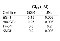
GI50 values for the PRMT5 inhibitors GSK3326595 and JNJ64619178 (Onametostat) in the indicated CCA cell lines.
Pemrametostat purchased from MedChemExpress. Usage Cited in: Gut. 2024 Sep 11:gutjnl-2024-332998. [Abstract]
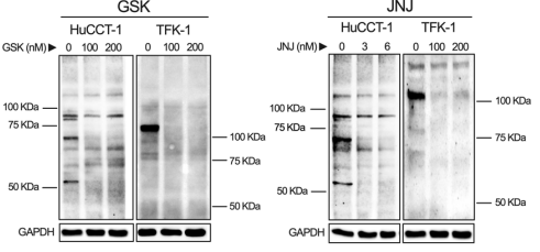
Immunoblot analysis of PRMT5-dependent symmetric dimethylarginine (SMDA) protein marks in control and GSK3326595 or JNJ64619178 (Onametostat) treated HuCCT-1 and TFK-1 cells at the indicated doses for 5 days.
Pemrametostat purchased from MedChemExpress. Usage Cited in: Gut. 2024 Sep 11:gutjnl-2024-332998. [Abstract]
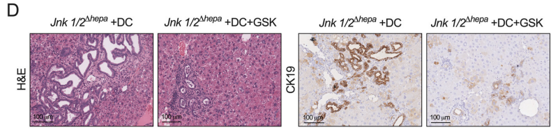
Immunohistochemical analysis of CK19 and H&E stainings showing the extent of CCA lesions in control JnkΔhepa+DEN+CCl4 and GSK3326595-treated mice. Representative images are shown.
-
Nat Commun
2025 Dec 3;16(1):10129. PMID: 41339334 -
Nat Commun
2021 Jun 8;12(1):3444. PMID: 34103528
Pemrametostat purchased from MedChemExpress. Usage Cited in: Nat Commun. 2021 Jun 8;12(1):3444. [Abstract]
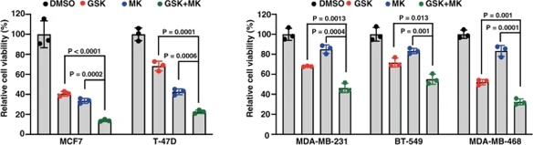
Breast cancer cells are treated with DMSO, 0.5 μM GSK3326595 (GSK) for 4 days before examining cell viability.
-
J Adv Res
The aryl hydrocarbon receptor inhibits antigen presentation to promote progression of pancreatic ductal adenocarcinoma. [Abstract]2025 Nov 5:S2090-1232(25)00874-4. PMID: 41203070 -
J Exp Clin Cancer Res
2022 Oct 5;41(1):293. PMID: 36199122 -
Adv Sci (Weinh)
2025 Sep;12(33):e08280. PMID: 40642900
Pemrametostat purchased from MedChemExpress. Usage Cited in: Adv Sci (Weinh). 2025 Sep;12(33):e08280. [Abstract]
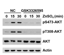
HCT116 cells were treated with GSK3326595 (10 μM) for 6 h,HCT116 cells were starved in amino acid-free medium for 1 h, and then supplemented with ZnSO4 for the indicated time. The level of pS473-AKT,binding
-
MedComm (2020)
SCR-7952, a highly selective MAT2A inhibitor, demonstrates synergistic antitumor activities in combination with the S-adenosylmethionine-competitive or the methylthioadenosine-cooperative protein arginine methyltransferase 5 inhibitors in methylthioadenosine phosphorylase-deleted tumors. [Abstract]2024 Sep 20;5(10):e705. PMID: 39309689 -
Cell Death Dis
Reversible arginine methylation regulates mitochondrial IDH2 activity: coordinated control by CARM1 and KDM3A/4A. [Abstract]2026 Feb 2;17(1):195. PMID: 41629283 -
Cancer Lett
DysUFMylation reprograms immunosuppressive neutrophils to potentiate anti-PD-1 therapy in hepatocellular carcinoma. [Abstract]2026 May 1:645:218395. PMID: 41780840 -
Clin Cancer Res
MTA-Cooperative PRMT5 Inhibitors are Efficacious in MTAP-Deleted Malignant Peripheral Nerve Sheath Tumor Models. [Abstract]2025 Jun 24. PMID: 40553452 -
Oncogene
2021 Apr;40(15):2711-2724. PMID: 33712705 -
EMBO Mol Med
2023 Aug 7;15(8):e17248. PMID: 37458145 -
Blood Adv
Loss of METTL3 attenuates blastic plasmacytoid dendritic cell neoplasm response to PRMT5 inhibition via IFN signaling. [Abstract]2022 Apr 28;bloodadvances.2021006306. PMID: 35482445 -
Cell Mol Life Sci
Opposing USP19 splice variants in TGF-β signaling and TGF-β-induced epithelial-mesenchymal transition of breast cancer cells. [Abstract]2023 Jan 17;80(2):43. PMID: 36646950 -
Biomed J
2026 Jun 10:101002. PMID: 42270077 -
EMBO Rep
2025 Oct 6. PMID: 41053315 -
Invest Ophthalmol Vis Sci
PRMT5 Regulates Senescence in Retinal Ganglion Cells by Targeting the Wnt/β-Catenin Signaling Cascade. [Abstract]2025 Jun 2;66(6):8. PMID: 40459496 -
Transl Oncol
TNG908 is a brain-penetrant, MTA-cooperative PRMT5 inhibitor developed for the treatment of MTAP-deleted cancers. [Abstract]2025 Feb:52:102264. PMID: 39756156 -
Sci Rep
2023 Jul 3;13(1):10752. PMID: 37400460 -
Cancers (Basel)
2023 Apr 27;15(9):2501. PMID: 37173967 -
J Cell Mol Med
2024 Mar;28(5):e17856. PMID: 37461162 -
J Biol Chem
Protein arginine methyltransferase 5 is essential for oncogene product EWSR1-ATF1-mediated gene transcription in clear cell sarcoma. [Abstract]2022 Oct;298(10):102434. PMID: 36041632 -
-
Breast Cancer (Dove Med Press)
Therapeutic Advantage of Targeting PRMT5 in Combination with Chemotherapies or EGFR/HER2 Inhibitors in Triple-Negative Breast Cancers. [Abstract]2023 Nov 6:15:785-799. PMID: 37954171 -
Mol Carcinog
PRMT1 inhibition promotes ferroptosis sensitivity via ACSL1 upregulation in acute myeloid leukemia. [Abstract]2023 Aug;62(8):1119-1135. PMID: 37144835 -
Glycobiology
Compromised CDK12 activity causes dependency on the high activity of O-GlcNAc transferase. [Abstract]2024 Dec 10;34(12):cwae081. PMID: 39361894 -
-
-
-
bioRxiv
Radiation synergizes with BET inhibition to stimulate durable, systemic anti-tumor immunity in murine cancer models. [Abstract]2026 Feb 18:2026.02.16.706212. PMID: 41757027 -
-
-
bioRxiv
2024 Aug 16:2024.08.15.608201. PMID: 39211199 -
-
-


Solvent & Solubility
In Vitro:
DMSO : 62.5 mg/mL (138.11 mM; Need ultrasonic; Hygroscopic DMSO has a significant impact on the solubility of product, please use newly opened DMSO)
Please refer to the solubility information to select the appropriate solvent. Once prepared, please aliquot and store the solution to prevent product inactivation from repeated freeze-thaw cycles.
Storage method and period of stock solution: -80°C, 2 years; -20°C, 1 year. When stored at -80°C, please use it within 2 years. When stored at -20°C, please use it within 1 year.
Please refer to the solubility information to select the appropriate solvent. Once prepared, please aliquot and store the solution to prevent product inactivation from repeated freeze-thaw cycles.
Storage method and period of stock solution: -80°C, 2 years; -20°C, 1 year. When stored at -80°C, please use it within 2 years. When stored at -20°C, please use it within 1 year.
Concentration (start) × Volume (start) = Concentration (final) × Volume (final)
In Vivo:
Select the appropriate dissolution method based on your experimental animal and administration route.
- For the following dissolution methods, please ensure to first prepare a clear stock solution using an In Vitro approach and then sequentially add co-solvents:
- To ensure reliable experimental results, the clarified stock solution can be appropriately stored based on storage conditions. As for the working solution for In Vivo experiments, it is recommended to prepare freshly and use it on the same day.
- The percentages shown for the solvents indicate their volumetric ratio in the final prepared solution. If precipitation or phase separation occurs during preparation, heat and/or sonication can be used to aid dissolution.
Add each solvent one by one: 10% DMSO 40% PEG300 5% Tween-80 45% Saline
Solubility: 2.08 mg/mL (4.60 mM); Clear solution; Need ultrasonic
This protocol yields a clear solution of 2.08 mg/mL.
Taking 1 mL working solution as an example, add 100 μL DMSO stock solution (20.8 mg/mL) to 400 μL PEG300, and mix evenly; then add 50 μL Tween-80 and mix evenly; then add 450 μL Saline to adjust the volume to 1 mL.
Preparation of Saline: Dissolve 0.9 g sodium chloride in ddH₂O and dilute to 100 mL to obtain a clear Saline solution.
Add each solvent one by one: 10% DMSO 90% (20% SBE-β-CD in Saline)
Solubility: 2.08 mg/mL (4.60 mM); Suspended solution; Need ultrasonic
This protocol yields a suspended solution of 2.08 mg/mL. Suspended solution can be used for oral and intraperitoneal injection.
Taking 1 mL working solution as an example, add 100 μL DMSO stock solution (20.8 mg/mL) to 900 μL 20% SBE-β-CD in Saline, and mix evenly.
Preparation of 20% SBE-β-CD in Saline (4°C, storage for one week): 2 g SBE-β-CD powder is dissolved in 10 mL Saline, completely dissolve until clear.
In Vivo Dissolution Calculator
Please enter the basic information of animal experiments:
-
-
-
-
Recommended: Prepare an additional quantity of animals to account for potential losses during experiments.
Please enter your animal formula composition:
-
%DMSO +
Recommended: Keep the proportion of DMSO in working solution below 2% if your animal is weak.
-
%+
-
+%Tween-80 + +
-
%Saline +
The co-solvents required include: DMSO, . All of co-solvents are available by MedChemExpress (MCE). , Tween 80. All of co-solvents are available by MedChemExpress (MCE).
Working solution concentration: 0.22 mg/mL
Method for preparing stock solution: mg drug dissolved in μL DMSO. Stock solution concentration: mg/mL.
1. Take μL DMSO stock solution;
2. Add μL .
μL , mix evenly;
3. Then add μL Tween 80, mix evenly;
4. Then add μL
Please ensure that the stock solution in the first step is dissolved to a clear state, and add co-solvents in sequence. You can use ultrasonic heating (ultrasonic cleaner, recommended frequency 20-40 kHz), vortexing, etc. to assist dissolution.


Purity & Documentation
-
Data Sheet (283 KB)
-
SDS (393 KB)
- English - EN (393 KB)
- Français - FR (393 KB)
- Deutsch - DE (393 KB)
- Norwegian - NO (393 KB)
- Español - ES (393 KB)
- Swedish - SV (393 KB)
- Italian - IT (393 KB)
- Korean - KR (393 KB)
- Portuguese - PT (393 KB)
-
Handling Instructions (2659 KB)
References
[1]. Che Y, et al. Exploiting PRMT5 as a target for combination therapy in mantle cell lymphoma characterized by frequent ATM and TP53 mutations [J]. Blood cancer journal, 2023, 13(1): 27. [Content Brief]
[5]. Luo Y, et al. Myelocytomatosis-protein arginine N-methyltransferase 5 Axis defines the tumorigenesis and immune response in hepatocellular carcinoma [J]. Hepatology, 2021, 74(4): 1932-1951. [Content Brief]


Complete Stock Solution Preparation Table
Please refer to the solubility information to select the appropriate solvent. Once prepared, please aliquot and store the solution to prevent product inactivation from repeated freeze-thaw cycles.
Storage method and period of stock solution: -80°C, 2 years; -20°C, 1 year. When stored at -80°C, please use it within 2 years. When stored at -20°C, please use it within 1 year.
| Optional Solvent | Concentration Solvent Mass | 1 mg | 5 mg | 10 mg | 25 mg |
|---|---|---|---|---|---|
| DMSO | 1 mM | 2.2097 mL | 11.0485 mL | 22.0970 mL | 55.2425 mL |
| 5 mM | 0.4419 mL | 2.2097 mL | 4.4194 mL | 11.0485 mL | |
| 10 mM | 0.2210 mL | 1.1049 mL | 2.2097 mL | 5.5243 mL | |
| 15 mM | 0.1473 mL | 0.7366 mL | 1.4731 mL | 3.6828 mL | |
| 20 mM | 0.1105 mL | 0.5524 mL | 1.1049 mL | 2.7621 mL | |
| 25 mM | 0.0884 mL | 0.4419 mL | 0.8839 mL | 2.2097 mL | |
| 30 mM | 0.0737 mL | 0.3683 mL | 0.7366 mL | 1.8414 mL | |
| 40 mM | 0.0552 mL | 0.2762 mL | 0.5524 mL | 1.3811 mL | |
| 50 mM | 0.0442 mL | 0.2210 mL | 0.4419 mL | 1.1049 mL | |
| 60 mM | 0.0368 mL | 0.1841 mL | 0.3683 mL | 0.9207 mL | |
| 80 mM | 0.0276 mL | 0.1381 mL | 0.2762 mL | 0.6905 mL | |
| 100 mM | 0.0221 mL | 0.1105 mL | 0.2210 mL | 0.5524 mL |


Pemrametostat Related Classifications
HY-101563 Related Classifications
 Powered by Bioz