



Grifolin
Based on 1 publication(s) in Google Scholar
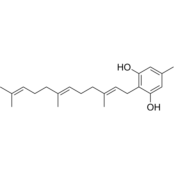
Grifolin is an antineoplastic agent can be isolated from the mushroom Albatrellus confluens and significantly induces apoptosis.
For research use only. We do not sell to patients.
- CAS No.: 6903-07-7
- Formula: C22H32O2
- Molecular Weight:328.49
-
Storage:
4°C, protect from light, stored under nitrogen
* In solvent : -80°C, 6 months; -20°C, 1 month (protect from light, stored under nitrogen)

To place orders, for customer services and technical support, please contact: MedChemExpress USA
Tel: 609-228-6898 E-mail: [email protected] [email protected]


-
 Biological Activity
-
Chemical Information
- Protocols
- Purity & Documentation
- References
-
 Help & FAQs


Publications Citing Use of MedChemExpress (MCE) Grifolin
More

Biological Activity
Description
Cellular Effect
|
Cell Line

|
Type | Value | Description | References |
|---|---|---|---|---|
| Mast cell | IC50 |
81.6 μL
Compound: 6
|
Inhibition of compound 48/80-induced histamine release in Wistar rat peritoneal mast cells
Inhibition of compound 48/80-induced histamine release in Wistar rat peritoneal mast cells
|
[PMID: 15270561] |


Chemical Information
-
CAS No. 6903-07-7
-
Appearance Oil
-
Molecular Weight 328.49
-
Formula C22H32O2
-
Color White to light yellow
-
SMILES
OC1=CC(C)=CC(O)=C1C/C=C(C)/CC/C=C(C)/CC/C=C(C)\C
-
Structure Classification
-
Initial Source
Albatrellus confluens
-
Shipping
Room temperature in continental US; may vary elsewhere.
-
Storage
4°C, protect from light, stored under nitrogen
* In solvent : -80°C, 6 months; -20°C, 1 month (protect from light, stored under nitrogen)


Publications (1)
-
Journal Impact Factor
-
Most Recent
-
Gut Microbes
Fusobacterium nucleatum induces chemoresistance in colorectal cancer by inhibiting pyroptosis via the Hippo pathway. [Abstract]2024 Jan-Dec;16(1):2333790. PMID: 38533566


Purity & Documentation
-
Data Sheet (264 KB)
-
SDS (252 KB)
- English - EN (252 KB)
- Français - FR (252 KB)
- Deutsch - DE (252 KB)
- Norwegian - NO (252 KB)
- Español - ES (252 KB)
- Swedish - SV (252 KB)
- Italian - IT (252 KB)
- Korean - KR (252 KB)
- Portuguese - PT (252 KB)
-
Handling Instructions (2659 KB)
References

Calculators
Concentration (start) × Volume (start) = Concentration (final) × Volume (final)


Grifolin Related Classifications
HY-N9271 Related Classifications
 Powered by Bioz