



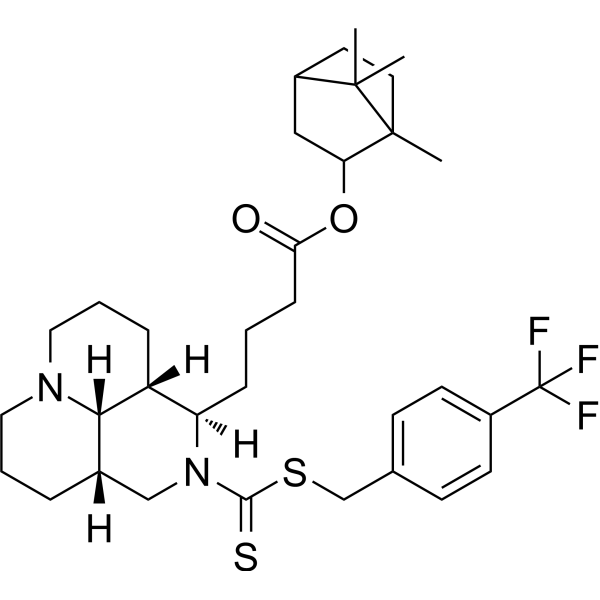
Anticancer agent 104
Anticancer agent 104 has anticancer activity, and induces cancer cell apoptosis.
商品は「研究用試薬」です。人や動物の医療用・臨床診断用・食品用の製品ではありません。
研究用途以外に使用した場合、当社は一切の責任を負いかねます。
- CAS 番号: 3047612-51-8
- 分子式: C34H47F3N2O2S2
- 分子量:636.87
-
保管条件:
Please store the product under the recommended conditions in the Certificate of Analysis.

To place orders, for customer services and technical support, please contact: MedChemExpress USA
Tel: 609-228-6898 E-mail: [email protected] [email protected]


-
 生物活性
生物活性
-
化学情報
- プロトコル
- 純度とドキュメンテーション
- 参考文献
-
 Help & FAQs




生物活性
製品説明
Cellular Effect
|
Cell Line

|
Type | Value | Description | References |
|---|---|---|---|---|
| 786-0 | IC50 |
91.67 μM
Compound: 4l
|
Anticancer activity against human 786-0 cells assessed as reduction in cell viability incubated for 48 hrs by MTT assay
Anticancer activity against human 786-0 cells assessed as reduction in cell viability incubated for 48 hrs by MTT assay
|
[PMID: 37084600] |
| Calu-1 | IC50 |
78.34 μM
Compound: 4l
|
Anticancer activity against human Calu-1 cells assessed as reduction in cell viability incubated for 48 hrs by MTT assay
Anticancer activity against human Calu-1 cells assessed as reduction in cell viability incubated for 48 hrs by MTT assay
|
[PMID: 37084600] |
| HEK-293T | IC50 |
195.17 μM
Compound: 4l
|
Cytotoxicity against human HEK293T cells assessed as reduction in cell viability incubated for 48 hrs by MTT assay
Cytotoxicity against human HEK293T cells assessed as reduction in cell viability incubated for 48 hrs by MTT assay
|
[PMID: 37084600] |
| HepG2 | IC50 |
31.39 μM
Compound: 4l
|
Anticancer activity against human HepG2 cells assessed as reduction in cell viability incubated for 48 hrs by MTT assay
Anticancer activity against human HepG2 cells assessed as reduction in cell viability incubated for 48 hrs by MTT assay
|
[PMID: 37084600] |
| Huh-7 | IC50 |
75.18 μM
Compound: 4l
|
Anticancer activity against human Huh-7 cells assessed as reduction in cell viability incubated for 48 hrs by MTT assay
Anticancer activity against human Huh-7 cells assessed as reduction in cell viability incubated for 48 hrs by MTT assay
|
[PMID: 37084600] |
| KGN | IC50 |
176.38 μM
Compound: 4l
|
Cytotoxicity against human KGN cells assessed as reduction in cell viability incubated for 48 hrs by MTT assay
Cytotoxicity against human KGN cells assessed as reduction in cell viability incubated for 48 hrs by MTT assay
|
[PMID: 37084600] |
| LX-2 | IC50 |
96.33 μM
Compound: 4l
|
Cytotoxicity against human LX2 cells assessed as reduction in cell viability incubated for 48 hrs by MTT assay
Cytotoxicity against human LX2 cells assessed as reduction in cell viability incubated for 48 hrs by MTT assay
|
[PMID: 37084600] |
| MCF-10A | IC50 |
21.06 μM
Compound: 4l
|
Cytotoxicity against human MCF-10A cells assessed as reduction in cell viability incubated for 48 hrs by MTT assay
Cytotoxicity against human MCF-10A cells assessed as reduction in cell viability incubated for 48 hrs by MTT assay
|
[PMID: 37084600] |
| SK-BR-3 | IC50 |
44.18 μM
Compound: 4l
|
Anticancer activity against human SK-BR-3 cells assessed as reduction in cell viability incubated for 48 hrs by MTT assay
Anticancer activity against human SK-BR-3 cells assessed as reduction in cell viability incubated for 48 hrs by MTT assay
|
[PMID: 37084600] |
| SK-OV-3 | IC50 |
112.19 μM
Compound: 4l
|
Anticancer activity against human SK-OV-3 cells assessed as reduction in cell viability incubated for 48 hrs by MTT assay
Anticancer activity against human SK-OV-3 cells assessed as reduction in cell viability incubated for 48 hrs by MTT assay
|
[PMID: 37084600] |
| WI-38 | IC50 |
81.48 μM
Compound: 4l
|
Cytotoxicity against human WI-38 cells assessed as reduction in cell viability incubated for 48 hrs by MTT assay
Cytotoxicity against human WI-38 cells assessed as reduction in cell viability incubated for 48 hrs by MTT assay
|
[PMID: 37084600] |
体外実験
Anticancer agent 104 (Compound 4l) (48 h) is toxic to HepG2 cells (IC50: 31.39 μM)[1].
Anticancer agent 104 (10-200 μg/mL, 48 h) exhibits volume reduction, chromatin margination, and eventually formed apoptotic vesicles[1].
Anticancer agent 104 (10-100 μg/mL, 48 h) induces HepG2 cell apoptosis[1].
MedChemExpress (MCE) has not independently confirmed the accuracy of these methods. They are for reference only.
-
Cell Line:Calu-1, SK-BR-3, HUH-7, 786-O, SK-OV-3 cells
-
Concentration:0-200 μM approximately
-
Incubation Time:48 h
-
Result:Shows cytotoxic activities with IC50s of 78.34, 44.18, 75.18, 91.67, 112.19 μM.
-
Cell Line:HepG2 cells
-
Concentration:10, 50 and 100 μg/mL
-
Incubation Time:48 h
-
Result:Showed apoptotic rate of 23.67%, 41.21% and 61.16% respectively.


化学情報
-
CAS 番号 3047612-51-8
-
分子量 636.87
-
分子式 C34H47F3N2O2S2
-
SMILES
C[C@@]1(C(C)([C@@](CC1)([H])C2)C)[C@@H]2OC(CCC[C@@H]3[C@]4([H])[C@]5([H])[C@](CCCN5CCC4)([H])CN3C(SCC6=CC=C(C=C6)C(F)(F)F)=S)=O
-
輸送条件
Room temperature in continental US; may vary elsewhere.
-
保管条件
Please store the product under the recommended conditions in the Certificate of Analysis.


純度とドキュメンテーション
参考文献

Calculators
濃度 (開始) × 体積 (開始) = 濃度 (終了) × 体積 (終了)


Anticancer agent 104 関連分類
HY-149231 関連分類
 Powered by Bioz