



KS100

Based on 1 Customer Validation
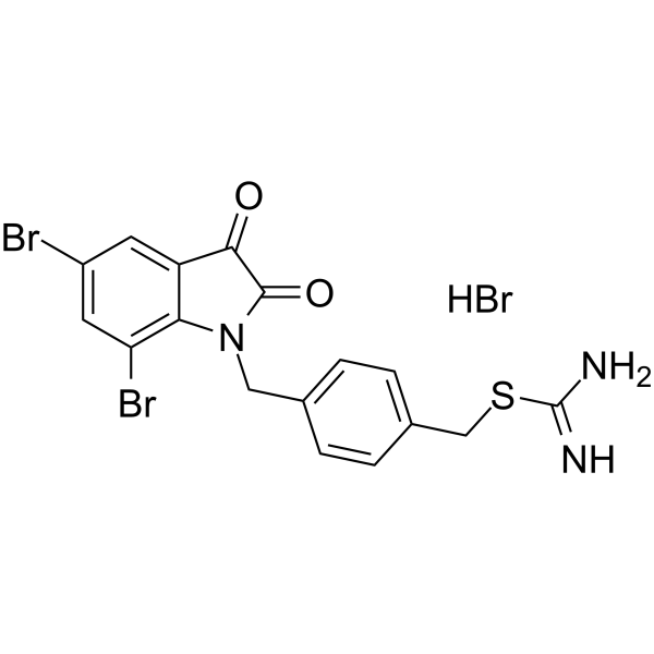
KS100 is a potent ALDH inhibitor with IC50s of 230, 1542, 193 nM for ALDH1A1, ALDH2, and ALDH3A1, respectively. KS100 shows antiproliferative and anticancer effects with low low toxic. KS100 significantly increases ROS activity, lipid peroxidation and toxic aldehyde accumulation. KS10600 induces apoptosis and cell cycle arrest at the G2/M phase.
Nur für Forschungszwecke. Wir verkaufen nicht an Patienten.
- Reinheit : 99.61%
- CAS. Nr.: 2408477-54-1
- Formel: C17H14Br3N3O2S
- Molecular Weight:564.09
-
Speicherung:
4°C, sealed storage, away from moisture
* The compound is unstable in solutions, freshly prepared is recommended.

To place orders, for customer services and technical support, please contact: MedChemExpress USA
Tel: 609-228-6898 E-mail: [email protected] [email protected]


-
 Biologische Aktivität
-
Chemical Information
-
Lösungsmittel & Löslichkeit
- Protokoll
- Reinheit & Dokumentation
- Verweise
-
 Help & FAQs


-
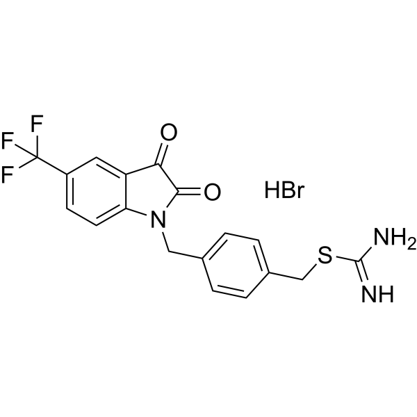
HY-146683
KS106ALDH Inhibitor -
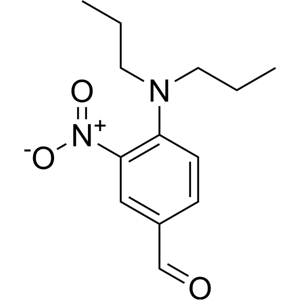
HY-144670
ALDH3A1-IN-1ALDH3A1 Inhibitor -
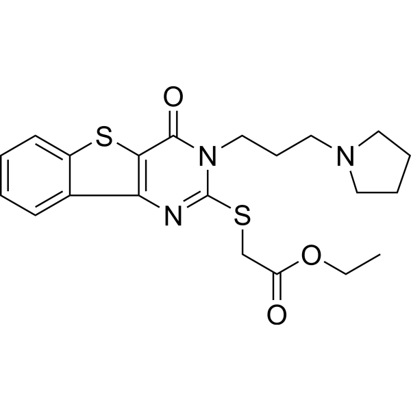
HY-110294
CM037ALDH1A1 Inhibitor -
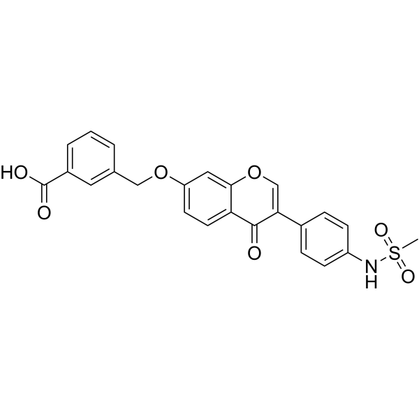
HY-19801
CVT-10216ALDH-2 Inhibitor -
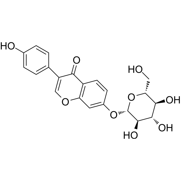
HY-N0018
DaidzinReverse Transcriptase Inhibitor -
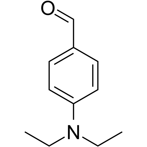
HY-W016645
4-DiethylaminobenzaldehydeALDH Inhibitor -
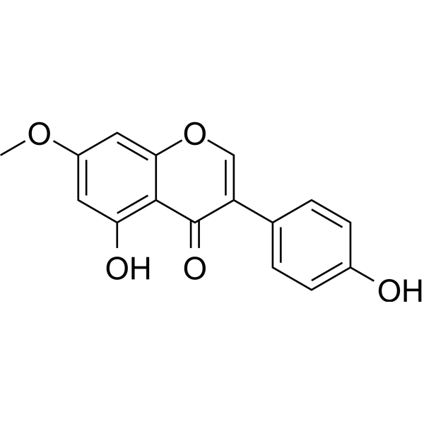
HY-N2597
PrunetinAldehyde Dehydrogenases Inhibitor -
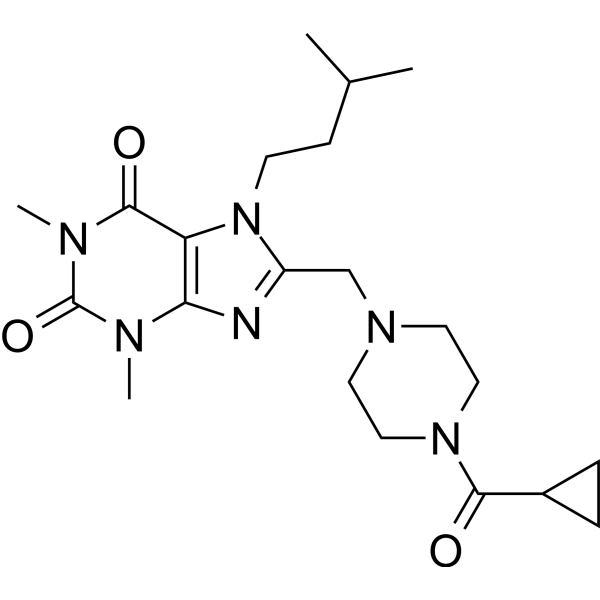
HY-18768
NCT-501ALDH1A1 Inhibitor -
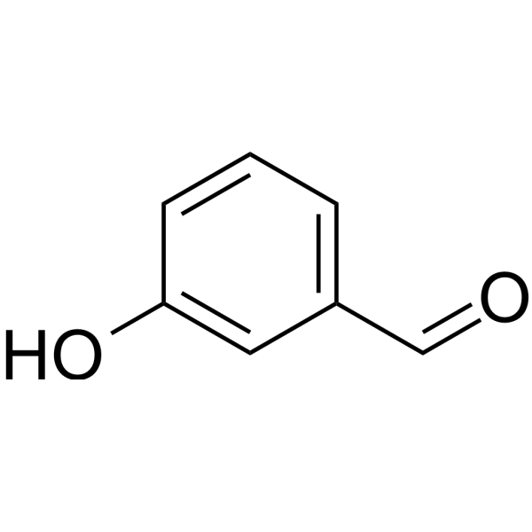
HY-76006
3-HydroxybenzaldehydeSubstrate For ALDH, Precursor Compound for Phenolic Compounds -
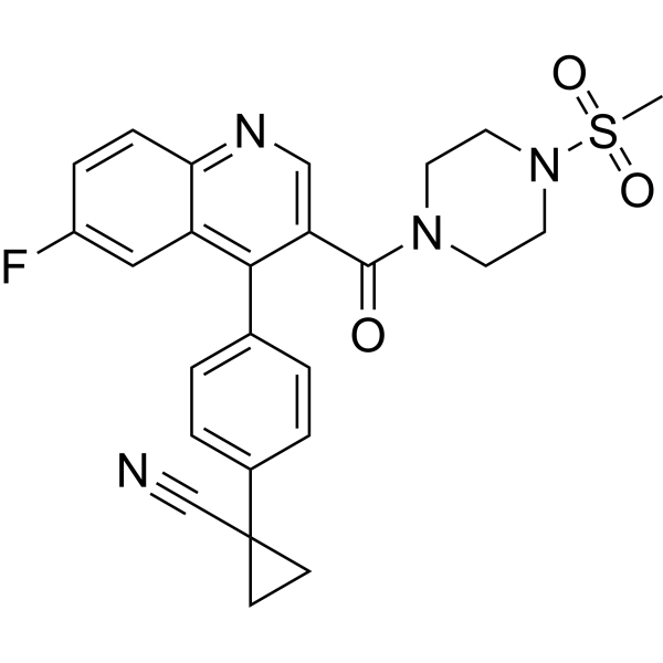
HY-112278
NCT-506ALDH Inhibitor -
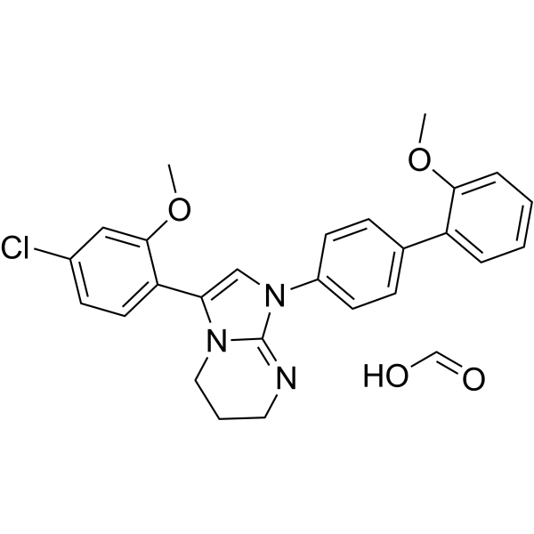
HY-148466
IGUANA-1ALDH1B1 Inhibitor -
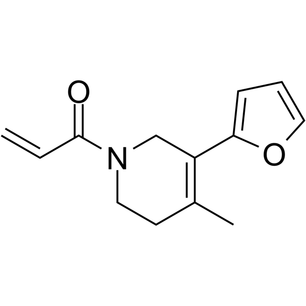
HY-122577
EN40ALDH3A1 Inhibitor


Biologische Aktivität
Beschreibung
IC50 & Target
IC50: 230 nM (ALDH1A1); 1542 nM (ALDH2); 193 nM (ALDH3A1)[1]
Cellular Effect
|
Cell Line

|
Type | Value | Description | References |
|---|---|---|---|---|
| HCT-116 | IC50 |
2.9 μM
Compound: 3j; KS100
|
Antiproliferative activity against human HCT116 cells measured after 72 hrs by MTS assay
Antiproliferative activity against human HCT116 cells measured after 72 hrs by MTS assay
|
[PMID: 31887569] |
| HT-29 | IC50 |
2.5 μM
Compound: 3j; KS100
|
Antiproliferative activity against human HT-29 cells measured after 72 hrs by MTS assay
Antiproliferative activity against human HT-29 cells measured after 72 hrs by MTS assay
|
[PMID: 31887569] |
| MM1.S | IC50 |
2.1 μM
Compound: 3j; KS100
|
Antiproliferative activity against human MM.1S cells measured after 72 hrs by MTS assay
Antiproliferative activity against human MM.1S cells measured after 72 hrs by MTS assay
|
[PMID: 31887569] |
| NCI-H929 | IC50 |
0.3 μM
Compound: 3j; KS100
|
Antiproliferative activity against human NCIH929 cells measured after 72 hrs by MTS assay
Antiproliferative activity against human NCIH929 cells measured after 72 hrs by MTS assay
|
[PMID: 31887569] |
| RPMI-8226 | IC50 |
1.2 μM
Compound: 3j; KS100
|
Antiproliferative activity against human RPMI8226 cells measured after 72 hrs by MTS assay
Antiproliferative activity against human RPMI8226 cells measured after 72 hrs by MTS assay
|
[PMID: 31887569] |
| U-266 | IC50 |
1 μM
Compound: 3j; KS100
|
Antiproliferative activity against human U266 cells measured after 72 hrs by MTS assay
Antiproliferative activity against human U266 cells measured after 72 hrs by MTS assay
|
[PMID: 31887569] |
| UACC-903 | IC50 |
3.7 μM
Compound: 3j; KS100
|
Antiproliferative activity against human UACC 903 cells measured after 72 hrs by MTS assay
Antiproliferative activity against human UACC 903 cells measured after 72 hrs by MTS assay
|
[PMID: 31887569] |
In Vitro
KS100 (compound 3j) (0-100 µM; 72 h) shows anti-proliferative activity with IC50s of 3.7, 2.1, 2.9, 2.5, 0.3, 1.0, 1.2, 1.3, 2.1, 9.8 µM for UACC 903, 1205 Lu, HCT116, HT29, NCIH929, U266, RPMI8226, MM.1R, MM.1S, FF2441 cells, respectively[1].
KS100 (5 µM, 24 h) induces apoptosis and cell cycle arrest at the G2/M phase[1].
MedChemExpress (MCE) has not independently confirmed the accuracy of these methods. They are for reference only.
- Detection of Apoptosis
- TUNEL staining for apoptotic DNA fragmentation
- Flow cytometric DNA-content cell-cycle staining
- Annexin V plus membrane-impermeant dye apoptosis staining
- ROS/oxidative-stress fluorescent staining
- BrdU Incorporation Assay
- Apoptosis Solutions
- Protocol for Cell Cycle
- Ferroptosis Solutions
- Cell Viability Determination by MTT Colorimetric Assay


Chemical Information
-
CAS. Nr. 2408477-54-1
-
Appearance Solid
-
Molecular Weight 564.09
-
Formel C17H14Br3N3O2S
-
Color Yellow to orange
-
SMILES
N=C(SCC1=CC=C(C=C1)CN2C(C(C3=CC(Br)=CC(Br)=C32)=O)=O)N.Br
-
Versand
Room temperature in continental US; may vary elsewhere.
-
Speicherung
4°C, sealed storage, away from moisture
* The compound is unstable in solutions, freshly prepared is recommended.


Lösungsmittel & Löslichkeit
In Vitro:
DMSO : 125 mg/mL (221.60 mM; Need ultrasonic; Hygroscopic DMSO has a significant impact on the solubility of product, please use newly opened DMSO)
Please refer to the solubility information to select the appropriate solvent. The compound is unstable in solutions, freshly prepared is recommended.
Please refer to the solubility information to select the appropriate solvent. The compound is unstable in solutions, freshly prepared is recommended.
Konzentration (Stammlösung) × Volumen (Stammlösung) = Konzentration (Ziellösung) × Volumen (Ziellösung)


Reinheit & Dokumentation
-
Data Sheet (273 KB)
-
SDS (252 KB)
- English - EN (252 KB)
- Français - FR (252 KB)
- Deutsch - DE (252 KB)
- Norwegian - NO (252 KB)
- Español - ES (252 KB)
- Swedish - SV (252 KB)
- Italian - IT (252 KB)
- Korean - KR (252 KB)
- Portuguese - PT (252 KB)
-
Handling Instructions (2659 KB)
Verweise


Complete Stock Solution Preparation Table
Please refer to the solubility information to select the appropriate solvent. The compound is unstable in solutions, freshly prepared is recommended.
| Optional Solvent | Concentration Solvent Mass | 1 mg | 5 mg | 10 mg | 25 mg |
|---|---|---|---|---|---|
| DMSO | 1 mM | 1.7728 mL | 8.8638 mL | 17.7277 mL | 44.3192 mL |
| 5 mM | 0.3546 mL | 1.7728 mL | 3.5455 mL | 8.8638 mL | |
| 10 mM | 0.1773 mL | 0.8864 mL | 1.7728 mL | 4.4319 mL | |
| 15 mM | 0.1182 mL | 0.5909 mL | 1.1818 mL | 2.9546 mL | |
| 20 mM | 0.0886 mL | 0.4432 mL | 0.8864 mL | 2.2160 mL | |
| 25 mM | 0.0709 mL | 0.3546 mL | 0.7091 mL | 1.7728 mL | |
| 30 mM | 0.0591 mL | 0.2955 mL | 0.5909 mL | 1.4773 mL | |
| 40 mM | 0.0443 mL | 0.2216 mL | 0.4432 mL | 1.1080 mL | |
| 50 mM | 0.0355 mL | 0.1773 mL | 0.3546 mL | 0.8864 mL | |
| 60 mM | 0.0295 mL | 0.1477 mL | 0.2955 mL | 0.7387 mL | |
| 80 mM | 0.0222 mL | 0.1108 mL | 0.2216 mL | 0.5540 mL | |
| 100 mM | 0.0177 mL | 0.0886 mL | 0.1773 mL | 0.4432 mL |


KS100 Related Classifications
HY-146682 Related Classifications
 Powered by Bioz