



Kumbicin C
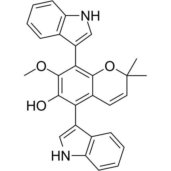
Kumbicin C is a bis-indolyl benzenoid compound from an Australian soil fungus, Aspergillus kumbius. Kumbicin C inhibits the growth of mouse myeloma cells and the Gram-positive bacterium Bacillus subtilis.
For research use only. We do not sell to patients.
- CAS No.: 1878151-58-6
- Formula: C28H24N2O3
- Molecular Weight:436.50
-
Storage:
Please store the product under the recommended conditions in the Certificate of Analysis.

To place orders, for customer services and technical support, please contact: MedChemExpress USA
Tel: 609-228-6898 E-mail: [email protected] [email protected]


-
 Biological Activity
-
Chemical Information
- Protocols
- Purity & Documentation
- References
-
 Help & FAQs




Biological Activity
Description
Cellular Effect
|
Cell Line

|
Type | Value | Description | References |
|---|---|---|---|---|
| L02 | IC50 |
11 μM
Compound: 6
|
Cytotoxicity against human L02 cells assessed as cell growth inhibition incubated for 72 hrs by Cell Titer-Glo assay
Cytotoxicity against human L02 cells assessed as cell growth inhibition incubated for 72 hrs by Cell Titer-Glo assay
|
[PMID: 37857311] |
| MV4-11 | IC50 |
1.2 μM
Compound: 6
|
Cytotoxicity against human MV4-11 cells assessed as cell growth inhibition incubated for 72 hrs by Cell Titer-Glo assay
Cytotoxicity against human MV4-11 cells assessed as cell growth inhibition incubated for 72 hrs by Cell Titer-Glo assay
|
[PMID: 37857311] |


Chemical Information
-
CAS No. 1878151-58-6
-
Molecular Weight 436.50
-
Formula C28H24N2O3
-
SMILES
CC1(C=CC2=C(C(O)=C(C(C3=CNC4=C3C=CC=C4)=C2O1)OC)C5=CNC6=C5C=CC=C6)C
-
Structure Classification
-
Initial Source
Aspergillus kumbius
-
Shipping
Room temperature in continental US; may vary elsewhere.
-
Storage
Please store the product under the recommended conditions in the Certificate of Analysis.


Purity & Documentation
References

Calculators
Concentration (start) × Volume (start) = Concentration (final) × Volume (final)


Kumbicin C Related Classifications
HY-122467 Related Classifications
 Powered by Bioz