



Kuwanon E

Based on 1 Customer Validation
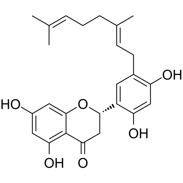
Kuwanon E is a flavonoid isolated from Morus alba, cytotoxic to human monocytic leukemic cell lines, and reduces the level of IL-1β.
For research use only. We do not sell to patients.
- Purity : 96.35%
- CAS No.: 68401-05-8
- Formula: C25H28O6
- Molecular Weight:424.49
-
Storage:
-20°C, protect from light
* In solvent : -80°C, 6 months; -20°C, 1 month (protect from light)

To place orders, for customer services and technical support, please contact: MedChemExpress USA
Tel: 609-228-6898 E-mail: [email protected] [email protected]


-
 Biological Activity
-
Chemical Information
- Protocols
- Purity & Documentation
- References
-
 Help & FAQs




Biological Activity
Description
IC50 & Target
Kuwanon E shows cytotoxic activity against THP-1 human monocytic leukemic cell line, with an IC50 of 4.0±0.08 μM[1].
Cellular Effect
|
Cell Line

|
Type | Value | Description | References |
|---|---|---|---|---|
| BJ | EC50 |
>10 μM
Compound: 13
|
Cytotoxicity against human BJ cells assessed as viable cells after 72 hrs by calcein AM assay
Cytotoxicity against human BJ cells assessed as viable cells after 72 hrs by calcein AM assay
|
[PMID: 20192247] |
| CCRF-CEM | EC50 |
>10 μM
Compound: 13
|
Cytotoxicity against human CEM cells assessed as viable cells after 72 hrs by calcein AM assay
Cytotoxicity against human CEM cells assessed as viable cells after 72 hrs by calcein AM assay
|
[PMID: 20192247] |
| HeLa | EC50 |
>10 μM
Compound: 13
|
Cytotoxicity against human HeLa cells assessed as viable cells after 72 hrs by calcein AM assay
Cytotoxicity against human HeLa cells assessed as viable cells after 72 hrs by calcein AM assay
|
[PMID: 20192247] |
| MCF7 | EC50 |
>10 μM
Compound: 13
|
Cytotoxicity against human MCF7 cells assessed as viable cells after 72 hrs by calcein AM assay
Cytotoxicity against human MCF7 cells assessed as viable cells after 72 hrs by calcein AM assay
|
[PMID: 20192247] |
| RPMI-8226 | EC50 |
>10 μM
Compound: 13
|
Cytotoxicity against human RPMI8226 cells assessed as viable cells after 72 hrs by calcein AM assay
Cytotoxicity against human RPMI8226 cells assessed as viable cells after 72 hrs by calcein AM assay
|
[PMID: 20192247] |
| SR | IC50 |
29.43 μM
Compound: Kuwanon E
|
Inhibition of SERCA1 in NewZealand rabbit skeletal muscle SR vesicles using ATP as substrate incubated for 2 mins followed by Ca2+ addition by NADH coupled enzyme assay
Inhibition of SERCA1 in NewZealand rabbit skeletal muscle SR vesicles using ATP as substrate incubated for 2 mins followed by Ca2+ addition by NADH coupled enzyme assay
|
[PMID: 32435418] |
| THP-1 | EC50 |
>10 μM
Compound: 13
|
Cytotoxicity against human THP1 cells after 72 hrs by erythosin B staining method
Cytotoxicity against human THP1 cells after 72 hrs by erythosin B staining method
|
[PMID: 20192247] |
| THP-1 | IC50 |
4 μM
Compound: 4
|
Cytotoxicity against human THP1 cells after 24 hrs by WST1 assay
Cytotoxicity against human THP1 cells after 24 hrs by WST1 assay
|
[PMID: 24901948] |
| U-266 | EC50 |
>10 μM
Compound: 13
|
Cytotoxicity against human U266 cells assessed as viable cells after 72 hrs by calcein AM assay
Cytotoxicity against human U266 cells assessed as viable cells after 72 hrs by calcein AM assay
|
[PMID: 20192247] |


Chemical Information
-
CAS No. 68401-05-8
-
Appearance Solid
-
Molecular Weight 424.49
-
Formula C25H28O6
-
Color Off-white to light brown
-
SMILES
O=C1C2=C(O)C=C(O)C=C2O[C@H](C3=CC(C/C=C(C)/CC/C=C(C)/C)=C(O)C=C3O)C1
-
Structure Classification
-
Initial Source
-
Shipping
Room temperature in continental US; may vary elsewhere.
-
Storage
-20°C, protect from light
* In solvent : -80°C, 6 months; -20°C, 1 month (protect from light)


Purity & Documentation
-
Data Sheet (271 KB)
-
SDS (394 KB)
- English - EN (394 KB)
- Français - FR (394 KB)
- Deutsch - DE (394 KB)
- Norwegian - NO (394 KB)
- Español - ES (394 KB)
- Swedish - SV (394 KB)
- Italian - IT (394 KB)
- Korean - KR (394 KB)
- Portuguese - PT (394 KB)
-
Handling Instructions (2659 KB)
References

Calculators
Concentration (start) × Volume (start) = Concentration (final) × Volume (final)


Kuwanon E Related Classifications
HY-N3514 Related Classifications
 Powered by Bioz