



LasB-IN-1
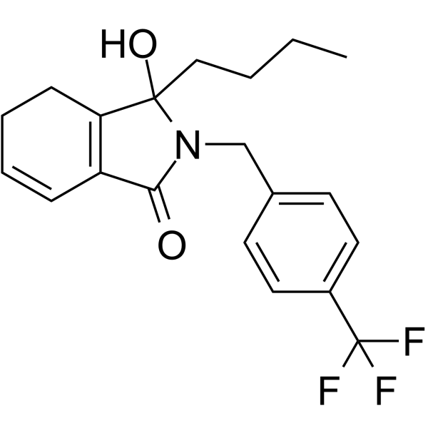
LasB-IN-1 (compound 5f) is a potent and orally active inhibitor of LasB (IC50 = 8.7 μM). LasB-IN-1 effectively attenuates elastase production and biofilm formation by P. aeruginosa while alleviating the inflammatory response through downregulating MAPK and NF-κB pathways. LasB-IN-1 is potential to be a novel anti-infective candidate against drug-resistant infections.
Nos produits utilisent uniquement pour la recherche. Nous ne vendons pas aux patients.
- Formule: C20H22F3NO2
- Masse moléculaire:365.39
-
Stockage:
Please store the product under the recommended conditions in the Certificate of Analysis.

To place orders, for customer services and technical support, please contact: MedChemExpress USA
Tel: 609-228-6898 E-mail: [email protected] [email protected]


-
 Activité biologique
-
Chemical Information
- Protocole
- Pureté et documentation
- Références
-
 Help & FAQs




Activité biologique
Description
IC50 & Target
IC50: 8.7 μM (LasB)[1]
Cellular Effect
|
Cell Line

|
Type | Value | Description | References |
|---|---|---|---|---|
| RAW264.7 | IC50 |
1.44 μM
Compound: 5f
|
Inhibition of LPS-induced ROS production in mouse RAW264.7 cells by DCFH-DA staining based fluorescence method
Inhibition of LPS-induced ROS production in mouse RAW264.7 cells by DCFH-DA staining based fluorescence method
|
[PMID: 37995562] |
| RAW264.7 | IC50 |
18.22 μM
Compound: 5f
|
Antiinflammatory activity in LPS-induced mouse RAW264.7 cells assessed as inhibition of IL-1 beta production pretreated for 3 hrs followed by LPS stimulation and measured after 24 hrs by ELISA method
Antiinflammatory activity in LPS-induced mouse RAW264.7 cells assessed as inhibition of IL-1 beta production pretreated for 3 hrs followed by LPS stimulation and measured after 24 hrs by ELISA method
|
[PMID: 37995562] |
| RAW264.7 | IC50 |
2.61 μM
Compound: 5f
|
Antiinflammatory activity in LPS-induced mouse RAW264.7 cells assessed as inhibition of IL-6 production pretreated for 3 hrs followed by LPS stimulation and measured after 24 hrs by ELISA method
Antiinflammatory activity in LPS-induced mouse RAW264.7 cells assessed as inhibition of IL-6 production pretreated for 3 hrs followed by LPS stimulation and measured after 24 hrs by ELISA method
|
[PMID: 37995562] |
| RAW264.7 | IC50 |
6.2 μM
Compound: 5f
|
Antiinflammatory activity in LPS-induced mouse RAW264.7 cells assessed as inhibition of TNF-alpha production pretreated for 3 hrs followed by LPS stimulation and measured after 24 hrs by ELISA method
Antiinflammatory activity in LPS-induced mouse RAW264.7 cells assessed as inhibition of TNF-alpha production pretreated for 3 hrs followed by LPS stimulation and measured after 24 hrs by ELISA method
|
[PMID: 37995562] |
In Vitro
LasB-IN-1 (5-10 μM, 10 h) suppresses the expression of LasB (IC50 = 8.7 μM), the elastase biosynthesis (IC50 = 7.3 μM) and the formation of biofilms (IC50 = 7.4 μM) in a dose-dependent manner in P. aeruginosa PAO1 strains[1].
LasB-IN-1 (1.25-20 μM, 24 h) reveals no cytotoxic effect in RAW264.7 cell and Vero cell. LasB-IN-1 (50-100 μM, 24 h) shows a slight cytotoxic effect. LasB-IN-1 (0.625-10 μM, 4 h) exerts negligible hemolytic effects on mouse and human erythrocytes[1].
LasB-IN-1 (10 μM, 4, 6 h) effectively inhibits the migration of macrophages to the site of injury in zebrafish larvae[1].
LasB-IN-1 (0.625-10 μM, 27 h) inhibits the production of IL-1β, TNF-α, and IL-6 in LPS (1 μg/mL, 24 h) treated RAW264.7 cells. LasB-IN-1 (1.25-10 μM, 27 h) also inhibits the mRNA expression of COX-2, iNOS, IL-1β, TNF-α, and IL-6[1].
LasB-IN-1 (1.25-10 μM, 27 h) significantly inhibits the phosphorylation of NF-κB p65, IκBα, JNK and ERK of LPS (1 μg/mL, 24 h) treated RAW264.7 cells in a concentration-dependent manner[1].
MedChemExpress (MCE) has not independently confirmed the accuracy of these methods. They are for reference only.
-
Cell Line:RAW264.7 cell and Vero cell
-
Concentration:1.25, 2.5, 5, 10, 20, 50, 100 μM
-
Incubation Time:24 h
-
Result:Revealed no cytotoxic effect at concentrations of lower than 20 μM, a slight cytotoxic effect was observed at a concentration of 50 μM, indicating the noncytotoxicity at concentrations effective against virulence and biofilm formation.
In Vivo
LasB-IN-1 (1, 2 mg/kg, i.g., 4 h) significantly downregulates expression levels of IL-1β, TNF-α, and IL-6 as well as reduces mRNA expression of COX-2, iNOS, IL-1β, TNF-α, and IL-6 in mice [1].
LasB-IN-1 (2, 4 mg/kg, i.g., once for 7 d) shows no obvious adverse reactions [1].
MedChemExpress (MCE) has not independently confirmed the accuracy of these methods. They are for reference only.
-
Animal Model:eight-week-old BALB/c wild-type mice (male, 18–22 g ), established a ALI model by using LPS (P. aeruginosa)[1]
-
Dosage:1 and 2 mg/kg
-
Administration:intragastric (i.g.), after treatment for 4 h, intraperitoneal injection of LPS (20 mg/kg), sacrificed by cervical dislocation after a 6 h treatment
-
Result:Reduced myeloperoxidase (MPO) activity in mice compared with that in LPS-only mice, indicating the inhibition of neutrophil infiltration in mouse lung tissues.
Exhibited significantly downregulated expression levels of IL-1β, TNF-α, and IL-6, with a concentration-dependent response while significantly reduced mRNA expression of COX-2, iNOS, IL-1β, TNF-α, and IL-6 in mice compared with LPS-only mice.
-
Animal Model:eight-week-old BALB/c wild-type mice (male, 18–22 g )[1]
-
Dosage:2 and 4 mg/kg
-
Administration:intragastric (i.g.) for 7 days
-
Result:Exhibited no adverse effects such as vomiting or diarrhea, had a negligible impact on the body weight of mice, did not induce any significant damage or histopathological alterations in vital organs.


Chemical Information
-
Masse moléculaire 365.39
-
Formule C20H22F3NO2
-
SMILES
O=C1N(CC2=CC=C(C(F)(F)F)C=C2)C(O)(CCCC)C3=C1C=CCC3
-
Livraison
Room temperature in continental US; may vary elsewhere.
-
Stockage
Please store the product under the recommended conditions in the Certificate of Analysis.


Pureté et documentation
Références

Calculators
Concentration (start) × Volume (start) = Concentration (final) × Volume (final)


LasB-IN-1 Related Classifications
HY-163030 Related Classifications
 Powered by Bioz