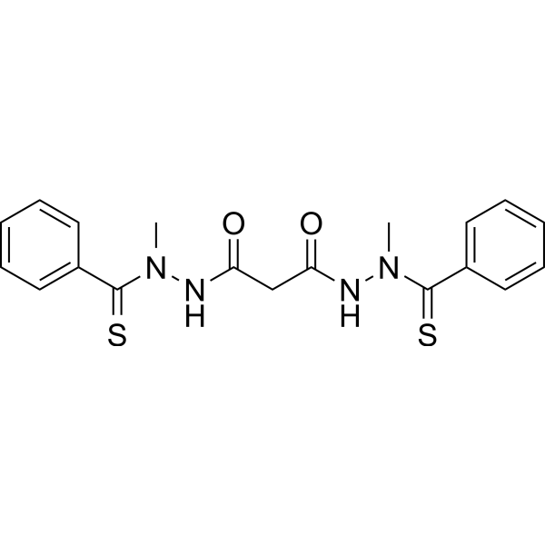
LCC-12 formate
Based on 1 Customer Validation
LCC-12 formate is a mitochondria-targeted Copper2+ -selective binder and metabolic/epigenetic reprogramming agent. LCC-12 formate inhibits NAD (H) redox cycling and reduces the NAD (H) pool. LCC-12 formate downregulates the expression of inflammatory genes and improves outcomes in mouse models of bacterial sepsis, endotoxemia, and SARS-CoV-2 infection. LCC-12 formate can be used in research related to acute inflammation, endotoxemia, sepsis, and SARS-CoV-2 infection.
For research use only. We do not sell to patients.
- Purity : 99.90%
- Formula: C17H38N10O2
- Molecular Weight:414.55
-
Storage:
-20°C, sealed storage, away from moisture
* In solvent : -80°C, 6 months; -20°C, 1 month (sealed storage, away from moisture)
Biological Activity
Description
In Vitro
LCC-12 (10 μM) formate reduces the levels of mitochondrial NAD+ and NADH pools in primary activated monocyte-derived macrophages (aMDM)[1].
LCC-12 formate selectively forms a stable monometallic complex with copper (II) in a cell-free system, while it shows no significant binding to most other divalent metal ions or copper (I)[2].
LCC-12 formate inhibits copper (II)-catalyzed redox cycling of NADH to NAD+ in a dose-dependent manner in a cell-free system mimicking the mitochondrial matrix[2].
LCC-12 formate induces epigenetic reprogramming in activated monocyte-derived macrophages, increases histone methylation levels, decreases histone acetylation levels, and downregulates pro-inflammatory gene expression and cytokine secretion[2].
LCC-12 formate inhibits the activation of immune cells with upregulated CD44, and exerts effects on CD44-related copper-dependent cellular plasticity[2].
LCC-12 formate potently interferes with the activation of monocyte-derived macrophages and inhibits CD44-associated epithelial-mesenchymal transition in cancer cells[4].
MedChemExpress (MCE) has not independently confirmed the accuracy of these methods. They are for reference only. Further protocols information, click here.
In Vivo
LCC-12 (i.p.) formate protects mice against LPS-induced death and prevents hypothermia[1].
MedChemExpress (MCE) has not independently confirmed the accuracy of these methods. They are for reference only.
Chemical Information
-
Appearance Solid
-
Molecular Weight 414.55
-
Formula C17H38N10O2
-
Color White to off-white
-
SMILES
NC(NC(NCCCCCCCCCCCCNC(NC(N)=N)=N)=N)=N.O=CO
-
Shipping
Room temperature in continental US; may vary elsewhere.
-
Storage
-20°C, sealed storage, away from moisture
* In solvent : -80°C, 6 months; -20°C, 1 month (sealed storage, away from moisture)
Protocols
-
RNA extraction experimental
By lysing cells, releasing RNA, and removing impurities such as proteins and DNA, high-purity RNA products are finally obtained. The commonly used traditional method is the guanidine isothiocyanate/phenol/chloroform method (Trizol), which is suitable for a variety of animal materials including animal tissues, microorganisms, cultured cells, etc., and most plant materials.
-
Research Protocol for Infectious Diseases
Infectious-disease experiments test how pathogens interact with host barriers, innate immune receptors, inflammatory signaling, pathogen replication, and tissue injury; pattern-recognition receptors such as TLRs, RIG-I-like receptors, NOD-like receptors, and inflammasomes detect microbial molecules and activate NF-κB, interferon, and cytokine responses. The central hypothesis is that infection severity reflects the balance between pathogen burden and host response: protective inflammation restricts pathogen growth, whereas excessive or mislocalized inflammation contributes to tissue damage and disease phenotype. Unresolved questions include which host pathways are protective versus pathogenic, why some infection models fail to translate to human disease, and which combined readouts best predict clinically relevant infection outcomes.
-
LPS-Induced Endotoxemia/Systemic Inflammation
Lipopolysaccharide (LPS)-induced endotoxemia is a widely used in vivo model of acute systemic inflammation in which LPS, a Gram-negative bacterial endotoxin, activates innate immune signaling primarily through TLR4, leading to rapid and transient induction of pro-inflammatory cytokines such as TNF-α, IL-6, and IL-1β in circulation and tissues. This cytokine surge is commonly used as a measurable readout of systemic inflammatory activation and immune dysregulation, and is typically assessed within hours after intraperitoneal LPS administration in mouse models of endotoxemia. The model captures key features of systemic inflammatory response syndrome, including cytokine release, immune cell activation, and downstream tissue responses, and has been used to evaluate anti-inflammatory interventions such as cytokine modulation, lipid mediators, and immune cell-targeting therapies.
-
ROS/oxidative-stress fluorescent staining
ROS/oxidative-stress fluorescent staining uses cell-permeant fluorogenic probes that become fluorescent after oxidation inside cells or tissues; commonly used examples include DCFH-DA/DCFDA for broad cellular oxidant detection, DHE for superoxide-related signal detection, MitoSOX for mitochondrial superoxide-related signal detection, and CellROX probes for oxidative-stress-associated fluorescence readouts. The assay detects probe oxidation rather than a single ROS species unless the probe and analysis method have been chemically validated for that species. DCFH-DA enters cells, is deacetylated by intracellular esterases to DCFH, and produces fluorescent DCF after oxidation, so the readout is used as an operational measure of total cellular oxidative stress rather than a species-specific ROS measurement. DHE and MitoSOX can report superoxide-related oxidation, but red fluorescence alone can include non-specific ethidium-like oxidation products; HPLC or optimized spectral approaches are
-
Carrageenan-Induced Paw Edema
Carrageenan-induced paw edema is an acute inflammation model in which intraplantar injection of carrageenan induces localized inflammatory swelling characterized by vascular permeability, leukocyte infiltration, and production of inflammatory mediators such as prostaglandins and cytokines, making it widely used to evaluate anti-inflammatory agents in vivo. The resulting paw volume or thickness increase is quantified over time as a direct readout of inflammatory intensity and drug efficacy, typically reflecting cyclooxygenase-mediated prostaglandin-driven edema formation and immune cell recruitment in peripheral tissue[20].
-
Zymosan-Induced Peritonitis
Zymosan-induced peritonitis is a sterile acute-inflammation model produced by intraperitoneal injection of zymosan, a yeast cell-wall particle preparation, followed by quantification of leukocyte recruitment and soluble inflammatory mediators in peritoneal lavage fluid. Low-dose zymosan peritonitis is commonly used as a self-resolving acute inflammation model in which neutrophil recruitment occurs early and monocyte/macrophage accumulation follows later. The assay readouts include total peritoneal leukocyte number, differential neutrophil and monocyte/macrophage counts, peritoneal cytokines and chemokines, plasma or peritoneal exudation, and optional lipidomic or metabolomic changes during inflammation and resolution. Early neutrophil recruitment after zymosan depends strongly on complement and mast-cell C5a receptor signaling, whereas later monocyte recruitment is linked to MCP-1/CCL2 production.
-
Research Protocol for Inflammation-related Diseases
The NLRP3 inflammasome is a cytosolic innate immune signaling platform that integrates priming signals and danger-signal activation to promote caspase-1 activation, maturation of IL-1β and IL-18, and gasdermin D-mediated pyroptotic cell death. The core experimental logic is to determine whether inflammatory disease phenotypes are driven by increased NLRP3 expression, ASC-containing inflammasome assembly, caspase-1 cleavage, GSDMD cleavage, and extracellular release of IL-1β/IL-18 rather than by nonspecific cell injury alone. The pathway is strongly linked to inflammation-related disease phenotypes because monosodium urate crystals activate NALP3/NLRP3 inflammasome signaling in gout-like crystal inflammation, cholesterol crystals activate NLRP3 inflammasomes in atherogenesis models, and DSS-induced intestinal inflammation has been reported to involve NLRP3 inflammasome activity. However, experimental colitis studies also show context-dependent protective effects of NLRP3 inflammasome co
Purity & Documentation
-
Data Sheet (267 KB)
-
SDS (252 KB)
- English - EN (252 KB)
- Français - FR (252 KB)
- Deutsch - DE (252 KB)
- Norwegian - NO (252 KB)
- Español - ES (252 KB)
- Swedish - SV (252 KB)
- Italian - IT (252 KB)
- Korean - KR (252 KB)
- Portuguese - PT (252 KB)
-
Handling Instructions (2659 KB)
References
[1]. Solier S, et al. A druggable copper-signalling pathway that drives inflammation. Nature. 2023 May;617(7960):386-394. [Content Brief]
[3]. Weiskirchen R. Targeting copper to combat macrophage-driven inflammation: a potential advanced therapeutic strategy. Signal Transduct Target Ther. 2023 Sep 11;8(1):339. [Content Brief]
[4]. Flemming A, et al. Copper boosts pro-inflammatory state of macrophages. Nature reviews. Immunology. 2023 Jun;23(6):344. [Content Brief]
Calculators
Concentration (start) × Volume (start) = Concentration (final) × Volume (final)