



Leucomalachite green
Based on 1 publication(s) in Google Scholar
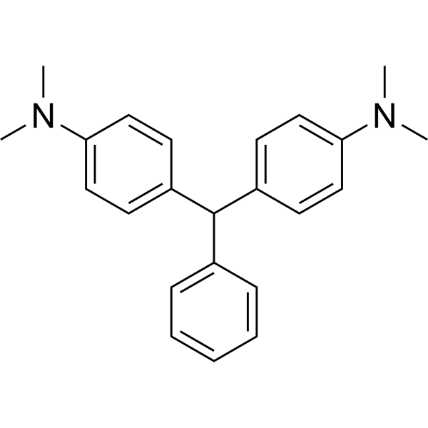
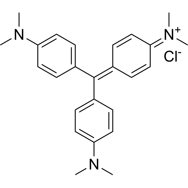
Leucomalachite green is the major reduced metabolite of malachite green (MG) and has lower cytotoxicity (such as HEp-2 and Caco-2) than malachite green. Leucomalachite green may be involved in interfering with cell metabolism or redox balance and can be used to evaluate its potential harm to human cells as a food contaminant.
For research use only. We do not sell to patients.
- Purity : 98.72%
- CAS No.: 129-73-7
- Formula: C23H26N2
- Molecular Weight:330.47
-
Storage:
4°C, protect from light
* In solvent : -80°C, 6 months; -20°C, 1 month (protect from light)

To place orders, for customer services and technical support, please contact: MedChemExpress USA
Tel: 609-228-6898 E-mail: [email protected] [email protected]


-
 Biological Activity
-
Chemical Information
-
Solvent & Solubility
- Protocols
- Purity & Documentation
- References
-
 Help & FAQs


-

HY-D0300R
Leucomalachite green (Standard)Malachite Green Metabolite -
HY-D0914
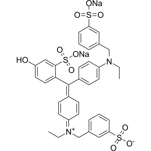
Fast Green FCFSea Green Triarylmethane Food Dye -
HY-B0324A
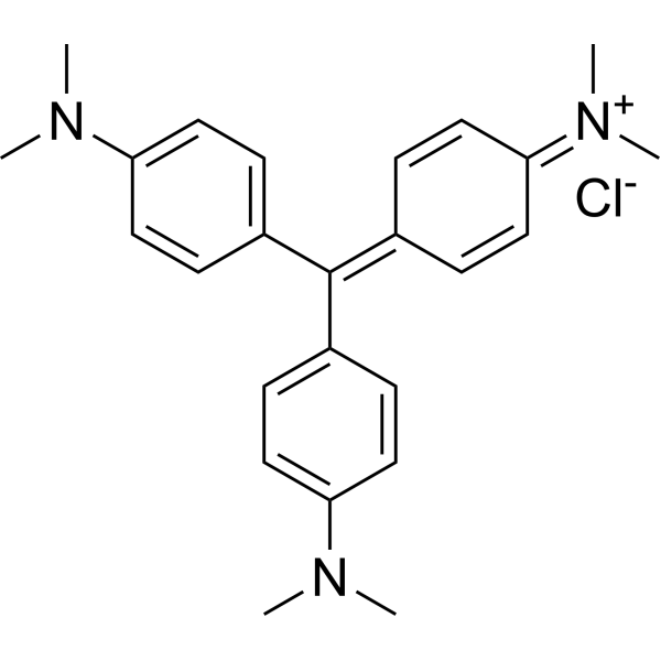
Crystal VioletFluorochrome -
HY-D0233
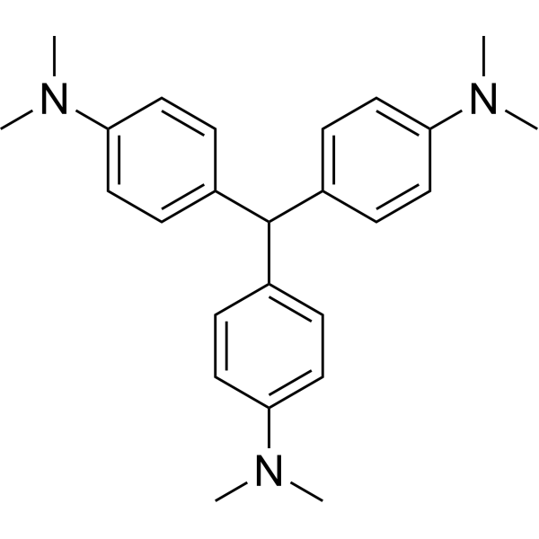
Leucocrystal violetTriphenylmethane Dye -
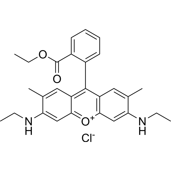
HY-D0309
Rhodamine 6GLaser Dye -

HY-DY2001
Crystal Violet (solution)Fluorochrome -
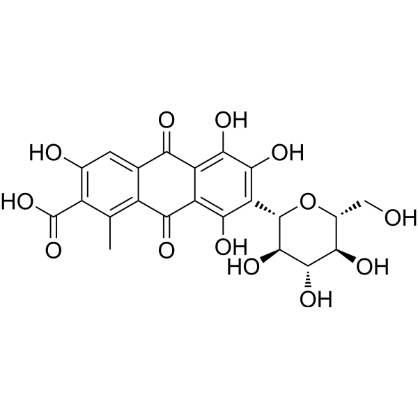
HY-N8407
Carminic acidRed Pigment -
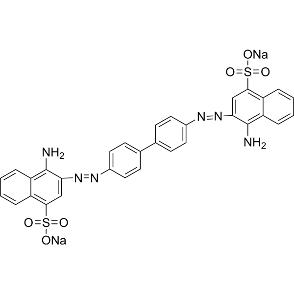
HY-D0236
Congo RedAzo Dye -
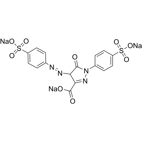
HY-D0257
TartrazineAzo Dye -
HY-W110794
Crystal violet, IND (0.0-2.0)Biochemical Assay Reagent -

HY-D0309R
Rhodamine 6G (Standard)Laser Dye -

HY-D0233R
Leucocrystal violet (Standard)Analytical Standard
Publications Citing Use of MedChemExpress (MCE) Leucomalachite green
More

Biological Activity
Description
In Vitro
Leucomalachite green (30-610 μM; 24 h) does not significantly inhibit cell viability (neutral red uptake assay, total protein content assay) and proliferation ability (colony formation assay) in HEp-2 human laryngeal cancer cell line, showing low cytotoxicity[2].
Leucomalachite green (25-100 μM; 24 h) does not show significant cytotoxicity in Caco-2 human colon cancer cell line experiments, as detected by MTT assay, LDH leakage assay and neutral red uptake assay[2].
MedChemExpress (MCE) has not independently confirmed the accuracy of these methods. They are for reference only. Further protocols information, click here.


Chemical Information
-
CAS No. 129-73-7
-
Appearance Solid
-
Molecular Weight 330.47
-
Formula C23H26N2
-
Color White to off-white
-
SMILES
CN(C)C1=CC=C(C(C2=CC=C(N(C)C)C=C2)C3=CC=CC=C3)C=C1
-
Shipping
Room temperature in continental US; may vary elsewhere.
-
Storage
4°C, protect from light
* In solvent : -80°C, 6 months; -20°C, 1 month (protect from light)


Publications (1)
-
Journal Impact Factor
-
Most Recent
-
Pharmaceuticals (Basel)
Identification of SARS-CoV-2 Main Protease Inhibitors Using Chemical Similarity Analysis Combined with Machine Learning. [Abstract]2024 Feb 12;17(2):240. PMID: 38399455


Solvent & Solubility
In Vitro:
DMSO : 100 mg/mL (302.60 mM; Need ultrasonic; Hygroscopic DMSO has a significant impact on the solubility of product, please use newly opened DMSO)
Please refer to the solubility information to select the appropriate solvent. Once prepared, please aliquot and store the solution to prevent product inactivation from repeated freeze-thaw cycles.
Storage method and period of stock solution: -80°C, 6 months; -20°C, 1 month (protect from light). When stored at -80°C, please use it within 6 months. When stored at -20°C, please use it within 1 month.
Please refer to the solubility information to select the appropriate solvent. Once prepared, please aliquot and store the solution to prevent product inactivation from repeated freeze-thaw cycles.
Storage method and period of stock solution: -80°C, 6 months; -20°C, 1 month (protect from light). When stored at -80°C, please use it within 6 months. When stored at -20°C, please use it within 1 month.
Concentration (start) × Volume (start) = Concentration (final) × Volume (final)

Protocols
-
Cell Cytotoxicity Assay
Cytotoxicity assays are usually based on the assessment of cell membrane damage, which can also be indirectly detected by measuring cell viability. Detection methods include MTT assay, CKK-8 assay, LDH assay and ATP assay, etc.
-
Mammalian live/dead viability and cytotoxicity staining
Live/dead viability and cytotoxicity staining assays are based on the simultaneous detection of intracellular esterase activity in metabolically active (viable) cells and membrane integrity loss in non-viable cells. In commonly used dual-staining approaches, membrane-permeant fluorogenic substrates are converted by intracellular esterases into fluorescent products in live cells, while impermeant DNA-binding dyes selectively enter cells with compromised plasma membranes and label nucleic acids in dead or dying cells, enabling discrimination between viable and non-viable populations by fluorescence microscopy or flow cytometry.
-
ROS/oxidative-stress fluorescent staining
ROS/oxidative-stress fluorescent staining uses cell-permeant fluorogenic probes that become fluorescent after oxidation inside cells or tissues; commonly used examples include DCFH-DA/DCFDA for broad cellular oxidant detection, DHE for superoxide-related signal detection, MitoSOX for mitochondrial superoxide-related signal detection, and CellROX probes for oxidative-stress-associated fluorescence readouts. The assay detects probe oxidation rather than a single ROS species unless the probe and analysis method have been chemically validated for that species. DCFH-DA enters cells, is deacetylated by intracellular esterases to DCFH, and produces fluorescent DCF after oxidation, so the readout is used as an operational measure of total cellular oxidative stress rather than a species-specific ROS measurement. DHE and MitoSOX can report superoxide-related oxidation, but red fluorescence alone can include non-specific ethidium-like oxidation products; HPLC or optimized spectral approaches are
-
Cell Viability Determination by MTT Colorimetric Assay
The following protocol uses the MTT colorimetric assay as a classic literature-established method for assessing cell viability/metabolic activity in cultured mammalian cells. MTT[3-(4,5-dimethylthiazol-2-yl)-2,5-diphenyltetrazolium bromide] is reduced by metabolically active cells to a colored formazan product; the amount of formazan is quantified spectrophotometrically and provides an indirect measure of metabolically active viable cells. Importantly, MTT reduction reflects cellular oxidoreductase/metabolic activity rather than an absolute direct count of living cells, so changes in cellular metabolism can alter the signal independently of cell number.


Purity & Documentation
-
Data Sheet (273 KB)
-
SDS (418 KB)
- English - EN (418 KB)
- Français - FR (418 KB)
- Deutsch - DE (418 KB)
- Norwegian - NO (418 KB)
- Español - ES (418 KB)
- Swedish - SV (418 KB)
- Italian - IT (418 KB)
- Korean - KR (418 KB)
- Portuguese - PT (418 KB)
-
Handling Instructions (2659 KB)
References
[1]. Ya-hui Li, et al. Development of a group selective molecularly imprinted polymers based solid phase extraction of malachite green from fish water and fish feed samples. Anal Chim Acta. 2008 Aug 29;624(2):317-25. [Content Brief]
[2]. Stammati A, et al. Effects of malachite green (MG) and its major metabolite, leucomalachite green (LMG), in two human cell lines. Toxicol In Vitro. 2005 Oct;19(7):853-8. [Content Brief]


Complete Stock Solution Preparation Table
Please refer to the solubility information to select the appropriate solvent. Once prepared, please aliquot and store the solution to prevent product inactivation from repeated freeze-thaw cycles.
Storage method and period of stock solution: -80°C, 6 months; -20°C, 1 month (protect from light). When stored at -80°C, please use it within 6 months. When stored at -20°C, please use it within 1 month.
| Optional Solvent | Concentration Solvent Mass | 1 mg | 5 mg | 10 mg | 25 mg |
|---|---|---|---|---|---|
| DMSO | 1 mM | 3.0260 mL | 15.1300 mL | 30.2599 mL | 75.6498 mL |
| 5 mM | 0.6052 mL | 3.0260 mL | 6.0520 mL | 15.1300 mL | |
| 10 mM | 0.3026 mL | 1.5130 mL | 3.0260 mL | 7.5650 mL | |
| 15 mM | 0.2017 mL | 1.0087 mL | 2.0173 mL | 5.0433 mL | |
| 20 mM | 0.1513 mL | 0.7565 mL | 1.5130 mL | 3.7825 mL | |
| 25 mM | 0.1210 mL | 0.6052 mL | 1.2104 mL | 3.0260 mL | |
| 30 mM | 0.1009 mL | 0.5043 mL | 1.0087 mL | 2.5217 mL | |
| 40 mM | 0.0756 mL | 0.3782 mL | 0.7565 mL | 1.8912 mL | |
| 50 mM | 0.0605 mL | 0.3026 mL | 0.6052 mL | 1.5130 mL | |
| 60 mM | 0.0504 mL | 0.2522 mL | 0.5043 mL | 1.2608 mL | |
| 80 mM | 0.0378 mL | 0.1891 mL | 0.3782 mL | 0.9456 mL | |
| 100 mM | 0.0303 mL | 0.1513 mL | 0.3026 mL | 0.7565 mL |


Leucomalachite green Related Classifications
HY-D0300 Related Classifications
 Powered by Bioz