




Engineered extracellular vesicles (EVs) surpass their natural counterparts by offering enhanced targeting capabilities and the ability to deliver specific therapeutic agents with high precision. These advances have unlocked new possibilities in treating a wide range of diseases, especially cancer, by overcoming traditional drug delivery limitations such as poor bioavailability and off-target effects. Additionally, engineered EVs can be tailored to carry genetic material, small molecules, or immunomodulatory agents, offering versatile treatment options[1-3].
In this issue, we provide an overview of engineered EVs, highlighting three key aspects: a general overview of EVs, methods for their construction, and their applications in cancer immunotherapy. These discussions aim to offer new insights into the development and therapeutic potential of engineered EVs.
- Overview of Extracellular Vesicles (EVs)
- Strategies for Preparation of Engineered EVs
- Applications of Engineered EVs in Cancer Immunotherapy
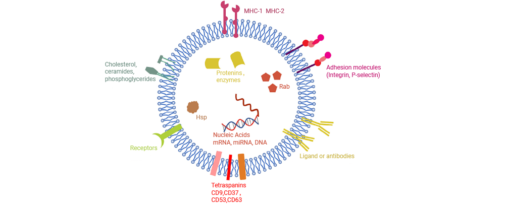
Figure 1. Composition of EVs[3].
EVs are nanoscale, lipid bilayer-encapsulated vesicles secreted by virtually all cell populations. The unique molecular composition of each EV reflects its cellular origin and dictates its function in intercellular communication.
Think of an EV as a tiny, biological "message bubble" sent from one cell to another. Its lipid bilayer membrane forms a communication interface, decorated with markers and functional molecules that guide selective uptake by recipient cells. Inside, EVs carry diverse molecular cargos—including nucleic acids (miRNA, mRNA, DNA), proteins, and other bioactive components—that can modulate signaling pathways and cellular processes in target cells. This natural delivery system underlies the role of EVs in intercellular communication and therapeutic applications.
Based on their origin and size, EVs are commonly classified into three primary subtypes: exosomes (Exos), microvesicles (MVs, alternatively termed ectosomes), and apoptotic bodies (ABs)[2-3]. These subtypes arise through distinct biogenetic pathways, a tightly regulated process that shapes their molecular composition and functional roles in intercellular communication (Fig. 2)[2].
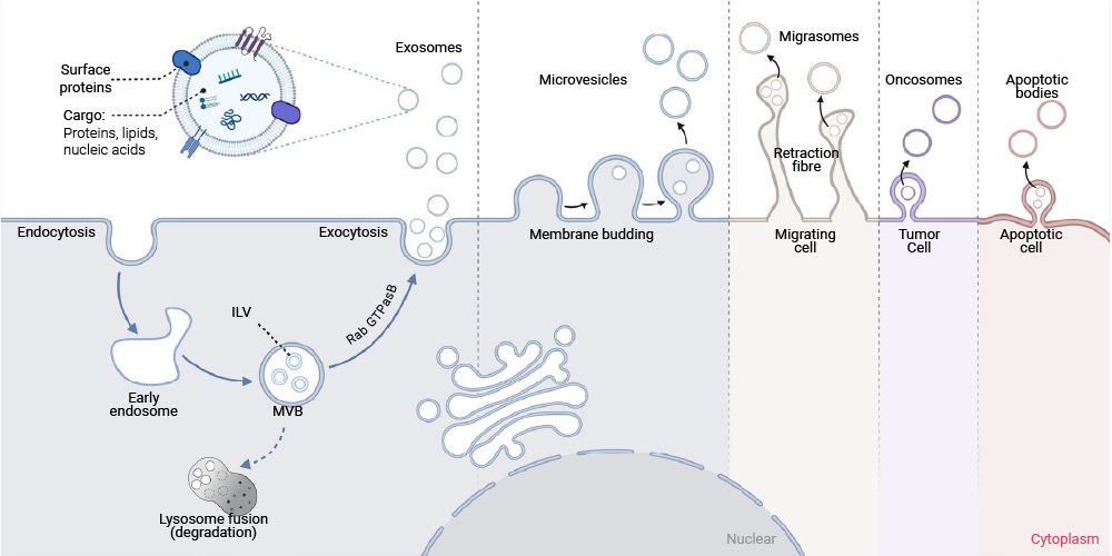
Figure 2. The biogenesis of EVs[2].
Exosomes (30-150 nm) are the most extensively studied EV subtype and are generated via the endosomal pathway. Their formation begins with inward budding of the plasma membrane to form early endosomes, which then mature into late endosomes. These late endosomes undergo further invagination to generate multivesicular bodies (MVBs) containing intraluminal vesicles (ILVs). Fusion of MVBs with the plasma membrane subsequently releases these ILVs into the extracellular space as exosomes. Importantly, the molecular cargo selectively packaged within exosomes reflects the physiological or pathological state of their parental cells and is dynamically influenced by intracellular signaling, cellular stress, and disease processes such as carcinogenesis.
Microvesicles (MVs, or Ectosomes; 100 nm - 1 μm) arise more directly from the plasma membrane. They are formed through outward budding and fission of the membrane, encapsulating portions of cytoplasm during this process.
Apoptotic bodies (ABs; 500 nm - several micrometers), the largest EVs, are produced during programmed cell death. They contain fragmented cellular components, including organelles and chromatin, and are released as the cell disassembles.
EVs can be categorized into natural and engineered subsets based on whether they have undergone artificial modification[3].
Natural EVs refer to vesicles isolated and purified from cell culture supernatants, biological fluids, or plant tissue lysates without subsequent artificial manipulation. Rich in bioactive molecules, these vesicles are capable of modulating cellular activities upon uptake by recipient cells or tissues. Despite their promising therapeutic potential across various diseases, their limited targeting specificity represents a major bottleneck that restricts clinical efficacy[1].
To address the limitations of natural EVs, targeted modifications can be introduced to enhance tissue- and cell-specific targeting, and exogenous therapeutic agents can be incorporated to improve their therapeutic efficacy. Engineered EVs have been extensively evaluated in preclinical studies for the treatment of a range of disorders. For example, surface modification of EVs with the cysteine-arginine-glutamate-lysine-alanine (CREKA) peptide produces vesicles that specifically recognize the fibrin-fibronectin complex. In rat models of femoral defects, CREKA-functionalized EVs demonstrated enhanced retention at the injury site, thereby promoting bone tissue regeneration. In another application, chronic myeloid leukemia (CML) cells, which overexpress interleukin-3 (IL-3) receptors, serve as ideal therapeutic targets. By fusing IL-3 with the EV-associated scaffold protein LAMP2B, LAMP2B-IL3 engineered EVs were generated. Co-loading these vesicles with oncogene-targeting small interfering RNAs (siRNAs) and the chemotherapeutic drug Imatinib enabled precise targeting of CML cells, resulting in robust inhibition of cancer cell proliferation and significant tumor regression[1].
Building on these advancements, a range of strategies has been developed to construct and optimize engineered EVs for therapeutic applications. Broadly, these approaches fall into two categories: "genetic engineering" and "chemical/mechanical engineering". In this section, we will discuss the principal methods employed in these two approaches and summarize the characteristics of each.
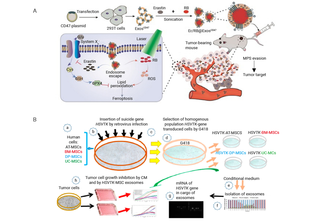
Figure 3. Method for genetically engineering EVs[3].
Genetic engineering of EVs enhances vesicle functionality through targeted genetic manipulation of donor cells genomes. This approach allows for the site-specific insertion, deletion, or modification of target genes within the donor cell genome, followed by isolation of EVs that encapsulate the desired genetic payload or therapeutic agents.
A common implementation involves transfecting donor cells with expression vectors—either plasmids or viral vectors—carrying the target gene. Due to the stability and well-characterized nature of the transfection systems, the modified donor cells secrete EVs displaying the target peptide on their surface.
Physical manipulation techniques can induce transient, reversible permeabilization of EV membranes—through processes such as pore formation or membrane reorganization—thereby enabling the direct encapsulation of small nucleic acid molecules (e.g., miRNAs and siRNAs) and therapeutic agents (e.g., anticancer drugs) into EVs. As a widely adopted EV engineering strategy, this membrane-based modification method is generally more straightforward than donor cell-targeted genetic engineering and has been extensively applied to fabricate EVs as carriers for drug and gene delivery. Common post-isolation techniques for direct EV engineering include electroporation, extrusion, sonication, incubation, bioconjugation, and click chemistry. Notably, the streamlined protocols of these membrane-targeted procedures offer greater suitability for clinical translation compared with donor cell-based genetic modification strategies.
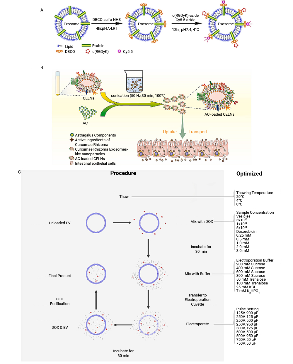
Figure 4. Methods for chemical and mechanical engineering of EVs[3].
Chemical modification of EVs serves a dual purpose: enhancing vesicle stability and maximizing targeting precision and delivery efficiency. This category primarily encompasses two technical routes: covalent conjugation and non-covalent binding.
EV modification can also be achieved via chemical transfection approaches. Specifically, transfection reagents or permeabilizing agents are used to facilitate the encapsulation of therapeutic agents into EVs while preserving the integrity of the vesicle lipid bilayer.
Co-incubation represents a straightforward and direct strategy for cargo loading. This method involves incubating the intended payload with pre-isolated EVs, allowing the cargo to diffuse into the vesicle lumen driven by concentration gradients.
Electroporation loads cargo by applying an external electric field to generate transient micropores in the EV membrane, thereby increasing its permeability. This technique can substantially enhance the loading efficiency of drug molecules into EVs.
A comparative analysis of the two strategies is conducted to identify the most suitable approach for practical applications. As summarized in Table 1, both the genetic engineering and chemical/mechanical engineering strategies possess distinct advantages as well as inherent limitations.
| Classification | Types of strategies | Principle | Advantages | Disadvantages |
|---|---|---|---|---|
| Genetic engineering | Genetic engineering | Through genetic modification technology, the donor cells are genetically manipulated to produce EVs containing the desired genetic material or drugs. | The expressed engineered EVs exhibit high stability | Cumbersome operation procedure |
| Chemical and mechanical engineering | Chemical coupling | Covalent binding and non-covalent binding | High stability of surface modification | Modifying or obscuring the active sites of surface proteins impairs function |
| Chemical transfection | Using transfectants to bind and encapsulate nucleic acids for transport into the EV. | Maintain the structure of EVs | High cytotoxicity and low efficiency | |
| Co-incubation | Diffusion across EV membrane | Easy operation | Low loading efficiency | |
| Electroporation | Creating micro-pores for diffusion by electric field | High loading efficiency | Influence EV Integrity; Cargo aggregation |
By constructing engineered EVs with enhanced stability and optimized delivery characteristics, these vesicles are well-suited for therapeutic applications. They can modulate immune signaling pathways and deliver therapeutic agents with high specificity and low toxicity in cancer immunotherapy. This section highlights several representative applications of engineered EVs in cancer immunotherapy[2].
Engineered EVs have emerged as transformative nanovaccine platforms, effectively overcoming key limitations of conventional tumor vaccines. Their inherent biocompatibility, targeted delivery potential, and ability to carry diverse immunogenic cargos make them ideal for eliciting robust and specific anti-tumor immune responses. Notably, dendritic cell-derived EVs (DC-EVs) function as natural adjuvants, enhancing antigen presentation and T-cell activation, while tumor-derived exosomes (TEXs) provide a ready source of tumor-associated antigens but often require engineering to mitigate their intrinsic immunosuppressive properties.
To further exploit these advantages, innovative engineering strategies have been developed. For example, TEXs have been modified with pH-sensitive fusogenic peptides to improve lysosomal escape and antigen cross-presentation. Other advanced platforms include hybrid nanovaccines that merge TEXs with DC membranes for enhanced lymph node targeting, as well as biomimetic systems combining EVs with photothermal agents such as black phosphorus quantum dots. These engineered EV-based vaccines not only improve antigen delivery but also actively remodel the tumor microenvironment, positioning them at the forefront of next-generation cancer immunotherapy.
Engineered EVs derived from cytotoxic immune cells offer a potent, cell-free alternative to adoptive cell therapies, replicating therapeutic functions while minimizing risks such as cytokine release syndrome. A prominent example is Natural Killer (NK) cell-derived EVs, which carry cytotoxic effectors like perforin and granzymes. This concept has been further advanced by creating NK-derived EV mimetics, termed NKsomes, which retain parental cell membrane proteins for enhanced tumor targeting and have demonstrated significant antitumor efficacy in preclinical models.
Beyond NK cells, EVs from other immune effectors show similar promise. Chimeric antigen receptor (CAR) T cell-derived EVs preserve both the target specificity and cytotoxic cargo of their parent cells, effectively inhibiting tumor growth. M1 macrophage-derived EVs also exhibit direct antitumor activity. The versatility of this platform is further illustrated by its capacity for precise molecular delivery; for instance, engineered small EVs can transport siRNA to silence immune checkpoints or deliver cytokines such as IL-2 to potentiate CD8⁺ T cell activity, underscoring their role as multifaceted and targeted therapeutic agents.
A major barrier in cancer immunotherapy is the immunosuppressive tumor microenvironment (TIME). Engineered EVs provide a multifaceted strategy to remodel the TIME by reprogramming resident immune cells and delivering targeted immunomodulators. A key approach involves switching tumor-associated macrophages (TAMs) from a pro-tumor (M2) to an anti-tumor (M1) phenotype. EVs derived from M1-polarized macrophages, or their synthetic biomimetic counterparts, have proven effective in driving this switch, an effect that is further enhanced when combined with immune checkpoint inhibitors.
In addition to cellular reprogramming, EVs serve as highly efficient delivery vehicles for innate immune agonists. They can be loaded with molecules such as RIG-I or STING agonists to directly activate these pathways within tumors, leading to robust interferon production and immune cell recruitment. Furthermore, EVs can be engineered to induce immunogenic cell death (ICD) by delivering chemotherapeutic agents, thereby converting immunologically "cold" tumors into "hot" ones. They also provide strategies to counteract specific immune evasion mechanisms, such as interfering with the packaging of immunosuppressive molecules like PD-L1 into tumor-derived vesicles. Collectively, these capabilities establish engineered EVs as powerful tools not merely for drug delivery, but for actively reshaping the tumor microenvironment to support effective immune attack.
| Classification | EVs type | Cancer Ttype | Biological role |
|---|---|---|---|
| Engineered EVs as nanovaccine platforms | DC-derived EVs | General cancer | Antigen delivery, immune adjuvant |
| Tumor-derived exosomes | General cancer | Tumor antigen delivery | |
| Tumor-derived exosomes | Breast cancer | Immune activation and immune response promotion | |
| Engineered EVs with cytotoxic effect | NK cell-derived EVs | General (tumors) | Cytotoxicity against tumor cells |
| NK-derived EV mimetics (NKsome) | General (tumors) | Tumor targeting and antitumor activity | |
| Engineered EVs for remodeling the tumor immune microenvironment | Exosomes delivering STING agonists | Not specified | Enhance antitumor immune response via cGAS-STING pathway |
| Exosomes from BM-MSCs delivering oxaliplatin and Gal-9 siRNA | Pancreatic ductal adenocarcinoma | Enhance immune reprogramming and recruit cytotoxic T cells |
EVs hold great promise as cell-free therapeutic tools in regenerative medicine. Engineered EVs, compared to natural EVs, exhibit enhanced tissue and cell targeting capabilities and can be loaded with specific drugs to improve their therapeutic effects. These vesicles can be constructed through two main strategies: genetic engineering and chemical/mechanical engineering (including chemical coupling, chemical transfection, co-incubation, and electroporation), each with distinct advantages and limitations. Notably, chemical/mechanical engineering approaches are simpler and more readily translatable to clinical settings. The demonstrated success of engineered EVs in cancer immunotherapy underscores their broad potential across diverse disease contexts and highlights their promise as next-generation therapeutic platforms.
| Product Name | Cat. No. | Bioactivity | Category |
|---|---|---|---|
| Inclisiran sodium | HY-132591A | A double-stranded siRNA molecule that inhibits the transcription of PCSK9. | siRNA Research Tools |
| SiRNA Negative Control | HY-150150 | siRNA used as a negative control in gene silencing studies. | |
| Camptothecin | HY-16560 | DNA topoisomerase I (Topo I) inhibitor that modulates HIF-1α activity by altering miRNA patterns in cancer cells. | miRNA Research Tools |
| Guanosine-5'-triphosphate disodium salt | HY-W010737 | G protein activator and nucleotide precursor; upregulates miRNAs (miR133a, miR133b) and induces exosome release. | |
| MicroRNA Antagomir Negative Control | HY-RI04602A | Chemically-modified oligonucleotide used as a negative control for miRNA antagomir studies. | |
| OTX-2002 | HY-185051 | A mRNA encoding two bifunctional fusion proteins (ZFBD-MQ1 and ZFBD-KRAB). | mRNA Cargo |
| Human IL18 mRNA | HY-174631 | mRNA encoding the human IL18 protein. | |
| IL-3 Protein, Mouse (HEK293, His) | HY-P70685 | Multilineage hematopoietic cytokine promoting platelet and neutrophil recovery in secondary hematopoietic failure. | Protein Cargo |
| Extracellular Vesicles (EVs) Compound Library | HY-L168 | Collection of 580+ small molecules related to EV biology for metabolism, cancer, and disease research. | EV / Exosome Research Libraries |
| Exosomes Compound Library | HY-L072 | Collection of 50+ compounds that inhibit or stimulate exosome secretion and biogenesis, useful for exosome research. |
Note: MCE can provide products for research use only. We do not sell to patients
 LinkedIn
 Researchgate
 Instagram
 X
X Facebook
 WhatsApp
 Reddit
 Mail
Mail
