



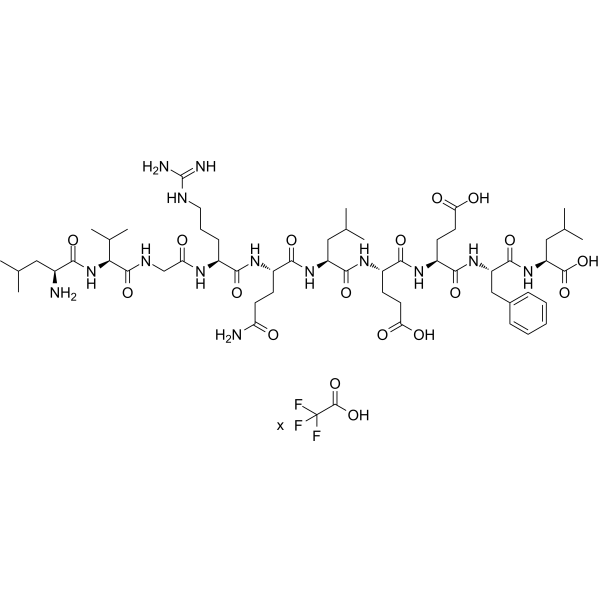
LVGRQLEEFL (mouse) TFA
LVGRQLEEFL (mouse) TFA can be named as G * peptide, corresponding to amino acids 113 to 122 in apolipoprotein J ([113,122] apoJ)}. LVGRQLEEFL (mouse) TFA exhibits anti-inflammatory and anti-atherogenic properties. LVGRQLEEFL (mouse) TFA can be added to an apoJ mimetic, to form HM-10/10 peptide, which is a mimetic peptide and a novel chimeric high density lipoprotein. HM-10/10 peptide protects retinal pigment epithelium (RPE) and photoreceptors from oxidant induced cell death.
For research use only. We do not sell to patients.
- Formula: C55H90N14O16.xC2HF3O2
- Molecular Weight:1203.39 (free acid)
-
Storage:
Sealed storage, away from moisture and light, under nitrogen.
Powder -80°C, 2 years , -20°C, 1 year* In solvent : -80°C, 6 months; -20°C, 1 month (sealed storage, away from moisture and light, under nitrogen)

To place orders, for customer services and technical support, please contact: MedChemExpress USA
Tel: 609-228-6898 E-mail: [email protected] [email protected]


-
 Biological Activity
-
Chemical Information
- Protocols
- Purity & Documentation
- References
-
 Help & FAQs




Biological Activity
Description


Chemical Information
-
Appearance Solid
-
Molecular Weight 1203.39 (free acid)
-
Formula C55H90N14O16.xC2HF3O2
-
Color White to off-white
-
Sequence
Leu-Val-Gly-Arg-Gln-Leu-Glu-Glu-Phe-Leu
-
Sequence Shortening
LVGRQLEEFL
-
Shipping
Room temperature in continental US; may vary elsewhere.
-
Storage
Sealed storage, away from moisture and light, under nitrogen
Powder -80°C 2 years -20°C 1 year * In solvent : -80°C, 6 months; -20°C, 1 month (sealed storage, away from moisture and light, under nitrogen)


Purity & Documentation
-
Data Sheet (269 KB)
-
SDS (252 KB)
- English - EN (252 KB)
- Français - FR (252 KB)
- Deutsch - DE (252 KB)
- Norwegian - NO (252 KB)
- Español - ES (252 KB)
- Swedish - SV (252 KB)
- Italian - IT (252 KB)
- Korean - KR (252 KB)
- Portuguese - PT (252 KB)
-
Handling Instructions (2659 KB)
References
[1]. Su F, et al. A Novel HDL-Mimetic Peptide HM-10/10 Protects RPE and Photoreceptors in Murine Models of Retinal Degeneration. Int J Mol Sci. 2019 Sep 27;20(19):4807. [Content Brief]
[2]. Mishra VK, et al. Structure and lipid interactions of an anti-inflammatory and anti-atherogenic 10-residue class G(*) apolipoprotein J peptide using solution NMR. Biochim Biophys Acta. 2011 Jan;1808(1):498-507. [Content Brief]

Calculators
Concentration (start) × Volume (start) = Concentration (final) × Volume (final)


LVGRQLEEFL (mouse) TFA Related Classifications
HY-P5169A Related Classifications
 Powered by Bioz