



Mapatumumab

Based on 1 Customer Validation
Mapatumumab (HGS-ETR1) is a fully human IgG1 agonistic monoclonal antibody that targets tumor necrosis factor-related apoptosis-inducing ligand receptor 1 (TRAIL-R1). Mapatumumab can be used for the research of cancer.
For research use only. We do not sell to patients.
- Purity : 97.85%
- CAS No.: 658052-09-6
- Molecular Weight:143.52 kDa
-
Storage:
Please store the product under the recommended conditions in the Certificate of Analysis.


To place orders, for customer services and technical support, please contact: MedChemExpress USA
Tel: 609-228-6898 E-mail: [email protected] [email protected]


-
 Biological Activity
-
Chemical Information
- Protocols
- Purity & Documentation
- References
-
 Help & FAQs








Biological Activity
Description
Isotype
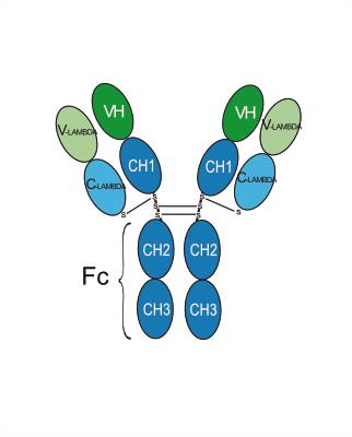
Human IgG1 lambda
Recommend Isotype Controls
Species Reactivity
Human
IC50 & Target
TNFRSF10A
In Vitro
Mapatumumab (0.01-100 μg/mL; 4 h) shows no growth inhibition to Pediatric Preclinical Testing Program (PPTP) cell lines[1]. Mapatumumab (1-100 ng/mL; 24 h) shows significant potentiation of cytotoxicity and synergy when combined with (epirubicin) EPI for bladder cancer cells[2]. Mapatumumab (100 ng/mL; 12 h) induces significant cell apoptosis of T24, 253J and J82 cells with the combination of EPI (1 μg/mL)[2]. Mapatumumab (100 ng/mL; 6-24 h) significantly activates caspase-8, -9 and -3 with the combination of EPI (1 μg/mL)[2].
MedChemExpress (MCE) has not independently confirmed the accuracy of these methods. They are for reference only. Further protocols information, click here.
In Vivo
MedChemExpress (MCE) has not independently confirmed the accuracy of these methods. They are for reference only.
-
Animal Model:Colo205-xenograft–bearing NMRI (nu/nu) nude mice[3]
-
Dosage:10 mg/kg
-
Administration:Intraperitoneal injection; 10 mg/kg; on days 1, 4, and 8
-
Result:Showed no significant differences when used alone, but when combined with irradiation (5×3 Gy, d1–5) showed a pronounced tumor regrowth-delay.
Clinical Trial
| NCT Number | Sponsor | Condition | Start Date |
Phase

|
|---|---|---|---|---|
| NCT01329991 | Plexxikon| | 2011-05 | PHASE1 |
Gene ID
Accession
Conjugated
Unconjugated
Reconsititution
The product can be reconstituted/diluted with sterile PBS or saline.
Format
-
Human IgG1 lambda
Application
ELISA, FACS, Functional assay
Verified Bioactivity
-
 Immobilized Human TNFRSF10A/TRAILR1, His tag can bind Mapatumumab. The EC50 for this effect is 17.21 ng/mL.
 -
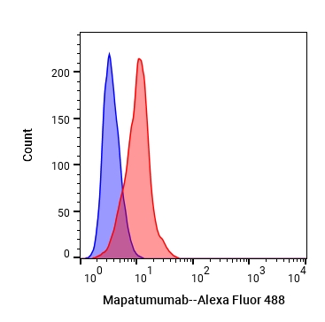 Flow cytometric analysis of 1X10⁶ A549 cells with Mapatumumab (HY-P99265, red). Cells were fixed with 4% paraformaldehyde. Then stained with the primary antibody at 1/200 dilution for an hour at 4℃. Alexa Fluor 488-conjugated AffiniPure Goat Anti-Human IgG H&L (AF488) (HY-P83776) was used as the secondary antibody at 1/1,000 dilution for 30 minutes at 4℃. Human IgG2 lambda (HY-P991206, blue) was used as the isotype control.


Chemical Information
-
CAS No. 658052-09-6
-
Appearance Liquid
-
Molecular Weight 143.52 kDa
-
Color Colorless to light yellow
-
SMILES
[Mapatumumab]
-
Synonyms
HGS-ETR1; Anti-Human TNFRSF10A Recombinant Antibody
-
Shipping
Shipping with dry ice.
-
Formulation
Please refer to the lot-specific COA for specific buffer information.
-
Storage
Please store the product under the recommended conditions in the Certificate of Analysis.

Protocols
-
Apoptosis
Apoptosis, also called programmed cell death, is generally characterized by distinct morphological characteristics.
-
TUNEL staining for apoptotic DNA fragmentation
TUNEL staining detects DNA strand breaks by using terminal deoxynucleotidyl transferase to add labeled nucleotides to exposed 3′-OH DNA termini, generating either microscopic staining in fixed cells or tissue sections, or fluorescence/cytometric signal in cell suspensions. TUNEL positivity reflects DNA fragmentation but should not be interpreted alone as definitive apoptosis, because TUNEL can also label necrotic, autolytic, mechanically damaged, or DNA-repair-associated DNA breaks.
-
Annexin V plus membrane-impermeant dye apoptosis staining
Annexin V-based apoptosis assays rely on the detection of phosphatidylserine (PS) externalization from the inner leaflet of the plasma membrane to the outer leaflet, an early biochemical hallmark of apoptosis. Fluorescently labeled Annexin V binds PS in a calcium-dependent manner, enabling identification of early apoptotic cells by flow cytometry or fluorescence microscopy. When combined with a membrane-impermeant DNA-binding dye (e. g. , propidium iodide), this approach allows discrimination between viable (Annexin V−/dye−), early apoptotic (Annexin V+/dye−), and late apoptotic or necrotic (Annexin V+/dye+) cell populations by assessing membrane integrity and PS exposure.
-
Apoptosis Solutions
Apoptosis is a regulated, generally non-lytic cell-death pathway that removes unwanted, damaged, infected, or abnormal cells through coordinated morphological changes, caspase activation, DNA fragmentation, and membrane remodeling. The intrinsic apoptosis pathway is controlled mainly by mitochondrial outer membrane permeabilization, BCL-2 family proteins, cytochrome c release, apoptosome formation, caspase-9 activation, and downstream executioner caspase-3/7 activation. The extrinsic apoptosis pathway is initiated by death receptors such as Fas, TNFR, and TRAIL receptors, which recruit adaptor proteins and activate caspase-8 before engaging executioner caspases or mitochondrial amplification through BID cleavage. Apoptosis is linked to many phenotypes, including cancer cell killing, tissue homeostasis, immune regulation, neurodegeneration, infection response, and treatment-induced cytotoxicity; unresolved questions include how apoptosis interacts with necroptosis, pyroptosis, ferroptos


Purity & Documentation
-
Data Sheet (260 KB)
-
SDS (251 KB)
- English - EN (251 KB)
- Français - FR (251 KB)
- Deutsch - DE (251 KB)
- Norwegian - NO (251 KB)
- Español - ES (251 KB)
- Swedish - SV (251 KB)
- Italian - IT (251 KB)
- Korean - KR (251 KB)
- Portuguese - PT (251 KB)
-
Inhibitory Antibodies User Guide (603 KB)
References
[2]. Ahmed SM, et al. Synergistic induction of apoptosis by mapatumumab and anthracyclines in human bladder cancer cells. Oncol Rep. 2015 Feb;33(2):566-72. [Content Brief]
[3]. Marini P, et al. Combination of the pro-apoptotic TRAIL-receptor antibody mapatumumab with ionizing radiation strongly increases long-term tumor control under ambient and hypoxic conditions. Int J Radiat Oncol Biol Phys. 2009 Sep 1;75(1):198-202. [Content Brief]

Calculators
Concentration (start) × Volume (start) = Concentration (final) × Volume (final)


Mapatumumab Related Classifications
HY-P99265 Related Classifications
 Powered by Bioz