



MAX-40279 hydrochloride
Based on 1 publication(s) in Google Scholar
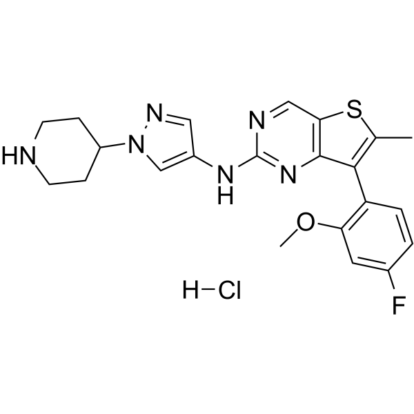
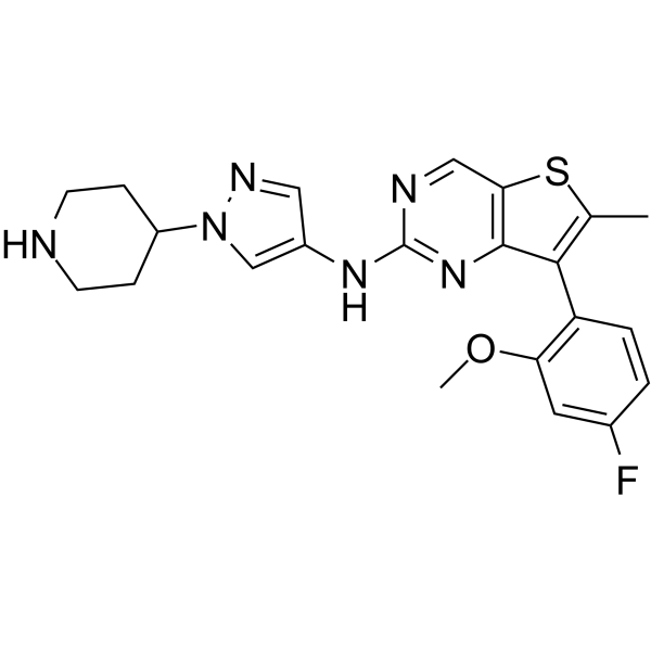
MAX-40279 hydrochloride is a dual and orally active inhibitor of FLT3 kinase and FGFR kinase. MAX-40279 hydrochloride is also effective against the FLT3 mutants such as FLT3D835Y, suggesting that it can overcome resistance to Quizartinib (HY-13001) and Sorafenib (HY-10201). MAX-40279 hydrochloride inhibits NDRG1 phosphorylation at Ser330 and suppresses endothelial-to-mesenchymal transition (EndMT). MAX-40279 hydrochloride can be used for the research of acute myelogenous leukemia (AML).
For research use only. We do not sell to patients.
- CAS No.: 2388506-51-0
- Formula: C22H24ClFN6OS
- Molecular Weight:474.98
-
Storage:
Please store the product under the recommended conditions in the Certificate of Analysis.

To place orders, for customer services and technical support, please contact: MedChemExpress USA
Tel: 609-228-6898 E-mail: [email protected] [email protected]


-
 Biological Activity
-
Chemical Information
- Protocols
- Purity & Documentation
- References
-
 Help & FAQs



Publications Citing Use of MedChemExpress (MCE) MAX-40279 hydrochloride
More

Biological Activity
Description
IC50 & Target
FLT3 and FGFR[1]
In Vitro
MAX-40279 (0.5-1 μM, 48 h) hydrochloride impedes EndMT by inhibiting NDRG1 phosphorylation at Ser-330 in HUVECs, MAECs, and MPLECs[3].
MedChemExpress (MCE) has not independently confirmed the accuracy of these methods. They are for reference only. Further protocols information, click here.
-
Cell Line:HUVECs
-
Concentration:0.5 and 1 μM
-
Incubation Time:48 h
-
Result:Attenuated migration of HUVEC induced by H2O2 and TGF-β.
-
Cell Line:HUVECs, MAECs, and MPLECs
-
Concentration:0.5 and 1 μM
-
Incubation Time:48 h
-
Result:Significantly inhibited the phosphorylation of NDRG1 at Ser-330.
Reduced the total protein level of NDRG1.
Inhibited the expression of mesenchymal markers such as ZEB1, α-SMA, Slug, Snail, and TAGLN.
In Vivo
MAX-40279 (12 mg/kg, p,o., twice daily for 21-28 days) hydrochloride significantly inhibited tumor growth in MV4-11 and KG-1 cells induced mice xenograft models[1].
MAX-40279 (7-15 mg/kg, p.o., twice daily for 2-3 weeks) hydrochloride significantly enhances the anti-PD-1 effect in mice breast cancer model[1].
MAX-40279 (p.o., single dose) hydrochloride has much higher drug concentration in bone marrow than in plasma in SD rats[2].
MedChemExpress (MCE) has not independently confirmed the accuracy of these methods. They are for reference only.
-
Animal Model:Micro patient-derived xenotransplantation model established in 5-week-old nu/nu mice[1]
-
Dosage:12 mg/kg
-
Administration:Oral administration (p.o.), once daily for 7 days
-
Result:Reduced the vitality of tumor cells.
The growth rates of tumour volume and weight were smaller but that the combined inhibition was more remarkable.
-
Animal Model:MV4-11 and KG-1 cells induced xenograft model established in immunodeficient mice[1]
-
Dosage:12 mg/kg
-
Administration:Oral administration (p.o.), once daily for 21-28 days
-
Result:Significantly inhibited tumor growth, with no significant weight loss or toxicity observed.
-
Animal Model:4T1 induced breast cancer model combination with anti-PD-1 established in mice[1]
-
Dosage:7, 10 and 15 mg/kg
-
Administration:Oral administration (p.o.), once daily for 2-3 weeks
-
Result:Significantly enhanced the efficacy of anti-PD-1 effect, and the combined treatment with anti-PD-1 antibodies yielded the best results.


Chemical Information
-
CAS No. 2388506-51-0
-
Molecular Weight 474.98
-
Formula C22H24ClFN6OS
-
SMILES
FC1=CC(OC)=C(C2=C(C)SC3=C2N=C(NC4=CN(C5CCNCC5)N=C4)N=C3)C=C1.[H]Cl
-
Shipping
Room temperature in continental US; may vary elsewhere.
-
Storage
Please store the product under the recommended conditions in the Certificate of Analysis.


Publications (1)
-
Journal Impact Factor
-
Most Recent
-
Theranostics
2025 Jan 1;15(2):745-765. PMID: 39744686

Protocols
-
Kinase activity and phosphorylation assays
Kinase activity assays measure the ability of kinases to transfer phosphate groups from ATP to specific substrates, while phosphorylation assays detect the presence and levels of phosphorylated proteins. Common methods include radiolabeled ATP incorporation (e. g. ,), ADP release detection via bioluminescence (e. g. ,[3]), enzyme-linked immunosorbent assays (ELISA) for phospho-specific epitopes (e. g. ,[6]), and microtiter-based formats for high-throughput screening (e. g. ,[8]). The ADP-Glo assay quantifies kinase activity by measuring ADP produced during phosphorylation using a luciferase-based system. Radiometric assays involve autoradiography or scintillation counting after incorporation of 32P-labeled ATP into substrate proteins. ELISA-based approaches rely on phospho-specific antibodies to detect activated kinases in cell lysates or purified samples.
-
Western Blot
Western blotting (WB) is a commonly used experimental method in molecular biology, biochemistry, and immunogenetics for identifying and quantifying target proteins. It combines gel electrophoresis with immunoassay, enabling researchers to analyze protein expression, post-translational modifications, and molecular weight.
-
Protocol for Kinase activity and phosphorylation assays
Kinase activity assays measure transfer of phosphate from ATP to a protein or peptide substrate, generating phosphorylated substrate, ADP, or incorporated radiolabeled phosphate as the readout; phosphorylation assays measure site-specific phosphorylation in cells or tissues as a proxy for kinase-pathway activation, inhibition, or substrate regulation. Phosphorylation can be detected by phospho-specific Western blot, immunoprecipitation kinase assay, phospho-immunofluorescence, phospho-flow cytometry, luminescent ADP detection, radiolabeled ATP incorporation, or reporter-based pathway assays, and these readouts can be applied to cancer cells, primary neurons, mouse tumors, organoids, inflammatory macrophages, ferroptosis studies, and mitophagy studies when the kinase target is biologically relevant.


Purity & Documentation
References

Calculators
Concentration (start) × Volume (start) = Concentration (final) × Volume (final)


MAX-40279 hydrochloride Related Classifications
HY-145723A Related Classifications
 Powered by Bioz