



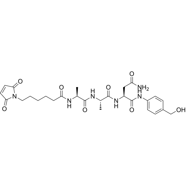
MC-Ala-Ala-Asn-PAB
MC-Ala-Ala-Asn-PAB is a linker extracted from patent CN104147612A, page 14. MC-Ala-Ala-Asn-PAB can be used to synthesis the tumor microenvironment specific activated micromolecular targeted conjugate.
For research use only. We do not sell to patients.
- CAS No.: 1638970-44-1
- Formula: C27H36N6O8
- Molecular Weight:572.61
-
Storage:
Please store the product under the recommended conditions in the Certificate of Analysis.

To place orders, for customer services and technical support, please contact: MedChemExpress USA
Tel: 609-228-6898 E-mail: [email protected] [email protected]


-
 Biological Activity
-
Chemical Information
- Protocols
- Purity & Documentation
- References
-
 Help & FAQs




Biological Activity
Description


Chemical Information
-
CAS No. 1638970-44-1
-
Molecular Weight 572.61
-
Formula C27H36N6O8
-
SMILES
O=C(C=CC1=O)N1CCCCCC(N[C@@H](C)C(N[C@@H](C)C(N[C@@H](CC(N)=O)C(NC2=CC=C(C=C2)CO)=O)=O)=O)=O
-
Shipping
Room temperature in continental US; may vary elsewhere.
-
Storage
Please store the product under the recommended conditions in the Certificate of Analysis.

Protocols
-
Multiplex immunofluorescence IHC
Multiplex immunofluorescence IHC detects multiple protein biomarkers in one tissue section by sequential antibody staining, HRP-mediated tyramide fluorophore deposition, heat-mediated antibody stripping, nuclear counterstaining, multispectral imaging, spectral unmixing, and digital cell phenotyping; TSA deposits fluorophore near the antigen so the fluorescence signal remains after primary and secondary antibodies are removed, enabling repeated staining cycles, including with antibodies from the same host species. Classic FFPE tumor immune-profiling applications use panels such as CD3, CD8, CD68/CD163, FOXP3, PD-1, PD-L1, pancytokeratin, Ki67, and DAPI to identify tumor cells, immune-cell subsets, checkpoint-marker expression, co-expression phenotypes, cell density, and spatial relationships in the tumor microenvironment.
-
Multiplex immunohistochemistry
Multiplex immunohistochemistry (mIHC), also known as tyramide dignal amplification (TSA), is an enzymatic detection method that uses horseradish peroxidase (HRP) to perform high-density in-situ labeling of target proteins or nucleic acids.
-
Polyacrylamide gel electrophoresis (PAGE) (<1 kb)
Gel electrophoresis is a method for separating biological macromolecules (such as nucleic acids or proteins) by forcing them through a gel matrix under an electric field.
-
Protocol for Protein Electrophoresis
Protein electrophoresis separates proteins in a polyacrylamide gel under an electric field; in SDS-PAGE, sodium dodecyl sulfate denatures proteins and gives them a broadly similar negative charge-to-mass ratio, so migration mainly reflects apparent molecular mass through the gel matrix. The readout is a stained protein band pattern: band position estimates apparent molecular mass using protein standards, band intensity reflects relative protein abundance within the linear range of staining/detection, and changes in band pattern can reflect protein expression, degradation, purification, or sample composition.
-
Research Protocol for Cancer Immunology
Cancer immunology studies how the immune system recognizes, suppresses, edits, or fails to eliminate malignant cells through tumor antigen release, antigen presentation, T-cell priming, immune trafficking, tumor-cell killing, and feedback inhibition in the tumor microenvironment. The cancer-immunity cycle links tumor antigenicity, dendritic-cell priming, CD8+ T-cell infiltration, cytotoxic function, and immune-checkpoint regulation to tumor rejection or immune escape. Immune-checkpoint pathways such as PD-1/PD-L1 and CTLA-4 suppress antitumor T-cell activity and can be therapeutically blocked, but many tumors remain resistant because of poor antigen presentation, weak T-cell infiltration, suppressive myeloid cells, regulatory T cells, and tumor-intrinsic immune-exclusion programs. Unresolved questions include which immune-cell states predict response, how tumor-intrinsic pathways exclude immune cells, how myeloid suppression limits checkpoint blockade, and which combination strategies


Purity & Documentation
References

Calculators
Concentration (start) × Volume (start) = Concentration (final) × Volume (final)


MC-Ala-Ala-Asn-PAB Related Classifications
HY-139245 Related Classifications
 Powered by Bioz