



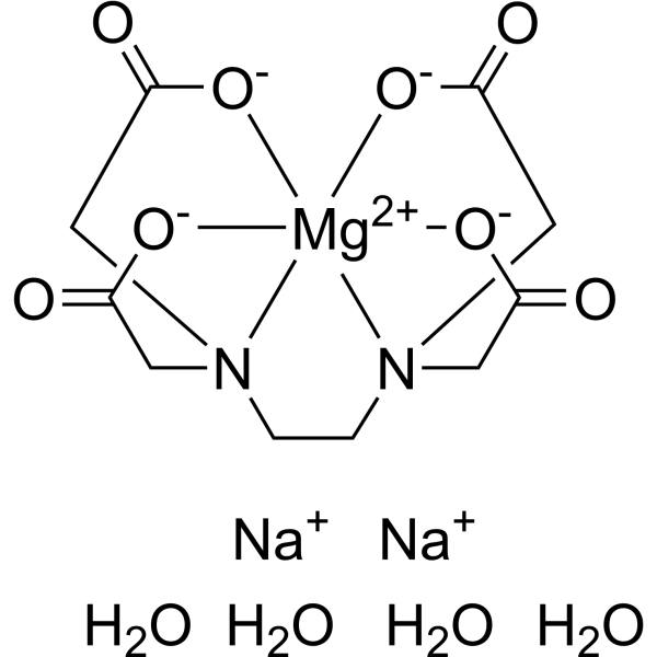
Mg(Ⅱ)-EDTA disodium tetrahydrate

Based on 1 Customer Validation
Mg(Ⅱ)-EDTA (Ethylenediaminetetraacetic acid magnesium) disodium tetrahydrate is a divalent metal chelator and ferroptosis inducer. Mg (Ⅱ)-EDTA disodium tetrahydrate reduces E-cadherin expression by chelating Ca2+ on the surface of tumor cells, thereby triggering cell dissociation and inducing epithelial-mesenchymal transition (EMT). Mg (Ⅱ)-EDTA disodium tetrahydrate further chelates lysosomal iron to form EDTA-Fe, induces ferroptosis through the Fenton reaction, and synergistically enhances immune responses to inhibit tumor metastasis. Mg (Ⅱ)-EDTA (disodium tetrahydrate) can be used in tumor-related research.
For research use only. We do not sell to patients.
- Purity : 99.0%
- CAS No.: 29932-54-5
- Formula: C10H20MgN2Na2O12
- Molecular Weight:430.56
-
Storage:
4°C, sealed storage, away from moisture
* In solvent : -80°C, 6 months; -20°C, 1 month (sealed storage, away from moisture)

To place orders, for customer services and technical support, please contact: MedChemExpress USA
Tel: 609-228-6898 E-mail: [email protected] [email protected]


-
 Biological Activity
-
Chemical Information
-
Solvent & Solubility
- Protocols
- Purity & Documentation
- References
-
 Help & FAQs




Biological Activity
Description
In Vitro
Mg(Ⅱ)-EDTA disodium tetrahydrate (0-400 μg/mL; 6 s-15 min) rapidly and dose-dependently chelates Ca2+ while releasing Mg2+ in solution, and the resulting EDTA-Ca chelates Fe2+ with slower kinetics; EDTA-Fe generates hydroxyl radicals via Fenton reaction in a time- and concentration-dependent manner[1].
Mg(Ⅱ)-EDTA disodium tetrahydrate (500 μg/mL) exhibits selective cytotoxicity toward 4T1 tumor cells, reducing cell survival to 53.3% while sparing HUVECs (79.3% survival)[1].
Mg(Ⅱ)-EDTA disodium tetrahydrate (100 μM Fe2+) induces ferroptosis in 4T1 cells via iron deprivation, Fenton reaction-mediated ROS generation, and LPO, characterized by reduced GPX4 expression, increased ACSL4 expression, mitochondrial damage, and cell death; these effects are enhanced by exogenous Fe2+ and inhibited by DFO[1].
MedChemExpress (MCE) has not independently confirmed the accuracy of these methods. They are for reference only. Further protocols information, click here.
In Vivo
Mg(Ⅱ)-EDTA disodium tetrahydrate (orthotopic injection; on days 1, 3, 5, 7, 9 post-tumor implantation) significantly inhibits tumor growth and metastasis in a patient-derived xenograft model, reducing tumor volume to 41.0% of the control, while maintaining good biocompatibility[1].
MedChemExpress (MCE) has not independently confirmed the accuracy of these methods. They are for reference only.


Chemical Information
-
CAS No. 29932-54-5
-
Appearance Solid
-
Molecular Weight 430.56
-
Formula C10H20MgN2Na2O12
-
Color White to off-white
-
SMILES
O=C1[O-][Mg+2]([N]2(C1)CC3=O)([O-]3)([O-]C4=O)([O-]5)[N](C4)(CC2)CC5=O.O.[Na+].O.O.O.[Na+]
-
Synonyms
Ethylenediaminetetraacetic acid magnesium disodium tetrahydrate
-
Shipping
Room temperature in continental US; may vary elsewhere.
-
Storage
4°C, sealed storage, away from moisture
* In solvent : -80°C, 6 months; -20°C, 1 month (sealed storage, away from moisture)


Solvent & Solubility
In Vitro:
H2O : 100 mg/mL (232.26 mM; Need ultrasonic)
Please refer to the solubility information to select the appropriate solvent. Once prepared, please aliquot and store the solution to prevent product inactivation from repeated freeze-thaw cycles.
Storage method and period of stock solution: -80°C, 6 months; -20°C, 1 month (sealed storage, away from moisture). When stored at -80°C, please use it within 6 months. When stored at -20°C, please use it within 1 month.
* Note: If you choose water as the stock solution, please dilute it to the working solution, then filter and sterilize it with a 0.22 μm filter before use.
Please refer to the solubility information to select the appropriate solvent. Once prepared, please aliquot and store the solution to prevent product inactivation from repeated freeze-thaw cycles.
Storage method and period of stock solution: -80°C, 6 months; -20°C, 1 month (sealed storage, away from moisture). When stored at -80°C, please use it within 6 months. When stored at -20°C, please use it within 1 month.
* Note: If you choose water as the stock solution, please dilute it to the working solution, then filter and sterilize it with a 0.22 μm filter before use.
Concentration (start) × Volume (start) = Concentration (final) × Volume (final)

Protocols
-
RNA extraction experimental
By lysing cells, releasing RNA, and removing impurities such as proteins and DNA, high-purity RNA products are finally obtained. The commonly used traditional method is the guanidine isothiocyanate/phenol/chloroform method (Trizol), which is suitable for a variety of animal materials including animal tissues, microorganisms, cultured cells, etc., and most plant materials.
-
Ferroptosis Solutions
Ferroptosis is an iron-dependent, non-apoptotic form of regulated cell death characterized by lethal lipid peroxidation and sensitivity to suppression by iron chelators or lipophilic radical-trapping antioxidants. The core pathway links cystine uptake through system Xc−, glutathione availability, GPX4-dependent detoxification of phospholipid hydroperoxides, iron-dependent oxidative reactions, and polyunsaturated-phospholipid metabolism into a cell-death program that is biochemically and morphologically distinct from apoptosis, necrosis, and autophagy. The ferroptosis pathway is experimentally linked to phenotype through chemical and genetic perturbation. Erastin induces ferroptosis by inhibiting cystine uptake through system Xc− and weakening antioxidant defenses, while GPX4 inhibition or depletion causes lipid peroxide accumulation and ferroptotic cancer-cell death. ACSL4 and oxidizable arachidonoyl- or adrenoyl-containing phosphatidylethanolamines shape ferroptosis sensitivity by con
-
Ca2+ Staining Technique
Ca2+ staining is an experimental technique that utilizes specific fluorescent probes (such as Fluo-4 AM, Fura-2, etc.) to qualitatively or quantitatively detect dynamic changes in intracellular Ca2+ concentrations; this is achieved by monitoring the changes in fluorescent signals generated when these probes bind to free intracellular calcium ions. The underlying principle relies primarily on the presence of chelating groups within the probe's molecular structure that possess high affinity for calcium ions.
-
Lysosome and acidic-vesicle live-cell staining
Lysosome and acidic-vesicle live-cell staining detects acidic intracellular compartments by using membrane-permeant acidotropic probes that accumulate in low-pH vesicles, including lysosomes, late endosomes, autolysosomes, and acidic phagosomes. LysoTracker staining is commonly used as an intensity-based readout of acidic lysosomal compartment abundance or enlargement, while acridine orange produces green fluorescence in less concentrated compartments and red fluorescence after concentration-dependent accumulation in acidic vesicular organelles. Loss or reduction of acridine-orange red signal can be used as a readout of lysosomal membrane permeabilization or reduced acidic-vesicle integrity. This protocol is designed for live cultured cells and can be adapted for fluorescence microscopy, high-content imaging, plate-reader readout, or flow cytometry when the selected literature supports the readout. Because these dyes report acidotropic accumulation rather than lysosome identity alone,
-
Orthotopic Cell-Line Xenograft
Orthotopic cell-line xenograft models involve implantation of human cancer cell lines into the anatomically corresponding organ of immunodeficient mice to reproduce tumor growth within a native microenvironment, enabling more clinically relevant tumor behavior compared with subcutaneous models. These models are widely used because orthotopic placement better recapitulates tumor progression, including invasion and metastatic spread, which are often underrepresented in heterotopic implantation systems. Compared with conventional xenografts, orthotopic implantation is described as more technically complex but provides improved simulation of tumor-microenvironment interactions and metastatic behavior, making it particularly valuable for translational oncology research. Surgical orthotopic implantation approaches have been emphasized as enabling faithful reproduction of clinical cancer features, including metastasis and disease progression patterns that align with the tumor’s organ of origi


Purity & Documentation
-
Data Sheet (287 KB)
-
SDS (456 KB)
- English - EN (456 KB)
- Français - FR (456 KB)
- Deutsch - DE (456 KB)
- Norwegian - NO (456 KB)
- Español - ES (456 KB)
- Swedish - SV (456 KB)
- Italian - IT (456 KB)
- Korean - KR (456 KB)
- Portuguese - PT (456 KB)
-
Handling Instructions (2659 KB)
References


Complete Stock Solution Preparation Table
Please refer to the solubility information to select the appropriate solvent. Once prepared, please aliquot and store the solution to prevent product inactivation from repeated freeze-thaw cycles.
Storage method and period of stock solution: -80°C, 6 months; -20°C, 1 month (sealed storage, away from moisture). When stored at -80°C, please use it within 6 months. When stored at -20°C, please use it within 1 month.
| Optional Solvent | Concentration Solvent Mass | 1 mg | 5 mg | 10 mg | 25 mg |
|---|---|---|---|---|---|
| H2O | 1 mM | 2.3226 mL | 11.6128 mL | 23.2256 mL | 58.0639 mL |
| 5 mM | 0.4645 mL | 2.3226 mL | 4.6451 mL | 11.6128 mL | |
| 10 mM | 0.2323 mL | 1.1613 mL | 2.3226 mL | 5.8064 mL | |
| 15 mM | 0.1548 mL | 0.7742 mL | 1.5484 mL | 3.8709 mL | |
| 20 mM | 0.1161 mL | 0.5806 mL | 1.1613 mL | 2.9032 mL | |
| 25 mM | 0.0929 mL | 0.4645 mL | 0.9290 mL | 2.3226 mL | |
| 30 mM | 0.0774 mL | 0.3871 mL | 0.7742 mL | 1.9355 mL | |
| 40 mM | 0.0581 mL | 0.2903 mL | 0.5806 mL | 1.4516 mL | |
| 50 mM | 0.0465 mL | 0.2323 mL | 0.4645 mL | 1.1613 mL | |
| 60 mM | 0.0387 mL | 0.1935 mL | 0.3871 mL | 0.9677 mL | |
| 80 mM | 0.0290 mL | 0.1452 mL | 0.2903 mL | 0.7258 mL | |
| 100 mM | 0.0232 mL | 0.1161 mL | 0.2323 mL | 0.5806 mL |
* Note: If you choose water as the stock solution, please dilute it to the working solution, then filter and sterilize it with a 0.22 μm filter before use.


Mg(Ⅱ)-EDTA disodium tetrahydrate Related Classifications
HY-W134005 Related Classifications
 Powered by Bioz
Keywords