



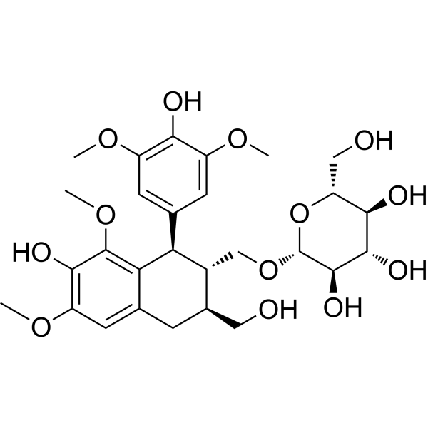
(-)-Lyoniresinol 9'-O-glucoside

Based on 1 Customer Validation
(-)-Lyoniresinol 9'-O-glucoside is an inhibitor of ROS. (-)-Lyoniresinol 9'-O-glucoside reduces lipid accumulation and lipid metabolic disorders in FFAs-exposed HepG2 cells. (-)-Lyoniresinol 9'-O-glucoside inhibits high glucose-induced reactive oxygen species production.
For research use only. We do not sell to patients.
- Purity : 99.54%
- CAS No.: 143236-02-6
- Formula: C28H38O13
- Molecular Weight:582.59
-
Storage:
4°C, sealed storage, away from moisture
* In solvent : -80°C, 6 months; -20°C, 1 month (sealed storage, away from moisture)

To place orders, for customer services and technical support, please contact: MedChemExpress USA
Tel: 609-228-6898 E-mail: [email protected] [email protected]


-
 Biological Activity
-
Chemical Information
- Protocols
- Purity & Documentation
- References
-
 Help & FAQs




Biological Activity
Description
Cellular Effect
|
Cell Line

|
Type | Value | Description | References |
|---|---|---|---|---|
| BMDC | IC50 |
>50 μM
Compound: 15
|
Antiinflammatory activity in C57BL/6 mouse BMDCs assessed as inhibition of LPS-stimulated IL12 production treated 1 hr before LPS challenge measured 18 hrs post stimulation by ELISA
Antiinflammatory activity in C57BL/6 mouse BMDCs assessed as inhibition of LPS-stimulated IL12 production treated 1 hr before LPS challenge measured 18 hrs post stimulation by ELISA
|
[PMID: 23484668] |
| BMDC | IC50 |
>50 μM
Compound: 15
|
Antiinflammatory activity in C57BL/6 mouse BMDCs assessed as inhibition of LPS-stimulated TNF-alpha production treated 1 hr before LPS challenge measured 18 hrs post stimulation by ELISA
Antiinflammatory activity in C57BL/6 mouse BMDCs assessed as inhibition of LPS-stimulated TNF-alpha production treated 1 hr before LPS challenge measured 18 hrs post stimulation by ELISA
|
[PMID: 23484668] |
| BMDC | IC50 |
14.55 μM
Compound: 15
|
Antiinflammatory activity in C57BL/6 mouse BMDCs assessed as inhibition of LPS-stimulated IL-12p40 production treated 1 hr before LPS challenge measured 18 hrs post stimulation by ELISA
Antiinflammatory activity in C57BL/6 mouse BMDCs assessed as inhibition of LPS-stimulated IL-12p40 production treated 1 hr before LPS challenge measured 18 hrs post stimulation by ELISA
|
[PMID: 23484668] |


Chemical Information
-
CAS No. 143236-02-6
-
Appearance Solid
-
Molecular Weight 582.59
-
Formula C28H38O13
-
Color White to off-white
-
SMILES
COC1=C(C(C2)=CC(OC)=C1O)[C@@H](C(C=C3OC)=CC(OC)=C3O)[C@@H]([C@H]2CO)CO[C@@H]([C@@H]([C@H]4O)O)O[C@@H]([C@H]4O)CO
-
Structure Classification
-
Initial Source
-
Shipping
Room temperature in continental US; may vary elsewhere.
-
Storage
4°C, sealed storage, away from moisture
* In solvent : -80°C, 6 months; -20°C, 1 month (sealed storage, away from moisture)

Protocols
-
3T3-L1 preadipocyte-to-adipocyte differentiation
3T3-L1 preadipocytes are induced to differentiate after growth arrest using adipogenic media containing insulin, dexamethasone, and IBMX; differentiation is assessed by lipid-droplet accumulation, triglyceride increase, Oil Red O staining, and adipocyte-marker induction such as PPARγ and C/EBPα.
-
ROS/oxidative-stress fluorescent staining
ROS/oxidative-stress fluorescent staining uses cell-permeant fluorogenic probes that become fluorescent after oxidation inside cells or tissues; commonly used examples include DCFH-DA/DCFDA for broad cellular oxidant detection, DHE for superoxide-related signal detection, MitoSOX for mitochondrial superoxide-related signal detection, and CellROX probes for oxidative-stress-associated fluorescence readouts. The assay detects probe oxidation rather than a single ROS species unless the probe and analysis method have been chemically validated for that species. DCFH-DA enters cells, is deacetylated by intracellular esterases to DCFH, and produces fluorescent DCF after oxidation, so the readout is used as an operational measure of total cellular oxidative stress rather than a species-specific ROS measurement. DHE and MitoSOX can report superoxide-related oxidation, but red fluorescence alone can include non-specific ethidium-like oxidation products; HPLC or optimized spectral approaches are
-
Lipid Droplets: Oil Red O/Sudan Dye Lipid Staining
Lipid droplets are intracellular organelles with a neutral-lipid core that stores triacylglycerols and sterol esters, and Oil Red O or Sudan dyes detect these hydrophobic lipid deposits by partitioning into retained lipids in fresh or frozen specimens. Oil Red O stains neutral triglycerides and lipids in frozen tissue sections or air-dried cytologic preparations, while Sudan Black B has also been used as a histochemical fat stain for lipid-rich tissue structures.


Purity & Documentation
-
Data Sheet (273 KB)
-
SDS (252 KB)
- English - EN (252 KB)
- Français - FR (252 KB)
- Deutsch - DE (252 KB)
- Norwegian - NO (252 KB)
- Español - ES (252 KB)
- Swedish - SV (252 KB)
- Italian - IT (252 KB)
- Korean - KR (252 KB)
- Portuguese - PT (252 KB)
-
Handling Instructions (2659 KB)
References

Calculators
Concentration (start) × Volume (start) = Concentration (final) × Volume (final)


(-)-Lyoniresinol 9'-O-glucoside Related Classifications
HY-N8466 Related Classifications
 Powered by Bioz