



MitoBloCK-6
Based on 1 publication(s) in Google Scholar
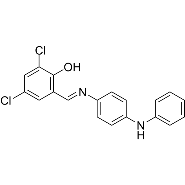
MitoBloCK-6 is a potent Erv1/ALR inhibitor, with an IC50 of 900 nM and 700 nM, respectively. MitoBloCK-6 also inhibits Erv2 (IC50=1.4 μM). MitoBloCK-6 can induce Apoptosis via cytochrome c release. MitoBloCK-6 inhibits growth of developing zebrafish motor neurons. MitoBloCK-6 has anticancer activity against liver cancer and leukemia.
For research use only. We do not sell to patients.
- Purity : 98.0%
- CAS No.: 303215-67-0
- Formula: C19H14Cl2N2O
- Molecular Weight:357.23
-
Storage:
4°C, protect from light
* In solvent : -80°C, 6 months; -20°C, 1 month (protect from light)

To place orders, for customer services and technical support, please contact: MedChemExpress USA
Tel: 609-228-6898 E-mail: [email protected] [email protected]


-
 Biological Activity
-
Chemical Information
-
Solvent & Solubility
- Protocols
- Purity & Documentation
- References
-
 Help & FAQs


-
HY-115467
MitoBloCK-10PAM complex Inhibitor -

HY-13259
MG-132Proteasome Inhibitor -
HY-N4000
DigitoninDetergent -
HY-Y0873A
PEG400 -

HY-Y0808
Dimethyl succinateSuccinate Analogue -

HY-10583
Y-27632 dihydrochlorideROCK Inhibitor -
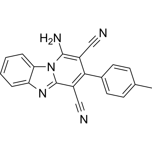
HY-136057
iFSP1FSP1 Inhibitor -
HY-K0206
Anti-c-Myc Magnetic Beads -
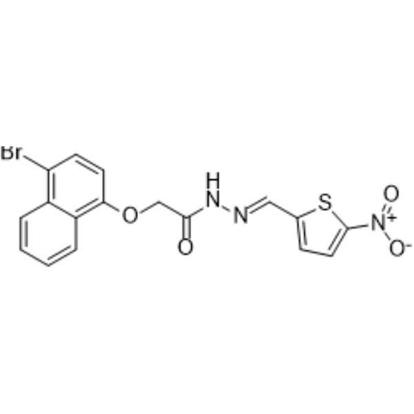
HY-125036
MitoBloCK-11Mitochondrial Protein Transport Inhibitors
Publications Citing Use of MedChemExpress (MCE) MitoBloCK-6
More

Biological Activity
Description
IC50 & Target
IC50: 900 nM (Erv1) 700 nM (ALR) 1.4 μM (Erv2)[1]
In Vitro
MitoBloCK-6 (20 μM, 8 h) induces apoptosis via cytochrome c release in human embryonic stem cells (hESCs)[1]. MitoBloCK-6 (20-40 μM, 72 h) inhibits proliferation of liver cancer cells McA-RH7777 via mitochondrial impairment[2]. MitoBloCK-6 kills leukemia cell lines (OCI-AML2, TEX, Jurkat and NB4) with an IC50 of 5-10 µM[3]. MitoBloCK-6 (0.078-5 μM) increases the differentiation of OCI-AML2 and NB4 cells via inhibition of ALR, as increased surface expression of the myeloid markers CD11b[4]. MitoBloCK-6 results in a significant decrease in the growth and branching of developing zebrafish motor neurons via inhibition of ALR[5].
MedChemExpress (MCE) has not independently confirmed the accuracy of these methods. They are for reference only. Further protocols information, click here.
-
Cell Line:human embryonic stem cells
-
Concentration:20 µM
-
Incubation Time:8 h
-
Result:Resulted in a shift in cytochrome c localization from mitochondria into the cytosol.
Showed similar number of cells releasing cytochrome c.
Showed downstream events in apoptosis (PARP and caspase-3 cleavage).
In Vivo
MitoBloCK-6 (80 mg/kg i.p. 5 of 7 days, 2 weeks) strongly reduces the engraftment of primary AML cells in mice[6].
MedChemExpress (MCE) has not independently confirmed the accuracy of these methods. They are for reference only.


Chemical Information
-
CAS No. 303215-67-0
-
Appearance Solid
-
Molecular Weight 357.23
-
Formula C19H14Cl2N2O
-
Color Yellow to orange
-
SMILES
OC1=C(/C=N/C2=CC=C(NC3=CC=CC=C3)C=C2)C=C(Cl)C=C1Cl
-
Shipping
Room temperature in continental US; may vary elsewhere.
-
Storage
4°C, protect from light
* In solvent : -80°C, 6 months; -20°C, 1 month (protect from light)


Publications (1)
-
Journal Impact Factor
-
Most Recent
-
bioRxiv
Mitochondrial degradation of metallothionein enables local zinc mobilization during zinc limitation. [Abstract]2026 Jun 10:2026.06.09.731205. PMID: 42327099


Solvent & Solubility
In Vitro:
DMSO : 100 mg/mL (279.93 mM; Need ultrasonic; Hygroscopic DMSO has a significant impact on the solubility of product, please use newly opened DMSO)
Please refer to the solubility information to select the appropriate solvent. Once prepared, please aliquot and store the solution to prevent product inactivation from repeated freeze-thaw cycles.
Storage method and period of stock solution: -80°C, 6 months; -20°C, 1 month (protect from light). When stored at -80°C, please use it within 6 months. When stored at -20°C, please use it within 1 month.
Please refer to the solubility information to select the appropriate solvent. Once prepared, please aliquot and store the solution to prevent product inactivation from repeated freeze-thaw cycles.
Storage method and period of stock solution: -80°C, 6 months; -20°C, 1 month (protect from light). When stored at -80°C, please use it within 6 months. When stored at -20°C, please use it within 1 month.
Concentration (start) × Volume (start) = Concentration (final) × Volume (final)

Protocols
-
Apoptosis
Apoptosis, also called programmed cell death, is generally characterized by distinct morphological characteristics.
-
TUNEL staining for apoptotic DNA fragmentation
TUNEL staining detects DNA strand breaks by using terminal deoxynucleotidyl transferase to add labeled nucleotides to exposed 3′-OH DNA termini, generating either microscopic staining in fixed cells or tissue sections, or fluorescence/cytometric signal in cell suspensions. TUNEL positivity reflects DNA fragmentation but should not be interpreted alone as definitive apoptosis, because TUNEL can also label necrotic, autolytic, mechanically damaged, or DNA-repair-associated DNA breaks.
-
Cell Cytotoxicity Assay
Cytotoxicity assays are usually based on the assessment of cell membrane damage, which can also be indirectly detected by measuring cell viability. Detection methods include MTT assay, CKK-8 assay, LDH assay and ATP assay, etc.
-
Annexin V plus membrane-impermeant dye apoptosis staining
Annexin V-based apoptosis assays rely on the detection of phosphatidylserine (PS) externalization from the inner leaflet of the plasma membrane to the outer leaflet, an early biochemical hallmark of apoptosis. Fluorescently labeled Annexin V binds PS in a calcium-dependent manner, enabling identification of early apoptotic cells by flow cytometry or fluorescence microscopy. When combined with a membrane-impermeant DNA-binding dye (e. g. , propidium iodide), this approach allows discrimination between viable (Annexin V−/dye−), early apoptotic (Annexin V+/dye−), and late apoptotic or necrotic (Annexin V+/dye+) cell populations by assessing membrane integrity and PS exposure.
-
Apoptosis Solutions
Apoptosis is a regulated, generally non-lytic cell-death pathway that removes unwanted, damaged, infected, or abnormal cells through coordinated morphological changes, caspase activation, DNA fragmentation, and membrane remodeling. The intrinsic apoptosis pathway is controlled mainly by mitochondrial outer membrane permeabilization, BCL-2 family proteins, cytochrome c release, apoptosome formation, caspase-9 activation, and downstream executioner caspase-3/7 activation. The extrinsic apoptosis pathway is initiated by death receptors such as Fas, TNFR, and TRAIL receptors, which recruit adaptor proteins and activate caspase-8 before engaging executioner caspases or mitochondrial amplification through BID cleavage. Apoptosis is linked to many phenotypes, including cancer cell killing, tissue homeostasis, immune regulation, neurodegeneration, infection response, and treatment-induced cytotoxicity; unresolved questions include how apoptosis interacts with necroptosis, pyroptosis, ferroptos
-
Liver Cancer Modeling
Liver cancer can be classified into primary liver cancer and secondary liver cancer. Secondary liver cancer is the metastatic liver cancer. Primary liver cancer includes hepatocellular carcinoma (HCC), intrahepatic cholangiocarcinoma (ICC) and fibrolamellar HCC, of which HCC is the most common form, accounting for approximately 90% of primary liver cancers[1]. HCC mouse models include chemical agent-induced models, transplanted tumor models, and genetic engineered models.


Purity & Documentation
-
Data Sheet (274 KB)
-
SDS (252 KB)
- English - EN (252 KB)
- Français - FR (252 KB)
- Deutsch - DE (252 KB)
- Norwegian - NO (252 KB)
- Español - ES (252 KB)
- Swedish - SV (252 KB)
- Italian - IT (252 KB)
- Korean - KR (252 KB)
- Portuguese - PT (252 KB)
-
Handling Instructions (2659 KB)
References
[1]. Dabir DV, et, al. A small molecule inhibitor of redox-regulated protein translocation into mitochondria. Dev Cell. 2013 Apr 15;25(1):81-92. [Content Brief]
[2]. Kabiri Y, et al. Mitochondrial Impairment by MitoBloCK-6 Inhibits Liver Cancer Cell Proliferation. Front Cell Dev Biol. 2021 Sep 20;9:725474. [Content Brief]
[4]. Singh RP, et al. Disrupting Mitochondrial Copper Distribution Inhibits Leukemic Stem Cell Self-Renewal. Cell Stem Cell. 2020 Jun 4;26(6):926-937.e10. [Content Brief]


Complete Stock Solution Preparation Table
Please refer to the solubility information to select the appropriate solvent. Once prepared, please aliquot and store the solution to prevent product inactivation from repeated freeze-thaw cycles.
Storage method and period of stock solution: -80°C, 6 months; -20°C, 1 month (protect from light). When stored at -80°C, please use it within 6 months. When stored at -20°C, please use it within 1 month.
| Optional Solvent | Concentration Solvent Mass | 1 mg | 5 mg | 10 mg | 25 mg |
|---|---|---|---|---|---|
| DMSO | 1 mM | 2.7993 mL | 13.9966 mL | 27.9932 mL | 69.9829 mL |
| 5 mM | 0.5599 mL | 2.7993 mL | 5.5986 mL | 13.9966 mL | |
| 10 mM | 0.2799 mL | 1.3997 mL | 2.7993 mL | 6.9983 mL | |
| 15 mM | 0.1866 mL | 0.9331 mL | 1.8662 mL | 4.6655 mL | |
| 20 mM | 0.1400 mL | 0.6998 mL | 1.3997 mL | 3.4991 mL | |
| 25 mM | 0.1120 mL | 0.5599 mL | 1.1197 mL | 2.7993 mL | |
| 30 mM | 0.0933 mL | 0.4666 mL | 0.9331 mL | 2.3328 mL | |
| 40 mM | 0.0700 mL | 0.3499 mL | 0.6998 mL | 1.7496 mL | |
| 50 mM | 0.0560 mL | 0.2799 mL | 0.5599 mL | 1.3997 mL | |
| 60 mM | 0.0467 mL | 0.2333 mL | 0.4666 mL | 1.1664 mL | |
| 80 mM | 0.0350 mL | 0.1750 mL | 0.3499 mL | 0.8748 mL | |
| 100 mM | 0.0280 mL | 0.1400 mL | 0.2799 mL | 0.6998 mL |


MitoBloCK-6 Related Classifications
HY-122652 Related Classifications
 Powered by Bioz