



Myoregulin TFA
Myoregulin (MLN peptide) TFA is a member of the regulin family. Myoregulin TFA regulates muscle performance by modulating intracellular calcium handling. Myoregulin TFA interactes directly with sarcoplasmic reticulum Ca2+-ATPase (SERCA) and impedinf Ca2+ uptake into the sarcoplasmic reticulum.
연구목적의 판매만을 진행합니다. 환자를 대상으로 한 판매는 하지 않습니다.
- 화학식: C239H391N53O67S3.xC2HF3O2
- 분자량:5175.17 (free base)
-
보관:
Please store the product under the recommended conditions in the Certificate of Analysis.

To place orders, for customer services and technical support, please contact: MedChemExpress USA
Tel: 609-228-6898 E-mail: [email protected] [email protected]


-
 Biological Activity
-
Chemical Information
- Protocol
- 순도&문서
- References
-
 Help & FAQs


All Calcium Channel Isoforms
More

Biological Activity
제품 설명


Chemical Information
-
분자량 5175.17 (free base)
-
화학식 C239H391N53O67S3.xC2HF3O2
-
Synonyms
MLN peptide TFA
-
Sequence
Met-Ser-Gly-Lys-Ser-Trp-Val-Leu-Ile-Ser-Thr-Thr-Ser-Pro-Gln-Ser-Leu-Glu-Asp-Glu-Ile-Leu-Gly-Arg-Leu-Leu-Lys-Ile-Leu-Phe-Val-Leu-Phe-Val-Asp-Leu-Met-Ser-Ile-Met-Tyr-Val-Val-Ile-Thr-Ser
-
Sequence Shortening
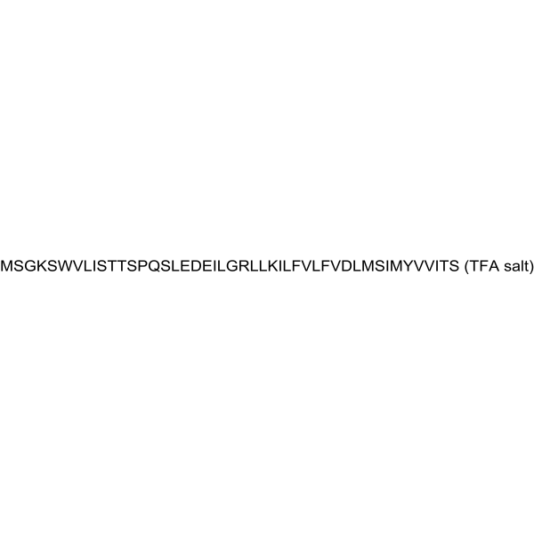
MSGKSWVLISTTSPQSLEDEILGRLLKILFVLFVDLMSIMYVVITS
-
선적
Room temperature in continental US; may vary elsewhere.
-
보관
Please store the product under the recommended conditions in the Certificate of Analysis.


순도&문서
References

Calculators
Concentration (start) × Volume (start) = Concentration (final) × Volume (final)


Myoregulin TFA Related Classifications
HY-P6008A Related Classifications
 Powered by Bioz