



N-Acetyl semax amidate

Based on 1 Customer Validation
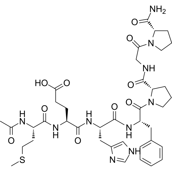
N-Acetyl semax amidate is a derivative of Semax (HY-P1146) (Met-Glu-His-Phe-Pro-Gly-Pro) obtained via N-terminal acetylation and C-terminal amidation, and it exhibits nootropic and neuroprotective activities. N-Acetyl semax amidate exerts neuroprotective effects by upregulating the expression of BDNF and NGF, inhibiting neuronal apoptosis, alleviating oxidative stress, and reducing glutamate excitotoxicity. N-Acetyl semax amidate can be used in research related to central nervous system diseases such as ischemic stroke, cognitive impairment, and neurodegenerative diseases.
For research use only. We do not sell to patients.
- Purity : 99.60%
- CAS No.: 2920938-90-3
- Formula: C39H54N10O10S
- Molecular Weight:854.97
-
Storage:
Sealed storage, away from moisture.
Powder -80°C, 2 years , -20°C, 1 year* In solvent : -80°C, 6 months; -20°C, 1 month (sealed storage, away from moisture)

To place orders, for customer services and technical support, please contact: MedChemExpress USA
Tel: 609-228-6898 E-mail: [email protected] [email protected]


-
 Biological Activity
-
Chemical Information
-
Solvent & Solubility
- Protocols
- Purity & Documentation
- References
-
 Help & FAQs




Biological Activity
Description
In Vitro
N-Acetyl semax amidate serves as the N-terminally acetylated and C-terminally amidated derivative of Met-Glu-His-Phe-Pro-Gly-Pro for the preparation of aqueous nasal formulations[1].
MedChemExpress (MCE) has not independently confirmed the accuracy of these methods. They are for reference only.
- PC12 NGF-Induced Neuronal Differentiation Culture
- RNA extraction experimental
- Detection of Apoptosis
- TUNEL staining for apoptotic DNA fragmentation
- Annexin V plus membrane-impermeant dye apoptosis staining
- ROS/oxidative-stress fluorescent staining
- Apoptosis Solutions
- PC12 NGF-induced neuronal-like differentiation
- Alzheimer’s Disease Modeling


Chemical Information
-
CAS No. 2920938-90-3
-
Appearance Solid
-
Molecular Weight 854.97
-
Formula C39H54N10O10S
-
Color White to off-white
-
Sequence Shortening
{Ac}-MEHFPGP-NH2
-
Shipping
Room temperature in continental US; may vary elsewhere.
-
Storage
Sealed storage, away from moisture
Powder -80°C 2 years -20°C 1 year * In solvent : -80°C, 6 months; -20°C, 1 month (sealed storage, away from moisture)


Solvent & Solubility
In Vitro:
DMSO : 100 mg/mL (116.96 mM; Need ultrasonic; Hygroscopic DMSO has a significant impact on the solubility of product, please use newly opened DMSO)
Please refer to the solubility information to select the appropriate solvent. Once prepared, please aliquot and store the solution to prevent product inactivation from repeated freeze-thaw cycles.
Storage method and period of stock solution: -80°C, 6 months; -20°C, 1 month (sealed storage, away from moisture). When stored at -80°C, please use it within 6 months. When stored at -20°C, please use it within 1 month.
Please refer to the solubility information to select the appropriate solvent. Once prepared, please aliquot and store the solution to prevent product inactivation from repeated freeze-thaw cycles.
Storage method and period of stock solution: -80°C, 6 months; -20°C, 1 month (sealed storage, away from moisture). When stored at -80°C, please use it within 6 months. When stored at -20°C, please use it within 1 month.
Concentration (start) × Volume (start) = Concentration (final) × Volume (final)


Purity & Documentation
-
Data Sheet (279 KB)
-
SDS (254 KB)
- English - EN (254 KB)
- Français - FR (254 KB)
- Deutsch - DE (254 KB)
- Norwegian - NO (254 KB)
- Español - ES (254 KB)
- Swedish - SV (254 KB)
- Italian - IT (254 KB)
- Korean - KR (254 KB)
- Portuguese - PT (254 KB)
-
Handling Instructions (2659 KB)
References


Complete Stock Solution Preparation Table
Please refer to the solubility information to select the appropriate solvent. Once prepared, please aliquot and store the solution to prevent product inactivation from repeated freeze-thaw cycles.
Storage method and period of stock solution: -80°C, 6 months; -20°C, 1 month (sealed storage, away from moisture). When stored at -80°C, please use it within 6 months. When stored at -20°C, please use it within 1 month.
| Optional Solvent | Concentration Solvent Mass | 1 mg | 5 mg | 10 mg | 25 mg |
|---|---|---|---|---|---|
| DMSO | 1 mM | 1.1696 mL | 5.8482 mL | 11.6963 mL | 29.2408 mL |
| 5 mM | 0.2339 mL | 1.1696 mL | 2.3393 mL | 5.8482 mL | |
| 10 mM | 0.1170 mL | 0.5848 mL | 1.1696 mL | 2.9241 mL | |
| 15 mM | 0.0780 mL | 0.3899 mL | 0.7798 mL | 1.9494 mL | |
| 20 mM | 0.0585 mL | 0.2924 mL | 0.5848 mL | 1.4620 mL | |
| 25 mM | 0.0468 mL | 0.2339 mL | 0.4679 mL | 1.1696 mL | |
| 30 mM | 0.0390 mL | 0.1949 mL | 0.3899 mL | 0.9747 mL | |
| 40 mM | 0.0292 mL | 0.1462 mL | 0.2924 mL | 0.7310 mL | |
| 50 mM | 0.0234 mL | 0.1170 mL | 0.2339 mL | 0.5848 mL | |
| 60 mM | 0.0195 mL | 0.0975 mL | 0.1949 mL | 0.4873 mL | |
| 80 mM | 0.0146 mL | 0.0731 mL | 0.1462 mL | 0.3655 mL | |
| 100 mM | 0.0117 mL | 0.0585 mL | 0.1170 mL | 0.2924 mL |


N-Acetyl semax amidate Related Classifications
HY-P3588 Related Classifications
 Powered by Bioz
Keywords
- N-Acetyl semax amidate
- 2920938-90-3
- Biochemical Assay Reagents
- transient ischemic attack
- glaucoma
- vascular malformations of the brain
- craniocerebral injury
- attention deficit hyperactivity disorder
- optic nerve disorders
- dyscirculatory encephalopathy
- narcosis
- neurotic disorders
- multiple sclerosis
- Inhibitor
- inhibitor
- inhibit