



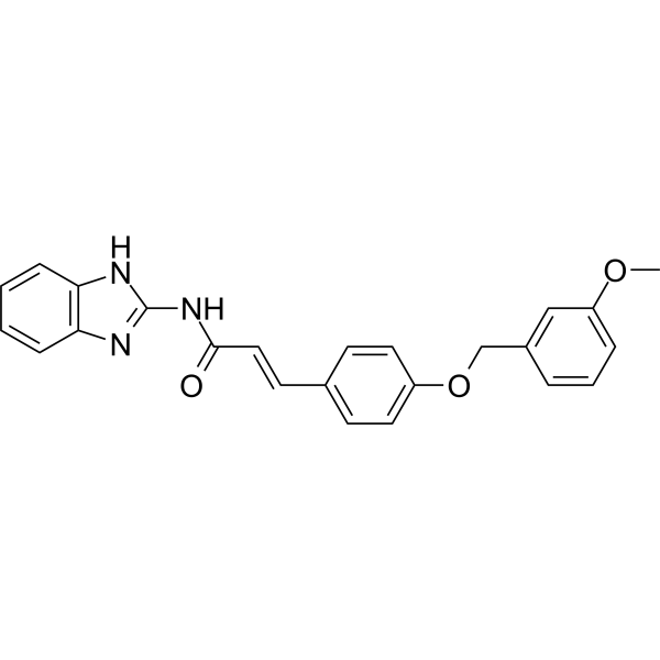
NF-κB-IN-8
NF-κB-IN-8 competitively antagonizes LPS binding to MD-2. NF-κB-IN-8 reduces the expression of inflammatory factors by binding to MD-2. NF-κB-IN-8 also inhibits ALP activity. NF-κB-IN-8 can be used for the research of inflammation such as acute lung injury (ALI).
For research use only. We do not sell to patients.
- CAS No.: 2924565-59-1
- Formula: C24H21N3O3
- Molecular Weight:399.44
-
Storage:
Please store the product under the recommended conditions in the Certificate of Analysis.

To place orders, for customer services and technical support, please contact: MedChemExpress USA
Tel: 609-228-6898 E-mail: [email protected] [email protected]


-
 Biological Activity
-
Chemical Information
- Protocols
- Purity & Documentation
- References
-
 Help & FAQs




Biological Activity
Description
Cellular Effect
|
Cell Line

|
Type | Value | Description | References |
|---|---|---|---|---|
| RAW264.7 | IC50 |
2.58 μM
Compound: L26
|
Antiinflammatory activity in LPS-induced mouse RAW264.7 cells assessed as decrease in LPS-induced IL-6 secretion pretreated for 30 mins followed by LPS-induction and measured after 24 hrs by ELISA analysis
Antiinflammatory activity in LPS-induced mouse RAW264.7 cells assessed as decrease in LPS-induced IL-6 secretion pretreated for 30 mins followed by LPS-induction and measured after 24 hrs by ELISA analysis
|
[PMID: 36963290] |
| RAW264.7 | IC50 |
8.41 μM
Compound: L26
|
Antiinflammatory activity in LPS-induced mouse RAW264.7 cells assessed as decrease in LPS-induced TNF-alpha secretion pretreated for 30 mins followed by LPS-induction and measured after 24 hrs by ELISA analysis
Antiinflammatory activity in LPS-induced mouse RAW264.7 cells assessed as decrease in LPS-induced TNF-alpha secretion pretreated for 30 mins followed by LPS-induction and measured after 24 hrs by ELISA analysis
|
[PMID: 36963290] |
In Vitro
NF-κB-IN-8 (Compound L26) (10 μM) inhibits ALP activity by 64.30%[1].
NF-κB-IN-8 (1-10 μM, 24 h) inhibits expression of IL-6 and TNF-α in RAW 264.7 cells[1].
NF-κB-IN-8 (0-50 μM) inhibits the binding of LPS to MD-2, determined by ELISA Asssay[1].
NF-κB-IN-8 (50 μM, overnight) inhibits the formation of LPS/MD-2/TLR4 polymers in RAW264.7 cells[1].
MedChemExpress (MCE) has not independently confirmed the accuracy of these methods. They are for reference only. Further protocols information, click here.
-
Cell Line:RAW264.7 cells
-
Concentration:50 μM
-
Incubation Time:Overnight
-
Result:Inhibited the expression of LPS/MD-2/TLR4 polymers.
In Vivo
NF-κB-IN-8 (1000 and 1500 mg/kg, intra-gastrically administration) is low in toxic and safe in mice[1].
NF-κB-IN-8 (10 mg/kg, rats) shows T1/2: 4.2 h, Cmax: 163.288 μg/L[1].
MedChemExpress (MCE) has not independently confirmed the accuracy of these methods. They are for reference only.
-
Animal Model:ALI mice induced by LPS[1]
-
Dosage:5 mg/kg
-
Administration:Intra-gastrically administration
-
Result:Downregulated the expression of pro-inflammatory factor Mrna (TNF-α).
Decreased ALP level in bronchoalveolar lavage fluid.
Inhibited the phosphorylation of p65 and increased the release of IκB-α. (WB assay, lung tissue sample)


Chemical Information
-
CAS No. 2924565-59-1
-
Molecular Weight 399.44
-
Formula C24H21N3O3
-
SMILES
COC1=CC=CC(COC2=CC=C(C=C2)/C=C/C(NC3=NC4=C(N3)C=CC=C4)=O)=C1
-
Shipping
Room temperature in continental US; may vary elsewhere.
-
Storage
Please store the product under the recommended conditions in the Certificate of Analysis.

Protocols
-
RNA extraction experimental
By lysing cells, releasing RNA, and removing impurities such as proteins and DNA, high-purity RNA products are finally obtained. The commonly used traditional method is the guanidine isothiocyanate/phenol/chloroform method (Trizol), which is suitable for a variety of animal materials including animal tissues, microorganisms, cultured cells, etc., and most plant materials.
-
Mesenchymal stromal/stem cell osteogenic differentiation
Mesenchymal stromal/stem cells can be induced toward an osteoblast-like lineage in vitro by culture in osteogenic medium containing dexamethasone, ascorbic acid or ascorbate-2-phosphate, and β-glycerophosphate; the differentiation process is commonly evaluated by alkaline phosphatase activity, osteogenic marker expression, collagenous matrix formation, and calcium-rich matrix mineralization. The main readouts are alkaline phosphatase activity as an early osteogenic marker and Alizarin Red S staining as a calcium-deposit readout for mineralized extracellular matrix; Alizarin Red S can be inspected microscopically or extracted and measured colorimetrically at 405 nm.
-
Research Protocol for Inflammation-related Diseases
The NLRP3 inflammasome is a cytosolic innate immune signaling platform that integrates priming signals and danger-signal activation to promote caspase-1 activation, maturation of IL-1β and IL-18, and gasdermin D-mediated pyroptotic cell death. The core experimental logic is to determine whether inflammatory disease phenotypes are driven by increased NLRP3 expression, ASC-containing inflammasome assembly, caspase-1 cleavage, GSDMD cleavage, and extracellular release of IL-1β/IL-18 rather than by nonspecific cell injury alone. The pathway is strongly linked to inflammation-related disease phenotypes because monosodium urate crystals activate NALP3/NLRP3 inflammasome signaling in gout-like crystal inflammation, cholesterol crystals activate NLRP3 inflammasomes in atherogenesis models, and DSS-induced intestinal inflammation has been reported to involve NLRP3 inflammasome activity. However, experimental colitis studies also show context-dependent protective effects of NLRP3 inflammasome co
-
Inhalation Toxicity Study
Inhalation toxicity studies expose rodents to a controlled aerosol, vapor, gas, or smoke atmosphere and assess respiratory and systemic toxicity using exposure-atmosphere characterization, clinical observations, body and organ weights, bronchoalveolar lavage fluid, histopathology, blood chemistry, hematology, and, when included, molecular endpoints such as transcriptomics, proteomics, lipidomics, or tissue burden analysis. The primary biological readouts are airway irritation, pulmonary inflammation, cytotoxicity, altered surfactant or lipid homeostasis, impaired particle clearance, and tissue remodeling, reflected by BALF cell differentials, BALF protein, LDH, phosphatase activities, cytokines, lung weight, microscopic respiratory-tract lesions, and retained lung burden.


Purity & Documentation
References

Calculators
Concentration (start) × Volume (start) = Concentration (final) × Volume (final)


NF-κB-IN-8 Related Classifications
HY-149248 Related Classifications
 Powered by Bioz