



Nrf2-IN-3
Based on 1 publication(s) in Google Scholar
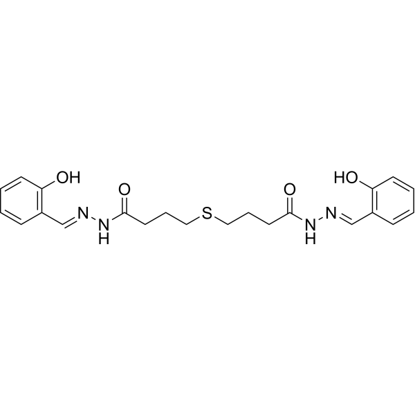
Nrf2-IN-3 (Compound R16) is a small-molecule NRF2 inhibitor and increases reactive oxygen species (ROS) production. Nrf2-IN-3 selectively binds KEAP1 mutants and restores their NRF2-inhibitory function by repairing the disrupted KEAP1/NRF2 interactions, leading to proteasome-dependent NRF2 degradation in cells. Nrf2-IN-3 sensitizes KEAP1-mutated tumor cells to Cisplatin (HY-17394), Gefitinib (HY-50895), and KEAP1 G333C-mutated xenograft to Cisplatin .
For research use only. We do not sell to patients.
- Purity : 98.10%
- CAS No.: 6325-13-9
- Formula: C22H26N4O4S
- Molecular Weight:442.53
-
Storage:Powder -20°C, 3 years ; In solvent -80°C, 6 months , -20°C, 1 month

To place orders, for customer services and technical support, please contact: MedChemExpress USA
Tel: 609-228-6898 E-mail: [email protected] [email protected]


-
 Biological Activity
-
Chemical Information
-
Solvent & Solubility
- Protocols
- Purity & Documentation
- References
-
 Help & FAQs


-
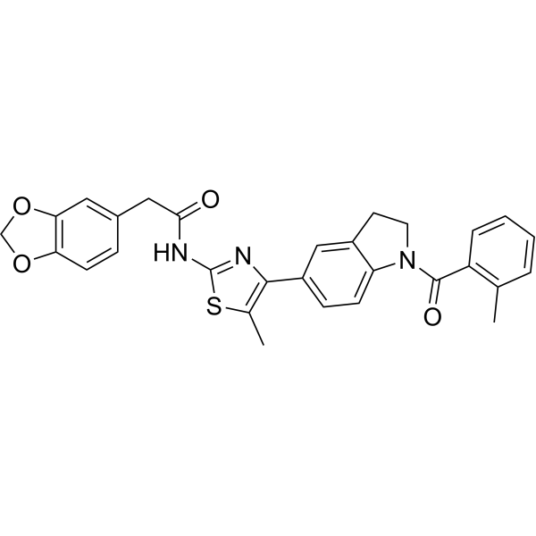
HY-100523
ML385Nrf2 Inhibitor -
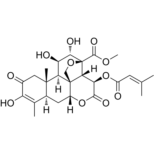
HY-19543
BrusatolNrf2 Inhibitor -
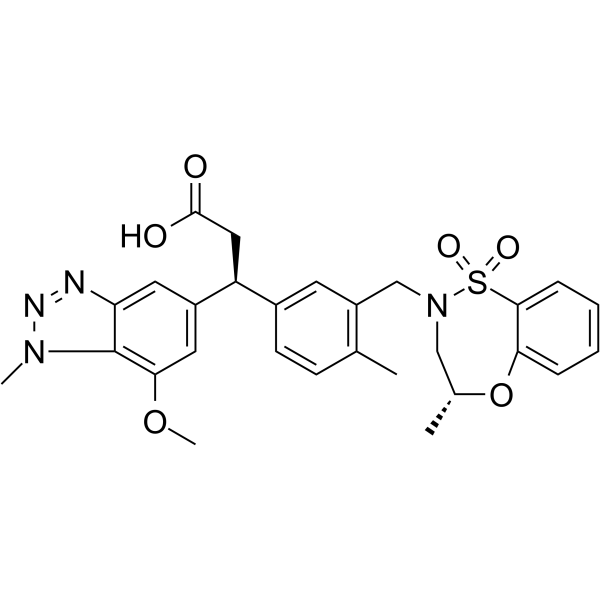
HY-101140
KI696KEAP1/NRF2 Protein-protein Interaction Inhibitor -
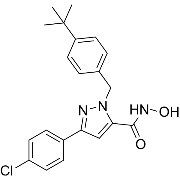
HY-101025
Nrf2-IN-1Nrf2 Inhibitor -
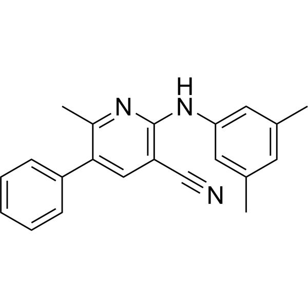
HY-153873
MSU38225Nrf2 Inhibitor -
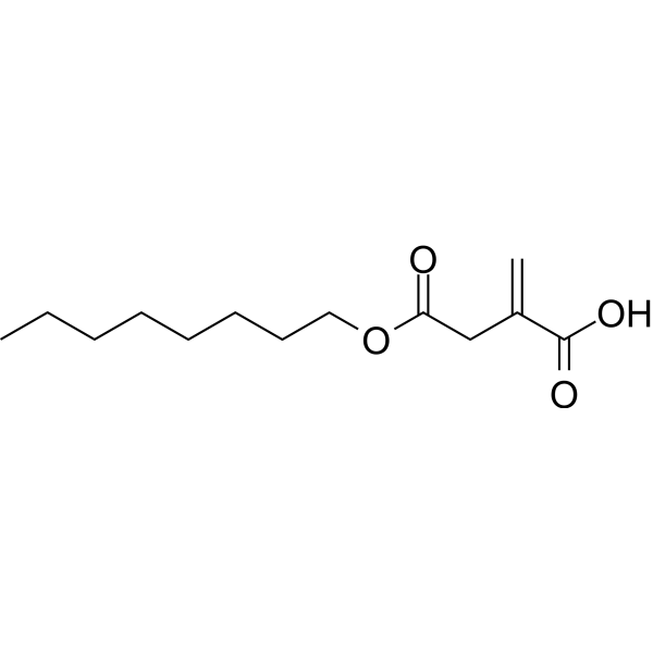
HY-112675
4-Octyl itaconateNrf2 Activator -
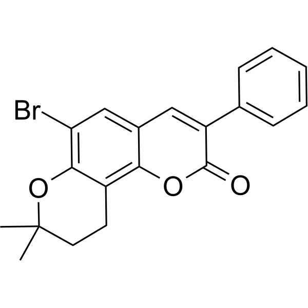
HY-145879
Nrf2 activator-2Nrf2 Activator -

HY-159641
VVD-130037NRF2 Glue Degrader -
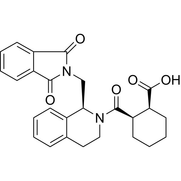
HY-110258
ML334NRF2 Activator -
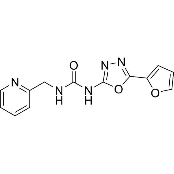
HY-19734
NK-252Nrf2 Activator -
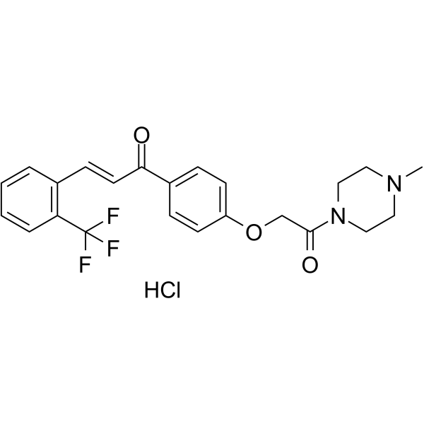
HY-146086
Nrf2 activator-4Nrf2 Activator -
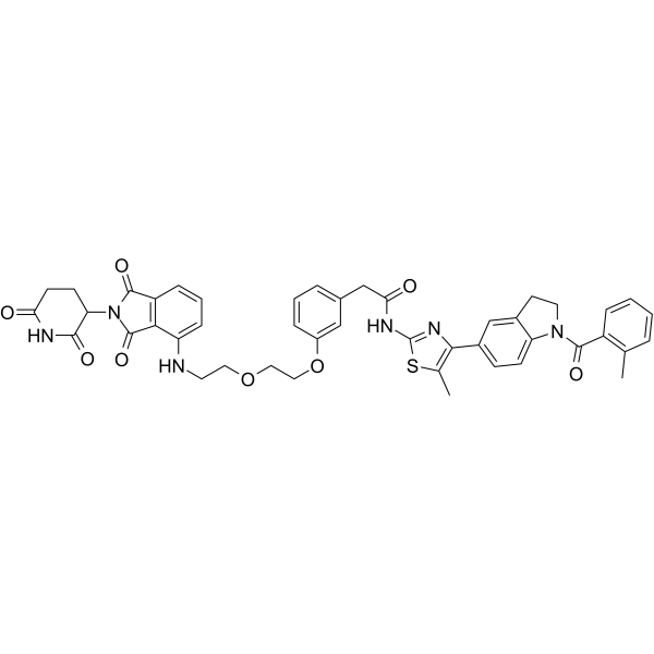
HY-162307
Nrf2 degrader 1Nrf2 PROTAC Degrader
Publications Citing Use of MedChemExpress (MCE) Nrf2-IN-3
More

Biological Activity
Description
In Vitro
Nrf2-IN-3 selectively sensitizes KEAP1-mutated cells, but not WT-KEAP1 cells, to Cisplatin (HY-17394) and Gefitinib (HY-50895) respectively [1].
Nrf2-IN-3 (24 h) selectively inhibits ARE-luc activity in KEAP1-mutated cells (A549, H1648, and H322 cells), with IC50s of 6.5 μM (A549), 7.3 μM (H1648), 5.7 μM (H322), respectively[1].
Nrf2-IN-3 (0-10 μM, 24 h) degrades NRF2 expression in A549, H1648, and H322 cells, which is mediated by the proteasome[1].
Nrf2-IN-3 binds KEAP1 mutants (G333C, G364C, and R460S mutants) and restores their capability to form mKEAP1/NRF2 complexes but has no effect on WT-KEAP1[1].
Nrf2-IN-3 (5, 10μM, 24 h) decreases NRF2 protein and increases reactive oxygen species (ROS) production in A549 (KEAP1 G333C mutation), but not A549 (WT-KEAP1) cells[1].
MedChemExpress (MCE) has not independently confirmed the accuracy of these methods. They are for reference only. Further protocols information, click here.
-
Cell Line:A549,H1648, H3229 cells
-
Concentration:0-10 μM
-
Incubation Time:72 h
-
Result:The IC50 (µM) of Cisplatin and Gefitinib in the presence and absence of Nrf2-IN-3 for 72 h.
A549 (G33C) H1648 (G364C) H322 (R460S) Cisplatin 0.81 1.33 4.6 Cisplatin + 0.1 µM Nrf2-IN-3 0.13 0.23 3.5 Cisplatin + 0.5 µM Nrf2-IN-3 0.07 0.17 0.9 Gefitinib 2.5 3.4 4.4 Gefitinib + 0.5 µM Nrf2-IN-3 0.36 0.67 0.92 Western Blot Analysis[1]-
Cell Line:A549,H1648, H3229 cells
-
Concentration:0,0.5, 1, 2, 5, 10 μM
-
Incubation Time:24 h
-
Result:Resulted in a decrease of NRF2 protein, and cells co-exposure to Epoxomicin (HY-13821) (1 μM) and Nrf2-IN-3 (5 μM) restored NRF2 levels.
Had no effect on NRF2 mRNA levels at 5 and 10 μM.
Reduced the mRNA levels of NQO1 and GCLM, two target genes of NRF2.
In Vivo
Nrf2-IN-3 (110 mg/kg i.p. daily for 5 days per week for six weeks) inhibits tumor growth and selectively sensitizes mKEAP1 A549ctl xenograft to Cisplatin (HY-17394)[1].MedChemExpress (MCE) has not independently confirmed the accuracy of these methods. They are for reference only.
-
Animal Model:Palpable tumors were generated by subcutaneous injection of a million A549ctl or A549(WT-KEAP1) cells in the flank of NOD-SCID mice[1].
-
Dosage:110 mg/kg for Nrf2-IN-3 and 2 mg/kg for Cisplatin.
-
Administration:Combination of Cisplatin (2 mg/kg) and Nrf2-IN-3 (110 mg/kg). The treatment started on day 18 after subcutaneous injection of A549ctl cells. Mice were treated i.p. with R16 at 110 mg/kg daily for 5 consecutive days per week, Cisplatin at 2 mg/kg twice per week, or the combination of both, or the vehicle
-
Result:Combination of Cisplatin and Nrf2-IN-3 strongly inhibited the growth of A549ctl xenograft, while Cisplatin or Nrf2-IN-3 alone has no effect[1].
Cisplatin alone or in combination with Nrf2-IN-3 significantly inhibited the growth of A549(WT-KEAP1) xenograft, but Nrf2-IN-3 did not significantly enhance the efficacy of cisplatin against this WT-KEAP1 tumor[1].
Cisplatin alone caused slight, but significant, weight loss, Nrf2-IN-3 did not enhance Cisplatin toxicity.
Nrf2-IN-3 alone or in combination with Cisplatin significantly decreased the levels of the NRF2 protein in the A549ctl tumors but not in the A549 (WT-KEAP1) tumors.


Chemical Information
-
CAS No. 6325-13-9
-
Appearance Solid
-
Molecular Weight 442.53
-
Formula C22H26N4O4S
-
Color White to off-white
-
SMILES
O=C(CCCSCCCC(N/N=C/C1=C(O)C=CC=C1)=O)N/N=C/C2=C(O)C=CC=C2
-
Shipping
Room temperature in continental US; may vary elsewhere.
-
Storage
Powder -20°C 3 years In solvent -80°C 6 months -20°C 1 month


Publications (1)
AllJournal Impact Factor-
Journal Impact Factor
-
Most Recent
-
Exp Mol Med
Machine learning-based integration of transcriptome and digital pathology for predicting chemoresistance in muscle-invasive bladder cancer. [Abstract]2026 May;58(5):1589-1607. PMID: 42104016


Solvent & Solubility
In Vitro:
DMSO : 116.67 mg/mL (263.64 mM; Need ultrasonic; Hygroscopic DMSO has a significant impact on the solubility of product, please use newly opened DMSO)
Please refer to the solubility information to select the appropriate solvent. Once prepared, please aliquot and store the solution to prevent product inactivation from repeated freeze-thaw cycles.
Storage method and period of stock solution: -80°C, 6 months; -20°C, 1 month. When stored at -80°C, please use it within 6 months. When stored at -20°C, please use it within 1 month.
View the Complete Stock Solution Preparation TablePreparing Stock SolutionsView the Complete Stock Solution Preparation TableConcentration Solvent Mass1 mg5 mg10 mgConcentrationSolventMass1 mM2.2597 mL11.2987 mL22.5973 mL5 mM0.4519 mL2.2597 mL4.5195 mL10 mM0.2260 mL1.1299 mL2.2597 mLPlease refer to the solubility information to select the appropriate solvent. Once prepared, please aliquot and store the solution to prevent product inactivation from repeated freeze-thaw cycles.
Storage method and period of stock solution: -80°C, 6 months; -20°C, 1 month. When stored at -80°C, please use it within 6 months. When stored at -20°C, please use it within 1 month.
Molarity CalculatorMass (g) = Concentration (mol/L) × Volume (L) × Molecular Weight (g/mol)= × ×Dilution CalculatorConcentration (start) × Volume (start) = Concentration (final) × Volume (final)
×=×
Protocols
All Cell Biology Animals and DiseasesMore-
ROS/oxidative-stress fluorescent staining
ROS/oxidative-stress fluorescent staining uses cell-permeant fluorogenic probes that become fluorescent after oxidation inside cells or tissues; commonly used examples include DCFH-DA/DCFDA for broad cellular oxidant detection, DHE for superoxide-related signal detection, MitoSOX for mitochondrial superoxide-related signal detection, and CellROX probes for oxidative-stress-associated fluorescence readouts. The assay detects probe oxidation rather than a single ROS species unless the probe and analysis method have been chemically validated for that species. DCFH-DA enters cells, is deacetylated by intracellular esterases to DCFH, and produces fluorescent DCF after oxidation, so the readout is used as an operational measure of total cellular oxidative stress rather than a species-specific ROS measurement. DHE and MitoSOX can report superoxide-related oxidation, but red fluorescence alone can include non-specific ethidium-like oxidation products; HPLC or optimized spectral approaches are
-
Patient-Derived Xenograft (PDX)
Patient-derived xenograft (PDX) models are generated by engrafting primary human tumor tissue directly into immunodeficient mice, allowing in vivo propagation of patient tumor biology without initial in vitro adaptation. These models are used to preserve key histopathological and molecular characteristics of the original tumor and enable assessment of tumor growth dynamics and therapeutic response in a living organism. The biological readout is tumor engraftment and subsequent growth in the murine host, which reflects the ability of human tumor cells to survive, vascularize, and expand in an immunocompromised microenvironment.
-
Splenic/Portal-Vein Liver Metastasis Xenograft
Splenic and portal-vein liver metastasis xenograft models deliver tumor cells into the portal circulation so that cells reach the liver first and form hepatic metastatic lesions; splenic injection uses the spleen as an access route to the portal system, while direct portal-vein injection introduces cells into the portal vein without requiring splenectomy. The assay detects liver colonization, intrahepatic tumor growth, tumor distribution, treatment response, survival, and liver-metastasis microenvironment changes; readouts include bioluminescence or fluorescence imaging, gross liver nodule counts, liver weight or tumor burden, histology, and survival.
-
Subcutaneous Cell-Line-Derived Xenograft
Subcutaneous cell-line-derived xenograft (CDX) models are established by implanting cultured human cancer cell lines into immunodeficient mice, where the injected cells form localized tumors that can be monitored in vivo as a measure of tumorigenic potential, growth kinetics, and treatment response. These models are widely used in oncology research because they allow reproducible tumor formation and enable comparative assessment of tumor growth between different cell lines or genetic manipulations in a controlled in vivo microenvironment. Subcutaneous implantation of cancer cells in immunodeficient mice is a standard approach for evaluating tumor growth behavior and therapeutic response across multiple cancer types, including prostate, esophageal, pancreatic, and colon cancer models.
-
Orthotopic Cell-Line Xenograft
Orthotopic cell-line xenograft models involve implantation of human cancer cell lines into the anatomically corresponding organ of immunodeficient mice to reproduce tumor growth within a native microenvironment, enabling more clinically relevant tumor behavior compared with subcutaneous models. These models are widely used because orthotopic placement better recapitulates tumor progression, including invasion and metastatic spread, which are often underrepresented in heterotopic implantation systems. Compared with conventional xenografts, orthotopic implantation is described as more technically complex but provides improved simulation of tumor-microenvironment interactions and metastatic behavior, making it particularly valuable for translational oncology research. Surgical orthotopic implantation approaches have been emphasized as enabling faithful reproduction of clinical cancer features, including metastasis and disease progression patterns that align with the tumor’s organ of origi
-
Intraperitoneal/Peritoneal Dissemination Xenograft
Intraperitoneal (IP) or peritoneal dissemination xenograft models are based on the introduction of human cancer cells into the peritoneal cavity of immunodeficient mice, where they attach to peritoneal surfaces, form multicellular aggregates or spheroids, and progressively generate disseminated tumor nodules that mimic advanced peritoneal metastatic disease. These models are widely used to study ovarian cancer progression, tumor-microenvironment interactions, and intraperitoneal therapeutic responses, often incorporating bioluminescence or fluorescence imaging to longitudinally monitor tumor burden in vivo. The biological principle relies on the capacity of tumor cells such as SKOV3 or related ovarian carcinoma lines to survive in suspension, aggregate within ascites-like fluid, adhere to mesothelial surfaces, and invade peritoneal organs, thereby recapitulating human peritoneal carcinomatosis patterns observed in advanced disease.


Purity & Documentation
Select Batch:Purity: 99.98%Assay: 99.98%ee.: 99.98%-
Data Sheet (275 KB)
-
SDS (252 KB)
- English - EN (252 KB)
- Français - FR (252 KB)
- Deutsch - DE (252 KB)
- Norwegian - NO (252 KB)
- Español - ES (252 KB)
- Swedish - SV (252 KB)
- Italian - IT (252 KB)
- Korean - KR (252 KB)
- Portuguese - PT (252 KB)
-
Handling Instructions (2659 KB)
References

Complete Stock Solution Preparation Table
Please refer to the solubility information to select the appropriate solvent. Once prepared, please aliquot and store the solution to prevent product inactivation from repeated freeze-thaw cycles.
Storage method and period of stock solution: -80°C, 6 months; -20°C, 1 month. When stored at -80°C, please use it within 6 months. When stored at -20°C, please use it within 1 month.
Optional Solvent Concentration Solvent Mass 1 mg 5 mg 10 mg 25 mg DMSO 1 mM 2.2597 mL 11.2987 mL 22.5973 mL 56.4933 mL 5 mM 0.4519 mL 2.2597 mL 4.5195 mL 11.2987 mL 10 mM 0.2260 mL 1.1299 mL 2.2597 mL 5.6493 mL 15 mM 0.1506 mL 0.7532 mL 1.5065 mL 3.7662 mL 20 mM 0.1130 mL 0.5649 mL 1.1299 mL 2.8247 mL 25 mM 0.0904 mL 0.4519 mL 0.9039 mL 2.2597 mL 30 mM 0.0753 mL 0.3766 mL 0.7532 mL 1.8831 mL 40 mM 0.0565 mL 0.2825 mL 0.5649 mL 1.4123 mL 50 mM 0.0452 mL 0.2260 mL 0.4519 mL 1.1299 mL 60 mM 0.0377 mL 0.1883 mL 0.3766 mL 0.9416 mL 80 mM 0.0282 mL 0.1412 mL 0.2825 mL 0.7062 mL 100 mM 0.0226 mL 0.1130 mL 0.2260 mL 0.5649 mL 

Nrf2-IN-3 Related Classifications
HY-149508 Related Classifications
Keywords
Assay Types -

 Powered by Bioz