



Pectinase, aspergillus niger

Based on 1 Customer Validation
Pectinase (EC 3.2.1.15) is a mixed enzymes that hydrolyze pectic substances, it mostly presents in microorganisms and higher plants. Pectinase is involved in the metabolism of the cell wall as well as in the growth of the cell, senescence, ripening of fruits, pathogenesis and abscission process.
For research use only. We do not sell to patients.
- CAS No.: 9032-75-1
-
Storage:
Please store the product under the recommended conditions in the Certificate of Analysis.


To place orders, for customer services and technical support, please contact: MedChemExpress USA
Tel: 609-228-6898 E-mail: [email protected] [email protected]


-
 Biological Activity
-
Chemical Information
-
Solvent & Solubility
- Protocols
- Purity & Documentation
- References
-
 Help & FAQs


-
HY-B2220
CellulaseEnzyme -
HY-B2220A
Cellulase R-10 from Trichoderma VrideHydrolase -

HY-126386B
Pectinase, rhizobiaEnzyme Mixture -
HY-P2875
HemicellulaseGlycoside Hydrolase -

HY-10182
LaduviglusibGSK-3 Inhibitor -
HY-100218A
RSL3GPX4 Inhibitor -
HY-P2902
Glucose oxidaseOxidoreductase Enzyme -
HY-F0001
NADH disodium saltReduced Coenzyme -
HY-111372
FinerenoneMineralocorticoid Receptor Antagonist -

HY-10583
Y-27632 dihydrochlorideROCK Inhibitor -
HY-12726
Liproxstatin-1Ferroptosis Inhibitor -
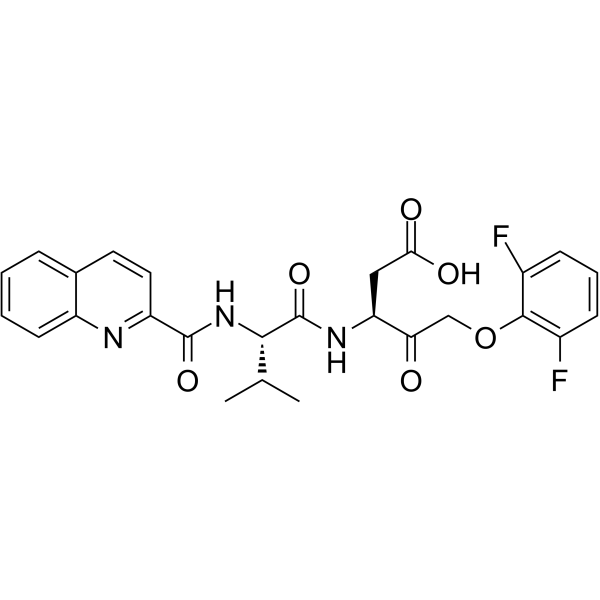
HY-12305
Q-VD-OPhPan-caspase Inhibitor


Biological Activity
Description
In Vitro
This product is derived fro M Aspergillus niger fermentation.
Temperature range: effective temperature range 25-65°C, optimal operating temperature 50-60°C
pH range: effective pH range 2.5-4.5, optimal pH 3.3-4.0.
Activators: Ca2+;Mg2+
Inhibitors: Cu2+;Fe3+
MedChemExpress (MCE) has not independently confirmed the accuracy of these methods. They are for reference only. Further protocols information, click here.
EC Number
3.2.1.15
Enzyme Activity
≥35 U/mg soild
Unit Definition
One unit is defined as the amount of enzyme that catalyzes the hydrolysis of pectin to produce 1 μg of galacturonic acid per hour using 1 g of enzyme powder or 1 mL of enzyme solution at 50°C and pH 3.5.


Chemical Information
-
CAS No. 9032-75-1
-
Appearance Solid
-
Color Light yellow to light brown
-
SMILES
[Pectinase, aspergillus niger]
-
Synonyms
EC 3.2.1.15
-
Shipping
Room temperature in continental US; may vary elsewhere.
-
Storage
Please store the product under the recommended conditions in the Certificate of Analysis.


Solvent & Solubility
In Vitro:
H2O : < 0.1 mg/mL (insoluble)

Protocols
-
Senescence-associated β-galactosidase staining
Senescence-associated β-galactosidase staining detects β-galactosidase activity that is histochemically visible at pH 6. 0 in senescent cells, where X-gal cleavage produces an insoluble blue precipitate observable by bright-field microscopy. This activity reflects increased lysosomal β-galactosidase/lysosomal mass rather than a senescence-essential enzyme, because GLB1 depletion or genetic lysosomal β-galactosidase deficiency can abolish SA-β-gal staining while cells still undergo senescence. SA-β-gal was originally reported in senescent but not presenescent fibroblasts and keratinocytes, absent from quiescent fibroblasts and terminally differentiated keratinocytes, and increased with donor age in human skin samples. Because SA-β-gal can also appear in some non-senescent or tissue-specific contexts, interpretation should be paired with experimental controls and, when possible, independent senescence markers.


Purity & Documentation
-
Data Sheet (264 KB)
-
SDS (418 KB)
- English - EN (418 KB)
- Français - FR (418 KB)
- Deutsch - DE (418 KB)
- Norwegian - NO (418 KB)
- Español - ES (418 KB)
- Swedish - SV (418 KB)
- Italian - IT (418 KB)
- Korean - KR (418 KB)
- Portuguese - PT (418 KB)
-
Handling Instructions (2659 KB)
References

Calculators
Concentration (start) × Volume (start) = Concentration (final) × Volume (final)


Pectinase, aspergillus niger Related Classifications
HY-126386 Related Classifications
 Powered by Bioz